

Optimization of improved motion-sensitized driven-equilibrium (iMSDE) blood suppression for carotid artery wall imaging
- PMID: 25160911
- PMCID: PMC4145260
- DOI: 10.1186/s12968-014-0061-5
Optimization of improved motion-sensitized driven-equilibrium (iMSDE) blood suppression for carotid artery wall imaging
Abstract
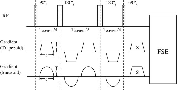
Background: Improved motion-sensitized driven-equilibrium (iMSDE) preparations have been successfully used in carotid artery wall imaging to achieve blood suppression, but it causes notable signal loss, mostly due to inherent T2 decay, eddy current effects and B1 + inhomogeneity. In this study, we investigate the signal to noise ratio (SNR) and blood suppression performance of iMSDE using composite RF pulses and sinusoidal gradients. Optimized first moment (m1) values for iMSDE prepared T1- and T2- weighted (T1- and T2-w) imaging are presented.
Methods: Twelve healthy volunteers and six patients with carotid artery disease underwent iMSDE and double inversion recovery (DIR) prepared T1- and T2-w fast spin echo (FSE) MRI of the carotid arteries. Modified iMSDE module using composite RF pulses and sinusoidal gradients were evaluated with a range of m1. SNR of adjacent muscle, vessel wall and the lumen were reported. The optimized iMSDE module was also tested in a 3D variable flip angle FSE (CUBE) acquisition.
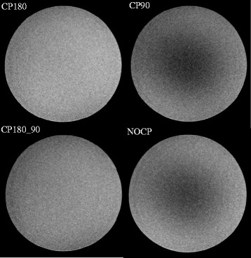
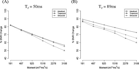
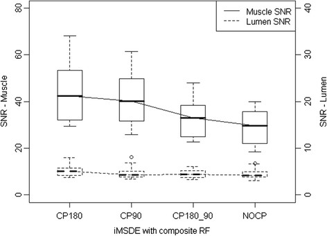
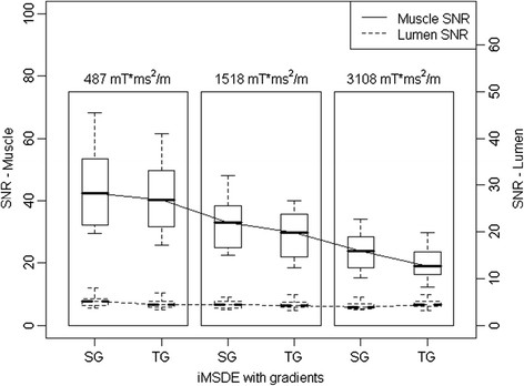
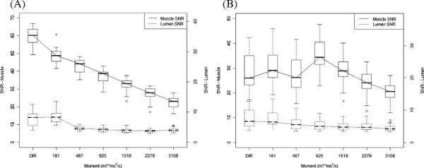
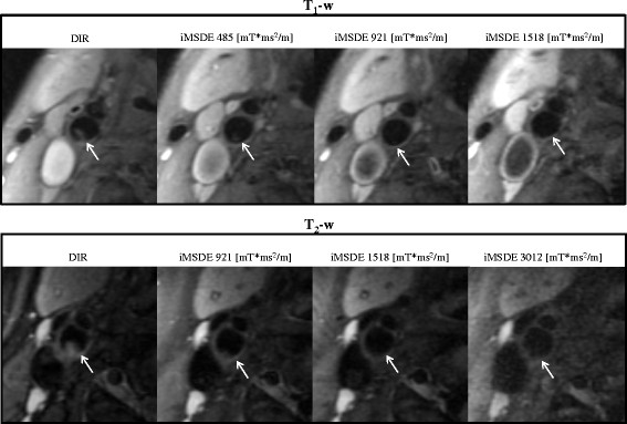
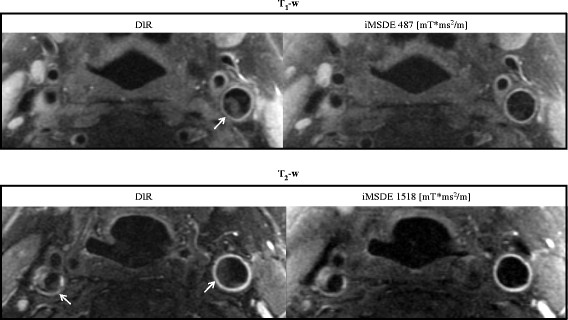
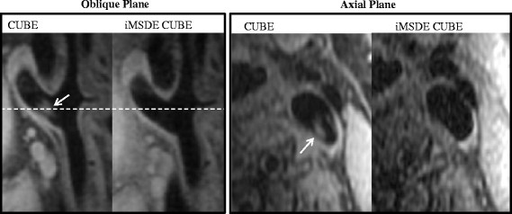
Results: The SNR of muscle was highest using sinusoidal gradients, and the relative improvement over the trapezoidal gradient increased with higher m1 (p<0.001). Optimal SNR was observed using an iMSDE preparation scheme containing two 180° composite pulses and standard 90° and -90° pulses (p=0.151). iMSDE produced better blood suppression relative to DIR preparations even with a small m1 of 487 mT*ms2/m (p<0.001). In T1-w iMSDE, there was a SNR decrease and an increased T2 weighting with increasing m1. In T2-w iMSDE, by matching the effective echo time (TE), the SNR was equivalent when m1 was <= 1518 mT*ms2/m, however, higher m1 values (2278 - 3108 mT*ms2/m) reduced the SNR. In the patient study, iMSDE improved blood suppression but reduced vessel wall CNR efficiency in both T1-w and T2-w imaging. iMSDE also effectively suppressed residual flow artifacts in the CUBE acquisition.
Conclusions: iMSDE preparation achieved better blood suppression than DIR preparation with reduced vessel wall CNR efficiency in T1-w and T2-w images. The optimized m1s are 487 mT*ms2/m for T1-w imaging and 1518 mT*ms2/m for T2-w imaging. Composite 180° refocusing pulses and sinusoidal gradients improve SNR performance. iMSDE further improves the inherent blood suppression of CUBE.
Figures
References
-
- Wang J, Yarnykh VL, Hatsukami T, Chu B, Balu N, Yuan C. Improved suppression of plaque-mimicking artifacts in black-blood carotid atherosclerosis imaging using a multislice motion-sensitized driven-equilibrium (MSDE) turbo spin-echo (TSE) sequence. Magn Reson Med. 2007;58:973–981. doi: 10.1002/mrm.21385. - DOI - PubMed
Publication types
MeSH terms
Grants and funding
LinkOut - more resources
Full Text Sources
Other Literature Sources
Medical
