. 2014 Aug 26;5(5):e01251-14.
doi: 10.1128/mBio.01251-14.

Human-specific bacterial pore-forming toxins induce programmed necrosis in erythrocytes

Affiliations

Human-specific bacterial pore-forming toxins induce programmed necrosis in erythrocytes

Timothy J LaRocca et al. mBio. .

Abstract

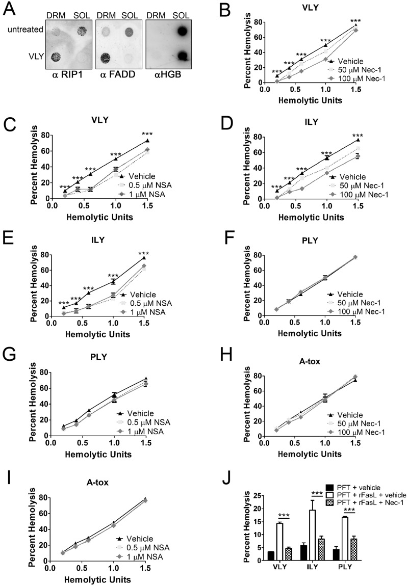
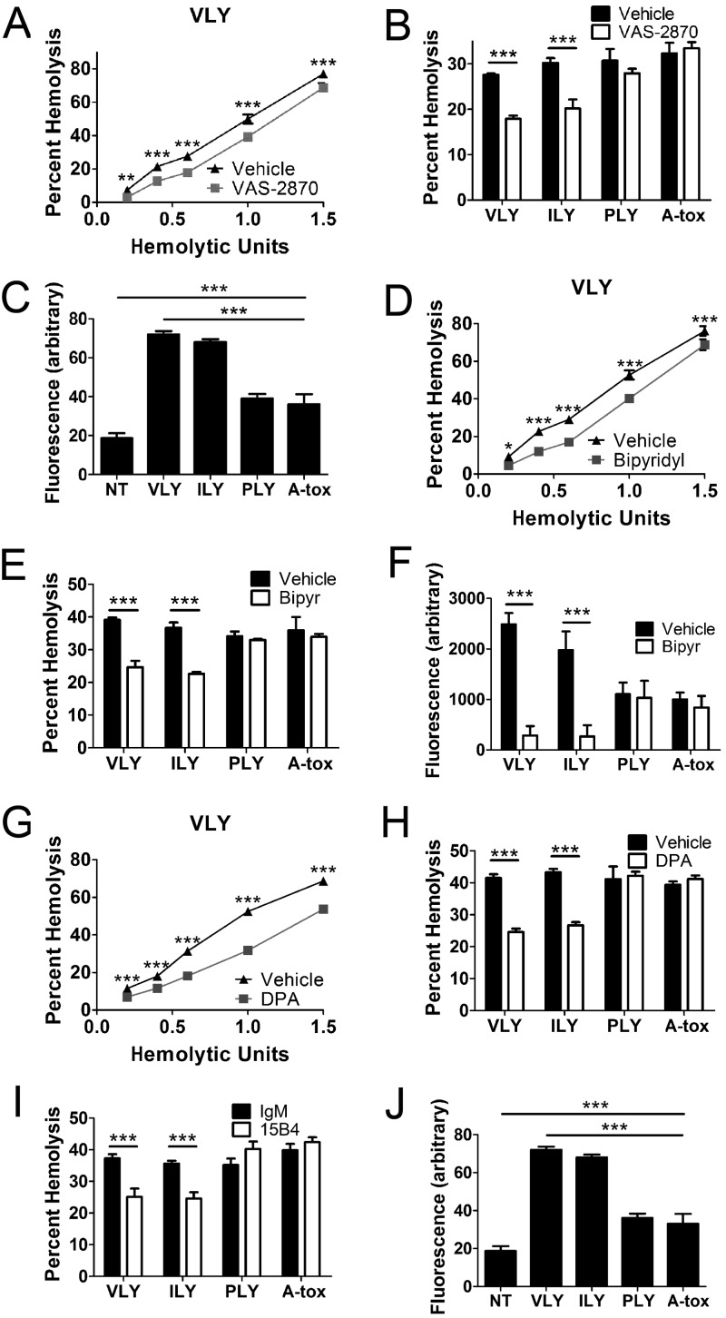
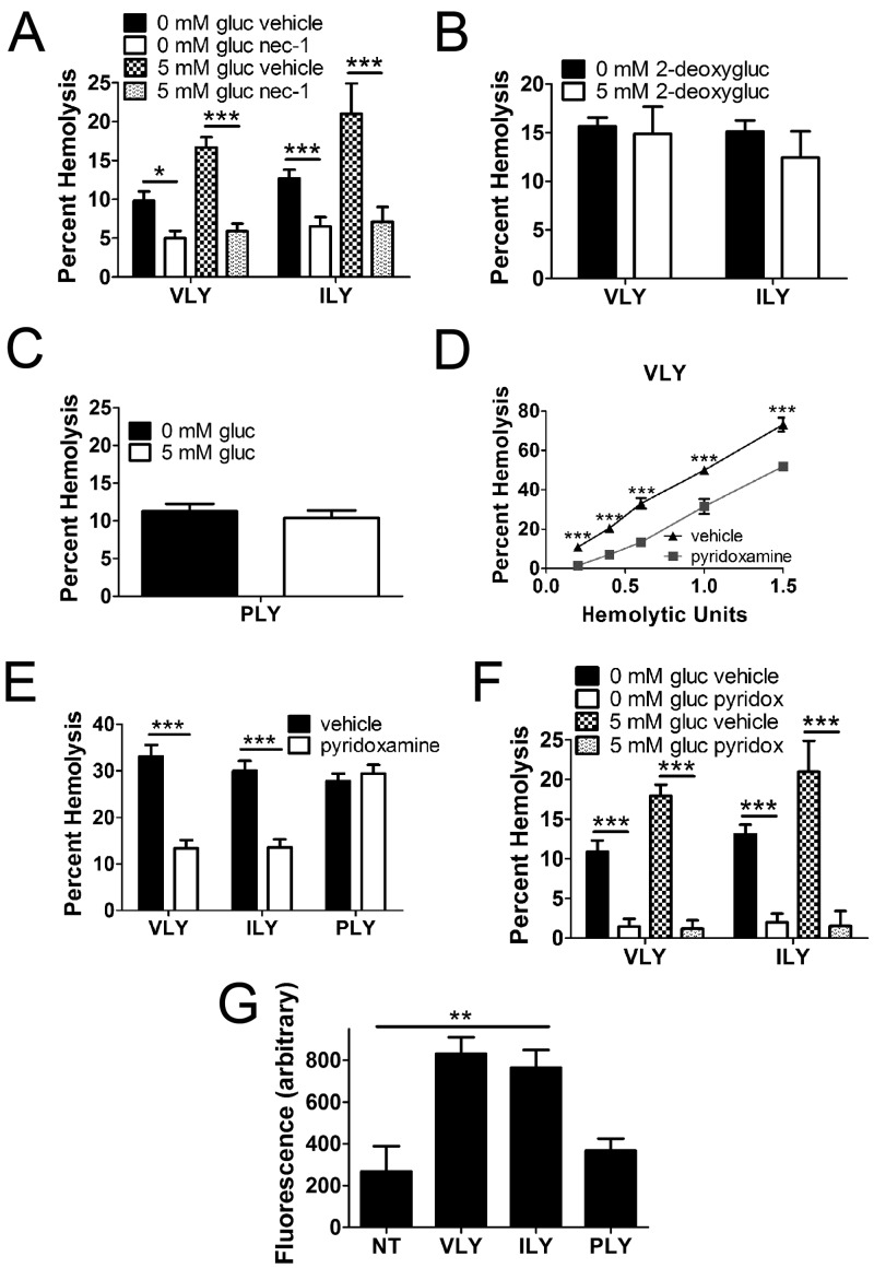
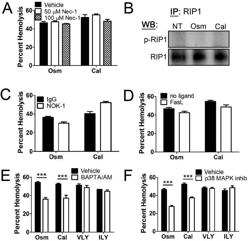
A subgroup of the cholesterol-dependent cytolysin (CDC) family of pore-forming toxins (PFTs) has an unusually narrow host range due to a requirement for binding to human CD59 (hCD59), a glycosylphosphatidylinositol (GPI)-linked complement regulatory molecule. hCD59-specific CDCs are produced by several organisms that inhabit human mucosal surfaces and can act as pathogens, including Gardnerella vaginalis and Streptococcus intermedius. The consequences and potential selective advantages of such PFT host limitation have remained unknown. Here, we demonstrate that, in addition to species restriction, PFT ligation of hCD59 triggers a previously unrecognized pathway for programmed necrosis in primary erythrocytes (red blood cells [RBCs]) from humans and transgenic mice expressing hCD59. Because they lack nuclei and mitochondria, RBCs have typically been thought to possess limited capacity to undergo programmed cell death. RBC programmed necrosis shares key molecular factors with nucleated cell necroptosis, including dependence on Fas/FasL signaling and RIP1 phosphorylation, necrosome assembly, and restriction by caspase-8. Death due to programmed necrosis in RBCs is executed by acid sphingomyelinase-dependent ceramide formation, NADPH oxidase- and iron-dependent reactive oxygen species formation, and glycolytic formation of advanced glycation end products. Bacterial PFTs that are hCD59 independent do not induce RBC programmed necrosis. RBC programmed necrosis is biochemically distinct from eryptosis, the only other known programmed cell death pathway in mature RBCs. Importantly, RBC programmed necrosis enhances the growth of PFT-producing pathogens during exposure to primary RBCs, consistent with a role for such signaling in microbial growth and pathogenesis.

Importance: In this work, we provide the first description of a new form of programmed cell death in erythrocytes (RBCs) that occurs as a consequence of cellular attack by human-specific bacterial toxins. By defining a new RBC death pathway that shares important components with necroptosis, a programmed necrosis module that occurs in nucleated cells, these findings expand our understanding of RBC biology and RBC-pathogen interactions. In addition, our work provides a link between cholesterol-dependent cytolysin (CDC) host restriction and promotion of bacterial growth in the presence of RBCs, which may provide a selective advantage to human-associated bacterial strains that elaborate such toxins and a potential explanation for the narrowing of host range observed in this toxin family.


Figures

FIG 1
FIG 1
VLY, an hCD59-specific PFT, induces lipid raft enlargement, including recruitment of Fas and FasL. (A) RBC detergent-resistant membranes (DRMs) increase in abundance in response to VLY as measured by DPH fluorescence. Cholesterol and proteins increase in DRMs after VLY treatment, with most proteins remaining soluble (SOL). (B) Dot blots showing that raft-associated proteins (CD59 and flotillin-1) and lipids (GM1 and GT1b) increase in DRMs in response to VLY. Hemoglobin (HGB) is excluded from DRMs. (C) Fas and FasL, normally excluded from rafts, are recruited to DRMs in response to VLY. ***, P < 0.001 (Student’s t test).
FIG 2
FIG 2
VLY and ILY, hCD59-specific PFTs, induce Fas/FasL-dependent death in human and hCD59-transgenic murine RBCs. (A) Neutralization of FasL with a MAb (NOK-1, 1 µg/ml) inhibits RBC death by VLY. (B) NOK-1 inhibits RBC death by VLY or ILY but not by hCD59-independent PLY or A-tox. (C and D) Exogenous rFasL (0.1 µg/ml) (C) or agonistic Fas MAb (2R2, 1 µg/ml) (D) enhances RBC death by VLY. (E and F) rFasL (E) or 2R2 (F) enhances RBC death caused by all PFTs tested. (G) C57BL/6J (nontransgenic) RBCs are resistant to VLY and ILY but sensitive to PLY. (H) Death of hCD59-transgenic murine RBCs by VLY or ILY is inhibited by NOK-1. IgG, irrelevant IgG. ***, P < 0.001; **, P < 0.01 (two-way analysis of variance, Bonferroni posttest).
FIG 3
FIG 3
VLY and ILY cause RIP1- and MLKL-dependent programmed necrosis in mature RBCs. (A) Dot blots showing recruitment of RIP1 and FADD to DRMs in response to VLY. Hemoglobin (HGB) remains in the soluble fraction (SOL). (B) Inhibition of RIP1 with nec-1 reduces RBC death by VLY relative to vehicle control. (C) Inhibition of MLKL with NSA reduces RBC death by VLY relative to vehicle control. (D to I) Inhibition of RIP1 (D) or MLKL (E) reduces RBC death by ILY and has no effect on death by PLY (F and G) or A-tox (H and I). (J) Enhanced RBC death caused by the combination of PFTs and rFasL is inhibited by nec-1 relative to vehicle control. ***, P < 0.001 (two-way analysis of variance, Bonferroni posttest).
FIG 4
FIG 4
VLY and ILY induce FasL-dependent phosphorylation of RIP1 and necrosome assembly in RBCs. (A) Coomassie blue-stained SDS-PAGE gel showing RIP1 IP. H, IgG heavy chain; L, IgG light chain. (B to F) Immunoblots from RIP1 IPs showing RIP1 phosphorylation (p-RIP1) in response to VLY or ILY (B), prevention of p-RIP1 with nec-1 (C), prevention of p-RIP1 with NOK-1 (D), p-RIP1 induced by rFasL (E), and coprecipitation of RIP3 and FADD with RIP1 in response to VLY or ILY that is prevented by RIP1 inhibition (F). NT, no treatment.
FIG 5
FIG 5
Caspase-8 is a necrosome component that antagonizes RBC programmed necrosis. (A) Coomassie blue-stained SDS-PAGE gel showing caspase-8 IP. H, IgG heavy chain; L, IgG light chain. (B) Immunoblots from caspase-8 IPs showing coprecipitation of RIP1 under all conditions and coprecipitation of FADD in response to VLY or ILY. RIP1 does not coprecipitate with caspase-8 in THP-1 cells. (C to E) Pan-caspase inhibition with 10 µM z-VAD-fmk enhanced RBC death by VLY or ILY (C and D, respectively) and was prevented by nec-1 (E). (F) Specific inhibition of caspase-8 with 10 µM z-IETD-fmk enhanced RBC death caused by VLY or ILY. Caspase-3 inhibition (10 µM z-DEVD-fmk) had no effect. (G) Levels of p-RIP1 increase in RBCs following inhibition of caspase-8 (z-IETD) relative to a caspase inhibitor negative control (z-FA). ***, P < 0.001 (two-way analysis of variance, Bonferroni posttest). z-FA-fmk, caspase inhibitor negative control.
FIG 6
FIG 6
RBC programmed necrosis is executed in part by aSMase-dependent ceramide formation and NOX/iron-dependent ROS. (A and B) NOX inhibition with 10 µM VAS-2870 reduces RBC death by VLY and ILY compared to vehicle control. (C) VLY and ILY induce ROS in RBCs as measured by DCFDA. (D and E) Iron chelation with 100 µM 2,2-bipyridyl reduces RBC death by VLY or ILY relative to vehicle control. (F) Iron chelation prevents ROS induction by VLY and ILY. (G and H) Inhibition of aSMase with 20 µM desipramine (DPA) reduced RBC death by VLY and ILY relative to vehicle control. (I) Ceramide neutralization with MAb 15B4 reduced death by VLY or ILY relative to irrelevant IgM (IgM). (J) VLY and ILY induce surface ceramide as determined by immunofluorescence. ***, P < 0.001 (two-way analysis of variance, Bonferroni posttest).
FIG 7
FIG 7
Glycolytic formation of AGEs is a component in the execution of RBC programmed necrosis. (A) Glucose (gluc) uptake by RBCs in a 5 mM solution enhanced death by VLY or ILY and was prevented by nec-1. (B) Uptake of the nonmetabolizable 2-deoxy-d-glucose (2-deoxygluc) had no effect on RBC death. (C) Glucose uptake had no effect on RBC death by the hCD59-independent PLY. (D and E) AGE inhibition with 1 mM pyridoxamine reduced death by VLY or ILY relative to vehicle control. (F) RBC death enhanced by glucose uptake is prevented by AGE inhibition. (G) VLY and ILY induce RBC AGE formation as determined by whole-cell immunofluorescence assay. ***, P < 0.001; **, P < 0.01; *, P < 0.05 (two-way analysis of variance, Bonferroni posttest).
FIG 8
FIG 8
RBC programmed necrosis differs from eryptosis. (A) Eryptosis by hyperosmotic stress (Osm) or excess calcium (Cal) is not inhibited by nec-1. (B) RIP1 IPs showing that eryptosis does not induce p-RIP1. (C and D) Eryptosis is unaffected by NOK-1 (C) or rFasL (D). (E and F) Eryptosis depends on intracellular Ca2+ as shown by chelation with 10 µM BAPTA/AM [1,2-bis(o-aminophenoxy)ethane-N,N,N′,N′-tetraacetic acid–acetoxymethyl ester] (E) and p38 mitogen-activated protein kinase (MAPK) as shown by inhibition with 10 µM p38 MAPK inhibitor III (Calbiochem) (F) while RBC programmed necrosis by VLY or ILY does not. ***, P < 0.001 (two-way analysis of variance, Bonferroni posttest).
FIG 9
FIG 9
RBC programmed necrosis enhances growth of PFT-producing pathogenic bacteria in vitro. (A and B) RIP1 IPs showing p-RIP1 in response to live G. vaginalis (GV) or S. intermedius (SI). A neutralizing PAb against CDCs (αCDC) prevents p-RIP1 formation relative to control prebleed (PB) serum. (C and D) Enhanced growth of 2 starting inocula (OD600 of 0.1 and OD600 of 0.01) of G. vaginalis (C) or S. intermedius (D) in the presence of human RBCs is partially inhibited by FasL neutralization with NOK-1 relative to irrelevant IgG. (E) Growth of S. pneumoniae, which produces the hCD59-independent PLY, is not affected by FasL neutralization. (F to H) Addition of rFasL to cultures including human RBCs and G. vaginalis (F), S. intermedius (G), or S. pneumoniae (H), inoculated at an OD600 of 0.1, enhanced the growth of all bacteria tested. ***, P < 0.001; **, P < 0.01 (two-way analysis of variance, Bonferroni posttest).

References

    1. Hod EA, Arinsburg SA, Francis RO, Hendrickson JE, Zimring JC, Spitalnik SL. 2010. Use of mouse models to study the mechanisms and consequences of RBC clearance. Vox Sang. 99:99–111. 10.1111/j.1423-0410.2010.01327.x - DOI - PMC - PubMed
    1. Alaarg A, Schiffelers RM, van Solinge WW, van Wijk R. 2013. Red blood cell vesiculation in hereditary hemolytic anemia. Front. Physiol. 4:365. 10.3389/fphys.2013.00365 - DOI - PMC - PubMed
    1. Ucar K. 2002. Clinical presentation and management of hemolytic anemias. Oncology (Williston Park) 16(9 Suppl 10):163–170 - PubMed
    1. Lang E, Qadri SM, Lang F. 2012. Killing me softly—suicidal erythrocyte death. Int. J. Biochem. Cell Biol. 44:1236–1243. 10.1016/j.biocel.2012.04.019 - DOI - PubMed
    1. Los FC, Randis TM, Aroian RV, Ratner AJ. 2013. Role of pore-forming toxins in bacterial infectious diseases. Microbiol. Mol. Biol. Rev. 77:173–207. 10.1128/MMBR.00052-12 - DOI - PMC - PubMed

MeSH terms

LinkOut - more resources