. 2014 Sep 16;111(37):13319-24.
doi: 10.1073/pnas.1321940111. Epub 2014 Aug 26.

Viral nanoparticle-encapsidated enzyme and restructured DNA for cell delivery and gene expression

Affiliations

Viral nanoparticle-encapsidated enzyme and restructured DNA for cell delivery and gene expression

Jinny L Liu et al. Proc Natl Acad Sci U S A. .

Abstract

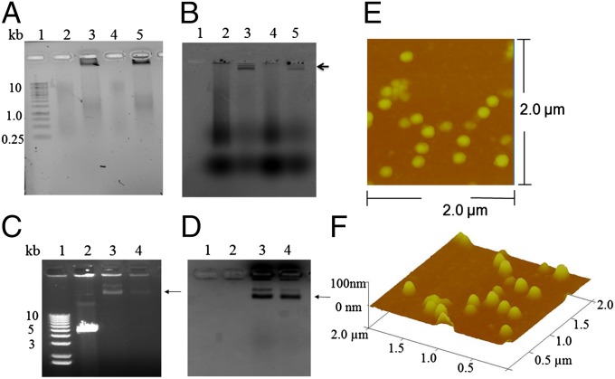
Packaging specific exogenous active proteins and DNAs together within a single viral-nanocontainer is challenging. The bacteriophage T4 capsid (100 × 70 nm) is well suited for this purpose, because it can hold a single long DNA or multiple short pieces of DNA up to 170 kb packed together with more than 1,000 protein molecules. Any linear DNA can be packaged in vitro into purified procapsids. The capsid-targeting sequence (CTS) directs virtually any protein into the procapsid. Procapsids are assembled with specific CTS-directed exogenous proteins that are encapsidated before the DNA. The capsid also can display on its surface high-affinity eukaryotic cell-binding peptides or proteins that are in fusion with small outer capsid and head outer capsid surface-decoration proteins that can be added in vivo or in vitro. In this study, we demonstrate that the site-specific recombinase cyclic recombination (Cre) targeted into the procapsid is enzymatically active within the procapsid and recircularizes linear plasmid DNA containing two terminal loxP recognition sites when packaged in vitro. mCherry expression driven by a cytomegalovirus promoter in the capsid containing Cre-circularized DNA is enhanced over linear DNA, as shown in recipient eukaryotic cells. The efficient and specific packaging into capsids and the unpackaging of both DNA and protein with release of the enzymatically altered protein-DNA complexes from the nanoparticles into cells have potential in numerous downstream drug and gene therapeutic applications.

Keywords: DNA packaging; Hoc; Soc; capsid decoration proteins; terminase.


Conflict of interest statement

The authors declare no conflict of interest.

Figures

Fig. 1.
Fig. 1.
The T4 capsid-derived specific exogenous DNA plus protein packaging and eukaryotic cell delivery scheme. (A) DNA encoding a 10-amino acid N-terminal CTS peptide fused to the phage P1 Cre allows synthesis of CTS-Cre and targeting of the enzyme into the early core-scaffold of the T4 procapsid in vivo. Procapsid assembly and maturation-specific viral protease stabilize the procapsid, remove most of the scaffolding core as peptides, and remove the CTS peptide from Cre. Mutations in the viral terminase block DNA packaging and allow a mature but DNA-empty large Cre-containing procapsid to be highly purified from viral-infected bacteria. (B) In vitro packaging into the mature capsid of plasmid DNA containing mCherry driven by a CMV promoter and two loxP sites flanking an SfiI restriction enzyme site that allow the linearization required for packaging. The DNA is packaged into the procapsid by the ATP-driven terminase motor protein (gp17) with high efficiency. (C) The packaged Cre enzyme recircularizes the packaged linear plasmid DNA between the two loxP sites. The DNA-containing capsid is taken up by eukaryotic cells, here without displaying a specific peptide target, or into eukaryotic cells specifically using Soc and Hoc displayed peptides that have high affinity for the RP1 and RP2 receptors, respectively.
Fig. 2.
Fig. 2.
Packaging of fluorescent dsDNAs into T4 procapsids and assessment of the integrity of T4 NPs. (A) Ethidium bromide staining viewed on a biospectrum imaging system (UVP). The gel was run for only a short time and adjusted to a bright background with less contrast to visualize the low molecular weight smear DNA. (B) The unstained gel viewed on a Typhoon imager using an Alexa 488 dye filter. The L-segment (3,320 bp) of ф6 dsRNA was reverse transcribed using aha-dCTP followed by conjugation with Alexa 488 NHS ester (Amersham), as shown in lane 2, and the dye-labeled L-segment dsDNA was packaged into control T4 procapsids lacking the Cre enzyme, as shown in lane 3 (arrow near the loading well). The S-segment (1,520 bp) of ф6 dsRNA (lane 4) was reverse transcribed by the same procedure. The dye-labeled S-segment dsDNA is shown in lane 5. Lane 1 shows the 1-kb DNA size marker (Thermo Scientific). The arrow indicates the position of packaged DNA retained by procapsids near the top of the agarose gel. (C and D) Images of the same agarose gel viewed on a Gel Doc EZ imaging system (Bio-Rad). (C) Ethidium bromide staining image with more contrast (dark background). The agarose gel was run for a longer time to allow the packaged DNA to migrate further for visualizing the packaged DNA on the top (indicated by the arrow) due to slow migration of the large procapsids. The doublet bands possibly result from two different charges of the procapsids. The unpackaged linear EGFP DNA with a size of ∼5.5 kb is shown in lane 2, and packaged EGFP DNA with a size >10 kb is shown in lane 3. Packaged Alexa 480-labeled L-segment dsDNA with a size >10 kb is shown in lane 4. (D) Coomassie blue staining image of C. T4 capsids in lanes 3 and 4 were stained with Coomassie blue as indicated by the arrow. (E and F) Purified T4 proheads for DNA packaging were visualized by AFM with a scanning area of 2.0 × 2.0 µm. A 3D view is shown in F.
Fig. 3.
Fig. 3.
Assessment of the cellular uptake of T4 procapsids containing packaged Alexa 488-labeled L-segment– and S-segment–derived ф6 dsDNA. (A and B) Histograms of the flow cytometry with varied concentrations of T4 procapsid-packaged Alexa 488-labeled L-segment (LL) and S-segment (SL) dsDNA, respectively. (C) The internalized Alexa 488-labeled M-segment (ML) dsDNA (∼2,000 bp, indicated by small arrows) within A549 cells was visualized by epifluorescent microscopy. (D) DAPI staining for nuclei (white arrow). (E) Overlay of C and D. (F) Bright-field image showing whole cells with nuclei indicated by the black arrow. (Scale bars, 10 µm.)
Fig. 4.
Fig. 4.
The recircularization of packaged linear mCherryC1–loxP plasmid DNA within procapsids containing Cre recombinase. (A) An immunoblot using anti-Cre antibody (Novagen) shows that the CTS–Cre recombinase is packaged into Cre+ T4 procapsids (lanes 2 and 3) but is not detected in a comparable amount of Cre T4 procapsids (lane 1). As shown in lane 3, DNase treatment did not affect the Cre+ content internalized within the procapsids. Lane 4 is a molecular size marker, and lanes 5–7 show increasing amounts of purified Cre recombinase (Novagen) as quantification standards and positive controls. (B) The mCherryC1–loxP plasmid was obtained by inserting two loxP sites linked by an SfiI restriction enzyme site (as indicated in the sequence box) into a commercial mCherry-C1 plasmid (Clontech). (C) Phenol-extracted packaged mCherry–loxP DNA within Cre and Cre+ procapsids. Lane 2 shows unpackaged SfiI-linearized pmCherry–loxP DNA. Lane 3 shows the slower-moving circular form of packaged pmCherryC1–loxP DNA isolated from Cre+ procapsids. The circular mCherry–loxP DNA (arrow) migrated more slowly than the packaged linear pmCherryC1–loxP DNA isolated from Cre procapsids in lane 4. (D) Packaged SfiI-linearized plasmid DNA from Cre and Cre+ T4 procapsids. Purified pmCherryC1–loxP DNA and phenol-extracted packaged DNA isolated from Cre and Cre+ T4 procapsids were assessed for E. coli transformation and compared by agarose electrophoresis. Lanes 2 and 3 show unpackaged linear pmCherryC1–loxP DNA cut by SfiI moving faster than the uncut circular pmCherryC1–loxP. Packaged linear pmCherryC1–loxP DNA remained linear in Cre T4 procapsids (lane 4), whereas the packaged linear pmCherryC1 DNA was circularized in Cre+ T4 procapsids (arrows in lane 3 in C and lane 5 in D). Lane 1 is the 1-kb+ DNA size marker (Life Technologies, Inc.). (E) An ∼40-fold increase in transformation efficiency was exhibited by the recircularized pmCherryC1–loxP DNA isolated from Cre+ T4 procapsids relative to Cre T4-packaged DNA (D, lane 4 vs. lane 5). Error bars represents SE; n = 2; *P < 0.05, t test.
Fig. 5.
Fig. 5.
Flow cytometry measurement of fluorescent protein expression in A549 cells from T4-packaged plasmid mCherryC1–loxP DNA. (A) A fluorescent control shows that treatment of the Alexa 546-labeled T4 NPs (6), which were obtained by conjugating the dyes to the capsid proteins, produced a concentration-dependent shift in the fluorescent population. The rightward shift of the curves (red and blue traces) indicates a higher cell fluorescent intensity with a larger portion of fluorescent cells. SFM without the addition of any T4 NPs (black trace) served as a negative control. (B and C) A larger fluorescent cell population was observed in A549 cells treated with the 4.0E+9 Cre+ T4 procapsids (red trace in B) and 8.0E+9 T4 procapsids containing circular pmCherryC1–loxP (red trace in C) relative to SFM (black traces in B and C), indicating the expression of fluorescent mCherry protein in A549 cells from the packaged circular pmCherryC1–loxP DNA. (D) A549 cells treated with the linear DNA packaged in Cre T4 NPs (red and blue traces) showed no difference in cell fluorescent intensity and population (i.e., no right shift) relative to cells treated with SFM (black trace).
Fig. 6.
Fig. 6.
Assessment of viability of A549 cells treated with Cre procapsids and of the cytotoxicity induced by the treatment of Cre T4 NPs. (A) The cell uptake of unlabeled Cre or Alexa 647-labeled Cre (A647-T4) procapsids showed no cytotoxicity for A549 cells, which appeared 100% viable. (BD) There is no apparent cytotoxicity in A549 cells treated with Cre T4 procapsids (B) relative to the untreated cells (C), whereas most of the cells appeared dead (i.e., as rounded opaque spots) after treatment with Cre+ T4 procapsids (D). All bright-field images of A549 cells were obtained using a 20× objective. (Scale bars, 50 µm.)

References

    1. Manchester M, Singh P. Virus-based nanoparticles (VNPs): Platform technologies for diagnostic imaging. Adv Drug Deliv Rev. 2006;58(14):1505–1522. - PubMed
    1. Robertson KL, Liu JL. Engineered viral nanoparticles for flow cytometry and fluorescence microscopy applications. Wiley Interdiscip Rev Nanomed Nanobiotechnol. 2012;4(5):511–524. - PubMed
    1. Manchester M, Steinmetz NF. Viruses and nanotechnology. Preface. Curr Top Microbiol Immunol. 2009;327:v–vi. - PubMed
    1. Wu Z, et al. Development of viral nanoparticles for efficient intracellular delivery. Nanoscale. 2012;4(11):3567–3576. - PMC - PubMed
    1. Ashley CE, et al. Cell-specific delivery of diverse cargos by bacteriophage MS2 virus-like particles. ACS Nano. 2011;5(7):5729–5745. - PMC - PubMed

Publication types

MeSH terms