

Successful venous angioplasty of superior vena cava syndrome after heart transplantation
- PMID: 25161772
- PMCID: PMC4137548
- DOI: 10.1155/2014/490276
Successful venous angioplasty of superior vena cava syndrome after heart transplantation
Abstract
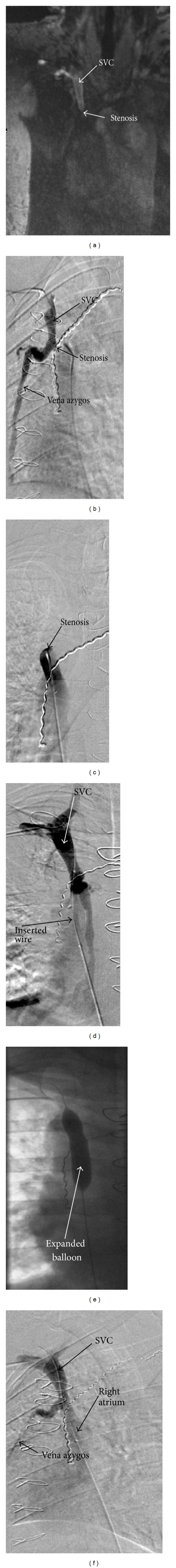
Introduction. For patients with terminal heart failure, heart transplantation (HTX) has become an established therapy. Before transplantation there are many repeated measurements with a pulmonary artery catheter (PAC) via the superior vena cava (SVC) necessary. After transplantation, endomyocardial biopsy (EMB) is recommended for routine surveillance of heart transplant rejection again through the SVC. Case Presentation. In this report, we present a HTX patient who developed a SVC syndrome as a possible complication of all these procedures via the SVC. This 35-year-old Caucasian male could be successfully treated by balloon dilatation/angioplasty. Conclusion. The SVC syndrome can lead to pressure increase in the venous system such as edema in the head and the upper part of the body and further serious complications like cerebral bleeding and ischemia, or respiratory problems. Balloon angioplasty and stent implantation are valid methods to treat stenoses of the SVC successfully.
Figures
Similar articles
-
[Superior vena cava thrombosis or stricture secondary to implanted central venous access: Six cases of endovascular and direct surgical treatment in cancer patients].J Med Vasc. 2018 Feb;43(1):20-28. doi: 10.1016/j.jdmv.2017.11.001. Epub 2017 Dec 26. J Med Vasc. 2018. PMID: 29425537 French.
-
Transcatheter stenting of superior vena cava to treat postoperative SVC syndrome in a child: a case report.Egypt Heart J. 2024 Aug 29;76(1):115. doi: 10.1186/s43044-024-00547-6. Egypt Heart J. 2024. PMID: 39210242 Free PMC article.
-
Sinus arrest following angioplasty and stenting for superior vena cava syndrome.J Invasive Cardiol. 2014 Feb;26(2):E21-3. J Invasive Cardiol. 2014. PMID: 24486673
-
Bleeding 'downhill' esophageal varices associated with benign superior vena cava obstruction: case report and literature review.BMC Gastroenterol. 2016 Oct 24;16(1):134. doi: 10.1186/s12876-016-0548-7. BMC Gastroenterol. 2016. PMID: 27776486 Free PMC article. Review.
-
Endovascular stenting for end-stage lung cancer patients with superior vena cava syndrome post first-line treatments - A single-center experience and literature review.J Chin Med Assoc. 2017 Aug;80(8):482-486. doi: 10.1016/j.jcma.2017.04.005. Epub 2017 May 10. J Chin Med Assoc. 2017. PMID: 28501315 Review.
References
-
- Boyle A. Current status of cardiac transplantation and mechanical circulatory support. Current Heart Failure Reports. 2009;6(1):28–33. - PubMed
-
- Gidwani UK, Mohanty B, Chatterjee K. The pulmonary artery catheter: a critical reappraisal. Cardiology Clinics. 2013;31(4):545–565. - PubMed
-
- Kogon BE, Plattner C, Jennings S, Lyle T, McConnell M, Book WM. Cyanosis produced by superior vena caval stenosis. Annals of Thoracic Surgery. 2008;85(3):1083–1085. - PubMed
LinkOut - more resources
Full Text Sources
Other Literature Sources
