

A peripheral ameloblastic fibro-odontoma in a 3-year-old girl: case report, immunohistochemical analysis, and literature review
- PMID: 25161776
- PMCID: PMC4100273
- DOI: 10.1155/2014/321671
A peripheral ameloblastic fibro-odontoma in a 3-year-old girl: case report, immunohistochemical analysis, and literature review
Abstract
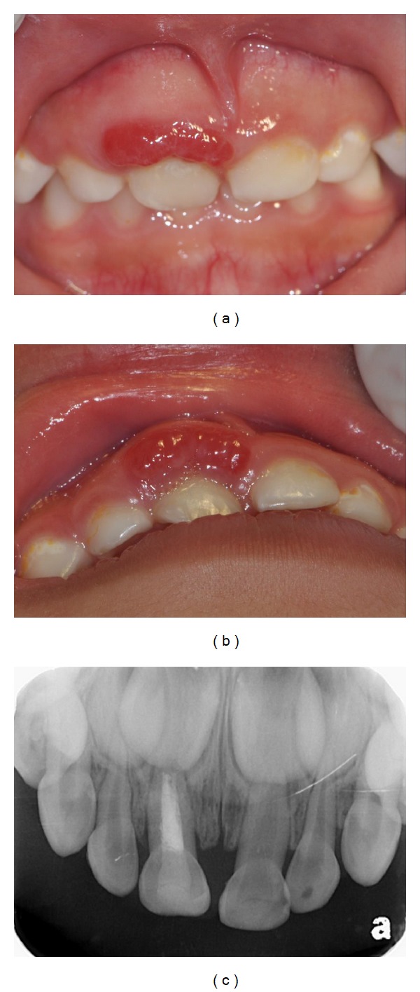
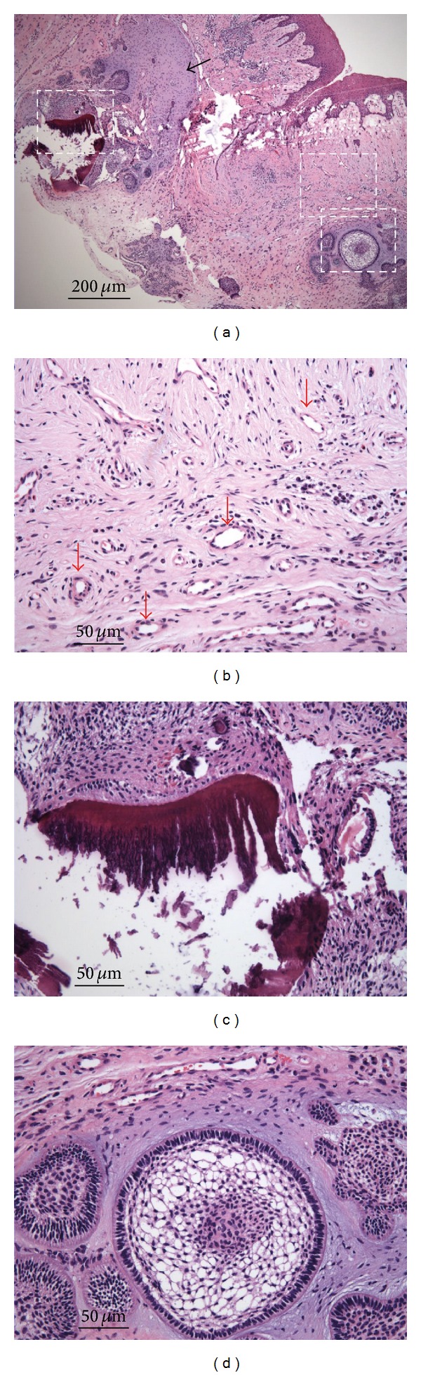
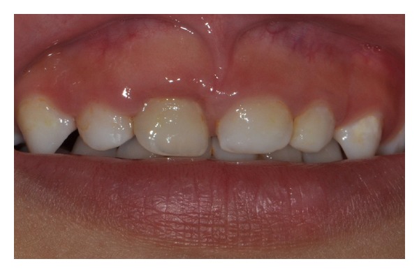
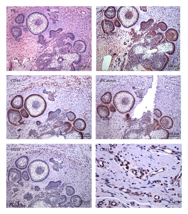
Ameloblastic fibro-odontoma (AFO) predominantly occurs in the jaw bones of children and young adults. Extraosseous AFO is extremely rare. We describe a peripheral ameloblastic fibro-odontoma in the maxillary gingiva of a 3-year-old girl. The clinical appearance resembled fiery red reactive gingival lesions. The histopathological examination of the excised lesion showed small islands and cords of odontogenic epithelium with cellular myxoid stroma in the subepithelial tissue. The mass contained calcified material and an enamel-like deposit. Many small blood vessels appeared in the connective tissue surrounding the odontogenic epithelium. The immunohistochemical assays showed strong reactivity for amelogenin, β-catenin, CD44, and CD31 in the tissue sections. There was no recurrence after the 1-year follow-up. Because this lesion clinically resembles other nonneoplastic lesions and is very rare in gingiva, establishing a correct diagnosis is achieved only based on specific histological characteristics. Conservative excision of the tumor is the treatment of choice.
Figures
References
-
- Takeda Y, Tomich CE. Pathology and Genetics of Head and Neck Tumors. Lyon, France: IARC Press; 2005. Ameloblastic fibro-odontoma; p. p. 309.
-
- de Riu G, Meloni SM, Contini M, Tullio A. Ameloblastic fibro-odontoma: case report and review of the literature. Journal of Cranio-Maxillofacial Surgery. 2010;38(2):141–144. - PubMed
-
- Buchner A. Peripheral odontogenic fibroma. Report of 5 cases. Journal of Cranio-Maxillo-Facial Surgery. 1989;17(3):134–138. - PubMed
-
- Kusama K, Masahiko M, Moro I. Peripheral ameloblastic fibroma of the mandible: report of a case. Journal of Oral and Maxillofacial Surgery. 1998;56(3):399–401. - PubMed
LinkOut - more resources
Full Text Sources
Other Literature Sources
Miscellaneous
