. 2014 Jun 26:4:611-6.
doi: 10.1016/j.fob.2014.06.004. eCollection 2014.

Analysis of aquaporin 9 expression in human epidermis and cultured keratinocytes

Affiliations

Analysis of aquaporin 9 expression in human epidermis and cultured keratinocytes

Yoshinori Sugiyama et al. FEBS Open Bio. .

Abstract

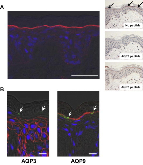
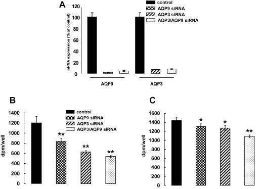
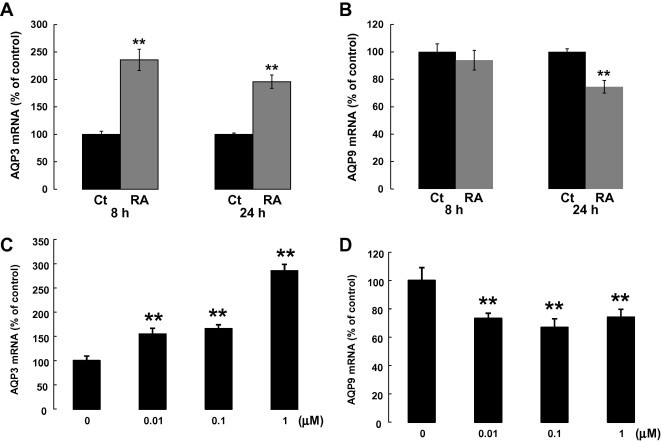
Aquaporin 9 (AQP9) is a member of the aquaglyceroporin family that transports glycerol, urea and other small solutes as well as water. Compared to the expression and function in epidermal keratinocytes of AQP3, another aquaglyceroporin, our knowledge of epidermal AQP9 remains elusive. In this study, we investigated the expression of AQP9 in the human epidermis and cultured keratinocytes. Immunofluorescence studies revealed that AQP9 expression is highly restricted to the stratum granulosum of the human epidermis, where occludin is also expressed at the tight junctions. Interestingly, the AQP3 staining decreased sharply below the cell layers in which AQP9 is expressed. In cultured normal human epidermal keratinocytes (NHEK), knock-down of AQP9 expression in the differentiated cells induced by RNA interference reduced glycerol uptake, which was not as pronounced as was the case with AQP3 knock-down cells. In contrast, similar reduction of urea uptake was detected in AQP9 and AQP3 knock-down cells. These findings suggested that AQP9 expression in NHEK facilitates at least the transport of glycerol and urea. Finally, we analyzed the effect of retinoic acid (RA), a potent stimulator of keratinocyte proliferation, on AQP3 and AQP9 mRNA expression in differentiated NHEK. Stimulation with RA at 1 μM for 24 h augmented AQP3 expression and down-regulated AQP9 expression. Collectively, these results indicate that AQP9 expression in epidermal keratinocytes is regulated in a different manner from that of AQP3.

Keywords: AQP3; AQP9; AQPs, aquaporins; DUOX1, dual oxidase I; Differentiated keratinocytes; Epidermis; GAPDH, glyceraldehyde 3-phosphate dehydrogenase; LXR, liver X receptor; NHEK, normal human epidermal keratinocytes; PPARγ, peroxisome proliferators-activated receptor gamma; RA, retinoic acid; SG, stratum granulosum; TJs, tight junctions; VD3, 1,25-dihydroxyvitamin D3.


Figures

Fig. 1
Fig. 1
Immunofluorescence staining for AQP9, AQP3 and occludin in the human epidermis. (A) Immunofluorescence studies were performed using abdominal skin sections (19 years of age). AQP9 expression was detected in the upper layer of the SG (left panel). The nucleus was stained with TO-PRO-3 iodide. Bar = 50 μm. The results of control experiments are shown in right panels. Immunohistochemical staining resulted in very similar localization of AQP9 (top panel). The signal detected in SG was not observed by antibody preabsorbed with antigen peptide (middle panel). The AQP9 staining was not affected in the presence of AQP3 peptide (bottom panel). (B) AQP3 and AQP9 expression sites were analyzed using sequential skin sections (32 years of age). To distinguish the localization of AQP9 from AQP3 in the epidermis, the tight junction component occludin was used as the staining marker. AQP3 was highly expressed from the basal layer to the stratum spinosum. In contrast, AQP9 staining was restricted to the upper stratum granulosum. The nucleus was stained with TO-PRO-3 iodide. Arrows: occludin expression sites. Bar = 10 μm.
Fig. 2
Fig. 2
VD3 upregulated AQP9 mRNA expression in NHEK. VD3 was added at 1 μM to NHEK at confluency in a low Ca2+ medium (0.15 mM) and the cells were further cultured for 3 days. As a control inducing cell differentiation, Ca2+ concentration of medium was elevated to 1.5 mM at the point of confluency. AQP9 mRNA expression was significantly induced by VD3, a keratinocyte differentiation reagent. The mRNA expression levels were normalized to that of GAPDH.
Fig. 3
Fig. 3
AQP9 siRNA transfection to NHEK reduced [14C]-glycerol and [14C]-urea uptake. NHEK samples mixed with AQP9 and/or AQP3 siRNA were seeded onto 12-well culture plates. On day 4 after culturing in a high Ca2+ medium, glycerol and urea permeability were analyzed. Real-time PCR analyses showed remarkable suppression of AQP9 and AQP3 mRNA expression by siRNA transfection (A). AQP9 and AQP3 siRNA transfection reduced [14C]-glycerol uptake compared to the cells transfected with non-target siRNA. Further decrease in uptake was detected with AQP3/AQP9 double siRNA transfection (B). [14C]-urea uptake was also reduced by both AQP9 and AQP3 siRNA transfection. This reduction was augmented by AQP3/AQP9 double siRNA transfection (C). The experiments were repeated three times. A representative data set is shown (n = 3; mean ± SD; p < 0.05, ∗∗p < 0.01).
Fig. 4
Fig. 4
RA upregulated AQP3 and downregulated AQP9 mRNA expression in NHEK. At confluence, the cells were cultured for 4 days in a high Ca2+ medium to induce cell differentiation. After that, the cells were treated with RA at 1 μM for 8 and 24 h. AQP3 mRNA expression was increased and AQP9 decreased by RA treatment (A, B). The change in expression of AQP3 (after 8 h) and AQP9 (after 24 h) was detected at doses as low as 0.01 μM (C, D). The mRNA expression levels were normalized to that of 18S rRNA. Values are means ± SD (n = 3; ∗∗p < 0.01).

References

    1. Borgnia M. Cellular and molecular biology of the aquaporin water channels. Annu. Rev. Biochem. 1999;68:425–458. - PubMed
    1. Takata K., Matsuzaki T., Tajika Y. Aquaporins: water channel proteins of the cell membrane. Prog. Histochem. Cytochem. 2004;39(1):1–83. - PubMed
    1. Ishibashi K. The evolutionary aspects of aquaporin family. Am. J. Physiol. Regul. Integr. Comp. Physiol. 2011;300(3):R566–R576. - PubMed
    1. Sougrat R. Functional expression of AQP3 in human skin epidermis and reconstructed epidermis. J. Invest. Dermatol. 2002;118(4):678–685. - PubMed
    1. Dumas M. Hydrating skin by stimulating biosynthesis of aquaporins. J. Drugs Dermatol. 2007;6(6 Suppl.):s20–s24. - PubMed