Role of wide-field autofluorescence imaging and scanning laser ophthalmoscopy in differentiation of choroidal pigmented lesions
- PMID: 25161946
- PMCID: PMC4137210
- DOI: 10.3980/j.issn.2222-3959.2014.04.21
Role of wide-field autofluorescence imaging and scanning laser ophthalmoscopy in differentiation of choroidal pigmented lesions
Abstract
Aim: To evaluate the diagnostic properties of wide-field fundus autofluorescence (FAF) scanning laser ophthalmoscope (SLO) imaging for differentiating choroidal pigmented lesions.
Methods: A consecutive series of 139 patients were included, 101 had established choroidal melanoma with 13 untreated lesions and 98 treated with radiotherapy. Thirty-eight had choroidal nevi. All patients underwent a full ophthalmological examination, undilated wide-field imaging, FAF and standardized US examination. FAF images and imaging characteristics from SLO were correlated with the structural findings in the two patient groups.
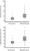
Results: Mean FAF intensity of melanomas was significantly lower than the FAF of choroidal nevi. Only 1 out of 38 included eyes with nevi touched the optic disc compared to 31 out of 101 eyes with melanomas. In 18 out of 101 melanomas subretinal fluid was seen at the pigmented lesion compared to none seen in eyes with confirmed choroidal nevi. In "green laser separation", a trend towards more mixed FAF appearance of melanomas compared to nevi was observed. The mean maximal and minimal transverse and longitudinal diameters of melanomas were significantly higher than those of nevi.
Conclusion: Wide-field SLO and FAF imaging may be an appropriate non-invasive diagnostic screening tool to differentiate benign from malign pigmented choroidal lesions.
Keywords: autofluorescence; choroidal lesion; imaging; melanoma; scanning laser ophthalmoscopy.
Figures


Similar articles
-
Comparative study between fundus autofluorescence and red reflectance imaging of choroidal nevi using ultra-wide-field scanning laser ophthalmoscopy.Retina. 2015 Jun;35(6):1202-10. doi: 10.1097/IAE.0000000000000463. Retina. 2015. PMID: 25650707
-
Choroidal pigmented lesions imaged by ultra-wide-field scanning laser ophthalmoscopy with two laser wavelengths (Optomap).Clin Ophthalmol. 2010 Jul 30;4:829-36. doi: 10.2147/opth.s11864. Clin Ophthalmol. 2010. PMID: 20689737 Free PMC article.
-
Retromode Scanning Laser Ophthalmoscopy for Choroidal Nevi: A Preliminary Study.Life (Basel). 2023 May 25;13(6):1253. doi: 10.3390/life13061253. Life (Basel). 2023. PMID: 37374036 Free PMC article.
-
Review of fundus autofluorescence in choroidal melanocytic lesions.Eye (Lond). 2009 Mar;23(3):497-503. doi: 10.1038/eye.2008.244. Epub 2008 Aug 1. Eye (Lond). 2009. PMID: 18670456 Review.
-
Fundus Autofluorescence Imaging in Patients with Choroidal Melanoma.Cancers (Basel). 2022 Apr 2;14(7):1809. doi: 10.3390/cancers14071809. Cancers (Basel). 2022. PMID: 35406581 Free PMC article. Review.
Cited by
-
The selective estrogen receptor modulator raloxifene mitigates the effect of all-trans-retinal toxicity in photoreceptor degeneration.J Biol Chem. 2019 Jun 14;294(24):9461-9475. doi: 10.1074/jbc.RA119.008697. Epub 2019 May 9. J Biol Chem. 2019. PMID: 31073029 Free PMC article.
-
Comparison of conventional color fundus photography and multicolor imaging in choroidal or retinal lesions.Graefes Arch Clin Exp Ophthalmol. 2018 Apr;256(4):643-649. doi: 10.1007/s00417-017-3884-6. Epub 2018 Feb 28. Graefes Arch Clin Exp Ophthalmol. 2018. PMID: 29492687 Free PMC article.
-
Update on wide- and ultra-widefield retinal imaging.Indian J Ophthalmol. 2015 Jul;63(7):575-81. doi: 10.4103/0301-4738.167122. Indian J Ophthalmol. 2015. PMID: 26458474 Free PMC article. Review.
-
Ultra-wide field retinal imaging: A wider clinical perspective.Indian J Ophthalmol. 2021 Apr;69(4):824-835. doi: 10.4103/ijo.IJO_1403_20. Indian J Ophthalmol. 2021. PMID: 33727441 Free PMC article. Review.
-
Widefield and Ultra-Widefield Retinal Imaging: A Geometrical Analysis.Life (Basel). 2023 Jan 10;13(1):202. doi: 10.3390/life13010202. Life (Basel). 2023. PMID: 36676151 Free PMC article.
References
-
- Bergman L, Seregard S, Nilsson B, Ringborg U, Lundell G, Ragnarsson-Olding B. Incidence of uveal melanoma in Sweden from 1960 to 1998. Invest Ophthalmol Vis Sci. 2002;43(8):2579–2583. - PubMed
-
- Wilkes S, Kurland L, Campbell RJ, Robertson DM. Incidence of uveal malignant melanoma. Am J Ophthalmol. 1979;88(3 Pt 2):629–630. - PubMed
-
- Bell DJ, Wilson MW. Choroidal melanoma: natural history and management options. Cancer Control. 2004;11(5):296–303. - PubMed
-
- Lorigan JG, Wallace S, Mavligit GM. The prevalence and location of metastases from ocular melanoma: imaging study in 110 patients. AJR Am J Roentgenol. 1991;157(6):1279–1281. - PubMed
-
- Shammas HF, Blodi FC. Peripapillary choroidal melanomas. Extension along the optic nerve and its sheaths. Arch Ophthalmol. 1978;96(3):440–445. - PubMed
LinkOut - more resources
Full Text Sources
