Coronary CT angiography in the quantitative assessment of coronary plaques
- PMID: 25162010
- PMCID: PMC4138793
- DOI: 10.1155/2014/346380
Coronary CT angiography in the quantitative assessment of coronary plaques
Abstract
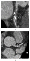
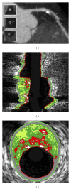
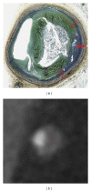
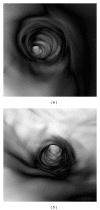
Coronary computed tomography angiography (CCTA) has been recently evaluated for its ability to assess coronary plaque characteristics, including plaque composition. Identification of the relationship between plaque composition by CCTA and patient clinical presentations may provide insight into the pathophysiology of coronary artery plaque, thus assisting identification of vulnerable plaques which are associated with the development of acute coronary syndrome. CCTA-generated 3D visualizations allow evaluation of both coronary lesions and lumen changes, which are considered to enhance the diagnostic performance of CCTA. The purpose of this review is to discuss the recent developments that have occurred in the field of CCTA with regard to its diagnostic accuracy in the quantitative assessment of coronary plaques, with a focus on the characterization of plaque components and identification of vulnerable plaques.
Figures


Similar articles
-
The napkin-ring sign indicates advanced atherosclerotic lesions in coronary CT angiography.JACC Cardiovasc Imaging. 2012 Dec;5(12):1243-52. doi: 10.1016/j.jcmg.2012.03.019. JACC Cardiovasc Imaging. 2012. PMID: 23236975
-
Prognostic Value of Coronary CT Angiography for Predicting Poor Cardiac Outcome in Stroke Patients without Known Cardiac Disease or Chest Pain: The Assessment of Coronary Artery Disease in Stroke Patients Study.Korean J Radiol. 2020 Sep;21(9):1055-1064. doi: 10.3348/kjr.2020.0103. Korean J Radiol. 2020. PMID: 32691541 Free PMC article.
-
Coronary plaque characteristics in computed tomography and 2-year outcomes: The PREDICT study.J Cardiovasc Comput Tomogr. 2018 Sep-Oct;12(5):436-443. doi: 10.1016/j.jcct.2018.07.001. Epub 2018 Jul 7. J Cardiovasc Comput Tomogr. 2018. PMID: 30017608
-
Coronary CT angiography and high-risk plaque morphology.Cardiovasc Interv Ther. 2013 Jan;28(1):1-8. doi: 10.1007/s12928-012-0140-1. Epub 2012 Oct 30. Cardiovasc Interv Ther. 2013. PMID: 23108779 Review.
-
The challenge of coronary calcium on coronary computed tomographic angiography (CCTA) scans: effect on interpretation and possible solutions.Int J Cardiovasc Imaging. 2015 Dec;31 Suppl 2:145-57. doi: 10.1007/s10554-015-0773-0. Epub 2015 Sep 25. Int J Cardiovasc Imaging. 2015. PMID: 26408105 Review.
Cited by
-
Giant left main stem aneurysm in a patient with non-sustained ventricular tachycardia: A case report.Clin Case Rep. 2024 Aug 17;12(8):e9335. doi: 10.1002/ccr3.9335. eCollection 2024 Aug. Clin Case Rep. 2024. PMID: 39156204 Free PMC article.
-
Coronary computed tomography angiography using model-based iterative reconstruction algorithms in the detection of significant coronary stenosis: how the plaque type influences the diagnostic performance.Pol J Radiol. 2019 Dec 9;84:e522-e529. doi: 10.5114/pjr.2019.91259. eCollection 2019. Pol J Radiol. 2019. PMID: 32082450 Free PMC article.
-
Technical principles, benefits, challenges, and applications of photon counting computed tomography in coronary imaging: a narrative review.Cardiovasc Diagn Ther. 2024 Aug 31;14(4):698-724. doi: 10.21037/cdt-24-52. Epub 2024 Jul 31. Cardiovasc Diagn Ther. 2024. PMID: 39263472 Free PMC article. Review.
-
Molecular imaging of plaques in coronary arteries with PET and SPECT.J Geriatr Cardiol. 2014 Sep;11(3):259-73. doi: 10.11909/j.issn.1671-5411.2014.03.005. J Geriatr Cardiol. 2014. PMID: 25278976 Free PMC article. Review.
-
Association between serum N-terminal pro-B-type natriuretic peptide levels and characteristics of coronary atherosclerotic plaque detected by coronary computed tomography angiography.Exp Ther Med. 2016 Aug;12(2):667-675. doi: 10.3892/etm.2016.3371. Epub 2016 May 19. Exp Ther Med. 2016. PMID: 27446259 Free PMC article.
References
-
- Topol EJ, Nissen SE. Our preoccupation with coronary luminology: the dissociation between clinical and angiographic findings in ischemic heart disease. Circulation. 1995;92(8):2333–2342. - PubMed
-
- Little WC, Constantinescu M, Applegate RJ, et al. Can coronary angiography predict the site of a subsequent myocardial infarction in patients with mild-to-moderate coronary artery disease? Circulation. 1988;78(5):1157–1166. - PubMed
-
- Burke AP, Farb A, Malcom GT, Liang Y, Smialek J, Virmani R. Coronary risk factors and plaque morphology in men with coronary disease who died suddenly. The New England Journal of Medicine. 1997;336(18):1276–1282. - PubMed
-
- Davies MJ. The composition of coronary-artery plaques. The New England Journal of Medicine. 1997;336(18):1313–1314. - PubMed
-
- Virmani R, Burke AP, Farb A, Kolodgie FD. Pathology of the vulnerable plaque. Journal of the American College of Cardiology. 2006;47(8):C13–C18. - PubMed
Publication types
MeSH terms
LinkOut - more resources
Full Text Sources
Other Literature Sources
Medical

