. 2014 Sep 18;513(7518):426-30.
doi: 10.1038/nature13725. Epub 2014 Aug 27.

Bidirectional switch of the valence associated with a hippocampal contextual memory engram

Affiliations

Bidirectional switch of the valence associated with a hippocampal contextual memory engram

Roger L Redondo et al. Nature. .

Abstract

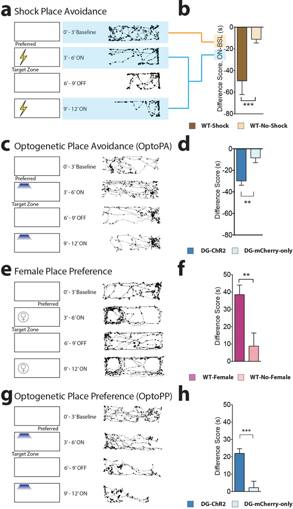
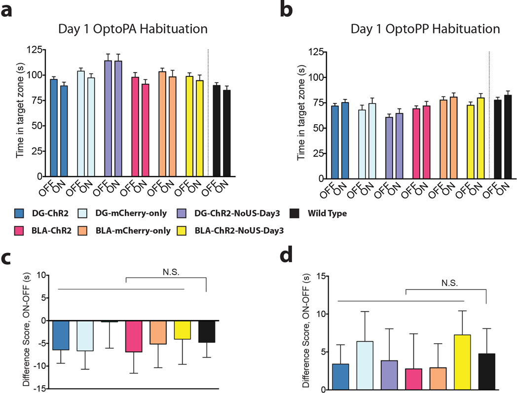
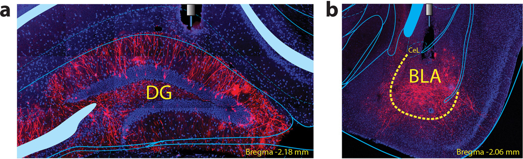
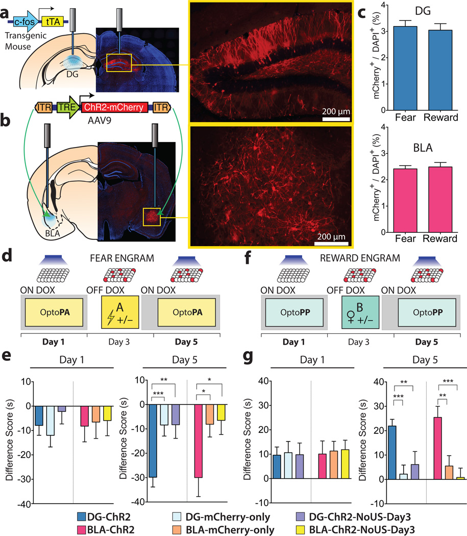
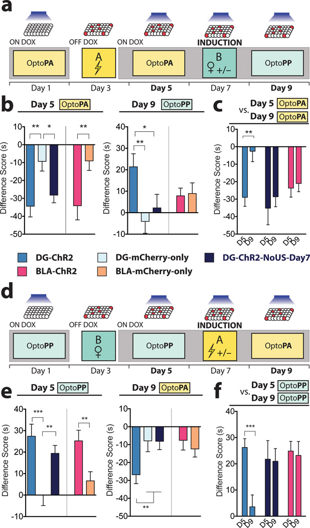
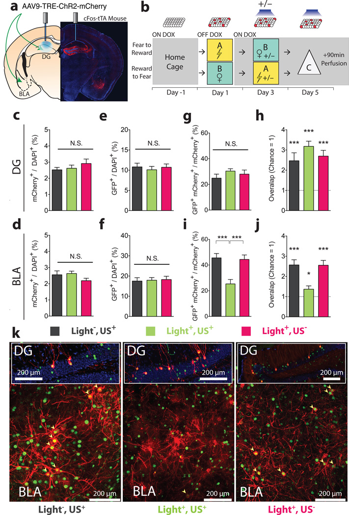
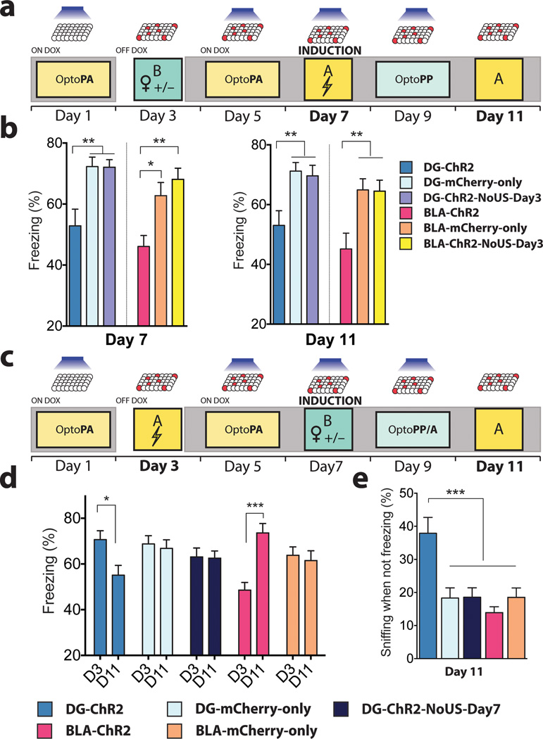
The valence of memories is malleable because of their intrinsic reconstructive property. This property of memory has been used clinically to treat maladaptive behaviours. However, the neuronal mechanisms and brain circuits that enable the switching of the valence of memories remain largely unknown. Here we investigated these mechanisms by applying the recently developed memory engram cell- manipulation technique. We labelled with channelrhodopsin-2 (ChR2) a population of cells in either the dorsal dentate gyrus (DG) of the hippocampus or the basolateral complex of the amygdala (BLA) that were specifically activated during contextual fear or reward conditioning. Both groups of fear-conditioned mice displayed aversive light-dependent responses in an optogenetic place avoidance test, whereas both DG- and BLA-labelled mice that underwent reward conditioning exhibited an appetitive response in an optogenetic place preference test. Next, in an attempt to reverse the valence of memory within a subject, mice whose DG or BLA engram had initially been labelled by contextual fear or reward conditioning were subjected to a second conditioning of the opposite valence while their original DG or BLA engram was reactivated by blue light. Subsequent optogenetic place avoidance and preference tests revealed that although the DG-engram group displayed a response indicating a switch of the memory valence, the BLA-engram group did not. This switch was also evident at the cellular level by a change in functional connectivity between DG engram-bearing cells and BLA engram-bearing cells. Thus, we found that in the DG, the neurons carrying the memory engram of a given neutral context have plasticity such that the valence of a conditioned response evoked by their reactivation can be reversed by re-associating this contextual memory engram with a new unconditioned stimulus of an opposite valence. Our present work provides new insight into the functional neural circuits underlying the malleability of emotional memory.


Figures

Extended Figure 1
Extended Figure 1. Light-induced avoidance and preference tests
a–b, Shock Place Avoidance test. a, After the 0–3min Baseline (BSL), the most preferred zone was established as the target zone. WT-Shock mice (n = 11) received foot shocks (0.15 mA DC, 2s duration every 5s) when entering the target zone during the ON phases (3–6 and 9–12 min). No shocks were delivered during the OFF phase (6–9 min). WT-No-Shock mice (n = 24) received no shocks. b, The difference score was lower in the WT-Shock mice compared to WT-No-Shock (t41 = 3.76, P < 0.001). c–d, Optogenetic Place Avoidance (OptoPA) test. c, Mice were allowed to explore the arena during BSL and the preferred side determined as the target zone. Light stimulation (20 Hz, 15 ms pulse width, 473 nm, >10 mW) was applied when the mice entered the target zone during the ON phases. Light stimulation was not delivered during the OFF phase. d, The difference score was lower in the DG-ChR2 mice (n = 48) compared to DG-mCherry-only mice (n = 39) (t85 = 3.55, P < 0.001). e–f, Female Place Preference test. e, After the 0–3min baseline (BSL), the less preferred zone was established as the target zone. During the ON phases (3–6 and 9–12 min), a corral containing a female was placed in the less preferred zone (target zone) and an empty corral was placed on the opposite side. Corrals, and the female, were removed during the OFF phase. f, Mice (n = 24) increased the time spent in the target zone compared to mice presented with two empty receptacles (n = 8) (t30 = 2.81, P < 0.01). g–h, Optogenetic Place Preference (OptoPP) test. g, Reward-labelled mice were allowed to explore the arena during BSL and the less preferred zone designated as the target zone. Light stimulation (20 Hz, 15 ms pulse width, 473 nm, >10 mW) was applied while the subject was in the target zone of the chamber at the 3–6 min and 9–12 min epochs (ON phase). h, , The difference score was increased in the DG-ChR2 mice (n = 54) compared to DG-mCherry-only mice (n = 31) (t88 = 4.361, P < 0.001). a,c,e,g (right panel), Representative tracks for experimental animals. Dots mark the position of the animal every 5 video frames and accumulate where the mice spend more time.
Extended Figure 2
Extended Figure 2. Light stimulation in the OptoPA and OptoPP Tests has no effect during habituation
a, Day 1 OptoPA Habituation. There are no within group differences in the average duration spent in the target zone during the average of the BSL and OFF phases (OFF) and the averages of the two light on phases (ON) during day 1 habituation in the OptoPA test, even though there is an overall difference between the two types of phases (F1,173 = 6.46, P < 0.05, 6 n.s. multiple comparisons. DG-ChR2 n = 48, DG-mCherry-only n = 39, DG-ChR2-NoUS-Day3 n = 17, BLA-ChR2 n = 32, BLA-mCherry-only n = 27, BLA-ChR2-NoUS-Day3 n = 27). b, Day 1 OptoPP Habituation. There are no within group differences in the average time duration spent in the target zone during the average of the BSL and OFF phases (OFF) and the average of the two light on phases (ON) during day 1 habituation in the OptoPP test, even though there is an overall difference between the two types of phases (F1,195 = 8.06, P < 0.01, 6 n.s. multiple comparisons. DG-ChR2 n = 54, DG-mCherry-only n = 36, DG-ChR2-NoUS-Day3 n = 24, BLA-ChR2 n = 35, BLA-mCherry-only n = 31, BLA-ChR2-NoUS-Day3 n = 21). c–d, There are no differences between experimental groups and wild type mice tested without light stimulation c, In the OptoPA test, the difference scores (ON-OFF) are similar between experimental groups (same as panel a) and wild type mice (n = 33) that did not receive light stimulation (F6,205 = 0.19, n.s.). d, In the OptoPP test, the difference scores (ON-OFF) are similar between experimental groups (same as panel b) and wild type mice (n = 33) that did not receive light stimulation (F6,227 = 0.21, n.s.).
Extended Figure 3
Extended Figure 3. Fibre positions in DG and BLA
a, Representative example of the fibre location and the expression of the ChR2-mCherry construct in the DG. b, Representative example of the fibre location and the expression of the ChR2-mCherry construct in the BLA.
Figure 1
Figure 1. Fear and reward engram reactivation, both in the DG and the BLA, drives place avoidance and place preference, respectively
a, b, c-fos tTA mice were injected with AAV9-TRE-ChR2-mCherry or TRE-mCherry and implanted with optical fibres bilaterally targeting the DG (a) or the BLA (b). c, Similar engram labelling in the DG (t33 = 0.42, n.s.) and BLA (t26 = 0.35, n.s.) after fear (DG n = 16; BLA n = 14) and reward (DG n = 19; BLA n = 14) conditioning d, Fear memory group experimental protocol. e, On Day 1, Difference Scores (Extended Fig. 1) were similar across all DG subgroups (F2,101 = 0.76, n.s.) and across all BLA subgroups (F2,72 = 0.03, n.s.). On day 5, Difference Scores were lower in DG-ChR2 (n = 48) and BLA-ChR2 mice (n = 21) compared to corresponding mCherry-only (DG n = 39; BLA n = 27) and DG- or BLA-ChR2-NoUS-Day3 mice (DG n = 17; BLA n = 27) (DG F2,101 = 7.99, P < 0.001; BLA F2,72 = 4.12, P < 0.05). f, Reward memory group experimental protocol. g, On day 1, Difference Scores were similar across all DG subgroups (F2,111 = 0.02, n.s.) and across all BLA subgroups (F2,83 = 0.04, n.s.). In the day 5 OptoPP test, Difference Scores were greater in DG-ChR2 (n = 54) and BLA-ChR2 mice (n = 35) compared to corresponding mCherry-only (DG n = 36; BLA n = 31) and DG- or BLA-ChR2-NoUS-Day3 mice (DG n = 24; BLA n = 21) (DG F2,111 = 9.76, P < 0.001; BLA F2,83 = 9.12, P < 0.001). Results show mean ± s.e.m.
Figure 2
Figure 2. The valence associated with the DG engram is reversed after induction with the US of opposite value
a, The Fear-to-Reward experimental protocol. b, On day 5, Difference Scores of DG-ChR2 (n = 16), DG-ChR2-NoUS-Day7 (n = 20), and BLA-ChR2 mice (n = 19) were lower compared to corresponding mCherry-only mice (DG n = 27; BLA n = 27) (DG F2,59 = 6.16, P < 0.01; BLA t44 = 2.73, P < 0.01). In the day 9 OptoPP test, Difference Scores of DG-ChR2 mice were greater than the control mice (F2,60 = 4.4, P < 0.05). Difference Scores of BLA-ChR2 mice were similar to those of BLA-mCherry-only mice (t44 = 0.16, n.s.). c, On day 9 OptoPA test, DG-ChR2 mice (n = 12) showed less aversive response compared to day 5 while both DG-ChR2-NoUS-Day7 (n = 16) and BLA-ChR2 (n = 17) mice showed similar Difference Scores on these days (F1,42 = 5.42, P < 0.05). d, Reward-to-Fear experimental protocol. e, On day 5 OptoPP test, Difference Scores of DG-ChR2 (n = 17), DG-ChR2-NoUS-Day7 (n = 29), and BLA-ChR2 mice (n =30) were greater compared to corresponding mCherry-only mice (DG n = 27; BLA n = 29) (DG F2,70 = 8.97, P < 0.001; BLA t57 = 2.85, P < 0.01) On day 9, Difference Scores of DG-ChR2 mice were lower compared to the control mice (F2,71 = 3.20, P < 0.05) and Difference Scores of BLA-ChR2 mice were similar to those of BLA-mCherry-only mice (t57 = 0.49, n.s.). f, On day 9 OptoPP test, DG-ChR2 mice (n =18) showed a reduced appetitive response compared to day 5 OptoPP test while both DG-ChR2-NoUS-Day7 (n = 21) and BLA-ChR2 mice (n = 18) showed similar Difference Scores on day 9 and day 5 (F1,54 = 6.58, P < 0.05). Results show mean ± s.e.m.
Figure 3
Figure 3. DG to BLA functional connectivity changes after induction
a, Injection sites and the optic fibre placement. b, Experimental protocol. On day 1, cells active during fear or reward experience were labelled. On day 3, on Dox, mice were randomly assigned to 3 groups: reward or fear conditioning without light reactivation (Light,US+) (n = 11); full induction protocol (Light+,US+) (n = 16); light stimulation but neither reward nor fear conditioning (Light+,US) (n = 16). On day 5, all animals received light stimulation in the DG for 12 minutes in a novel context (Context C) before brains were subjected to immunohistochemistry. Similar proportions of neurons were labelled by the fear or reward conditioning on day 3 in all groups, both in c, DG (F2,40 = 0.77, n.s.) and d, BLA (F2,40 = 2.40, n.s.). Light delivery to the DG on day 5 led to the activation (GFP+/DAPI+) of similar proportions of cells in the e, DG (F2,40 = 0.21, n.s.) and f, BLA (F2,40 = 0.06, n.s.). g, Levels of reactivation (GFP+mCherry+/mCherry+) in the DG were similar across all groups (F2,40 = 0.61, n.s.) and h, above levels of chance (one sample t-tests against chance overlap: –Light,+US t10 = 4.24, P < 0.01; Light+,US+ t15 = 8.56, P < 0.001; Light+,US t15 = 5.5, P < 0.001). i, Levels of reactivation (GFP+mCherry+ / mCherry+) in the BLA were lower in the Light+,US+ compared to Light,US+ and Light+,US (F2,40 = 11.82, P < 0.001) even though overlap levels j, remained above chance (one sample t-tests: Light,US+ t10 = 7.41, P < 0.001; Light+,US+ t15 = 2.33, P < 0.05; Light+,US t15 = 6.94, P < 0.001). k, Representative images of double immunofluorescence for GFP (green) and mCherry (red) in the DG and BLA. Results show mean ± s.e.m.
Figure 4
Figure 4. Memory induction alters naturally cued fear memory
a, Reward-to-Fear scheme with Context A fear memory tests on day 7 and day 11. b, On day 7, freezing levels during the last 3 minutes of the induction procedure were reduced in DG-ChR2 (n = 18) and BLA-ChR2 mice (n = 19) compared to corresponding mCherry-only (DG n = 21; BLA n = 20) and ChR2-NoUS-Day3 mice (DG n = 27; BLA n = 24) (DG F2, 63 = 8.768, P < 0.001; BLA F2,60 = 8.49, P < 0.001). These reduced freezing levels remained on day 11 (DG F2,63 = 6.25, P < 0.01; BLA F2,60 = 6.86, P < 0.01). c, Fear-to-Reward scheme with Context A fear memory tests on day 3 and day 11. d, Compared to the last three minutes of the fear conditioning on day 3, only DG-ChR2 mice (n =27) showed a reduction of freezing responses on day 11 (Interaction F4,151 = 8.48, P < 0.001). BLA-ChR2 mice (n = 29) showed increased levels of freezing on day 11 compared to day 3. DG-ChR2-NoUS-Day7 mice (n = 42) and mCherry-only mice (DG n = 32; BLA n = 29) did not show differences between day 3 and day 11. e, After correcting for the time spent freezing, DG-ChR2 mice spent a larger proportion of time sniffing than any other group in Context A on day 11 (F4,151 = 7.78, P < 0.001). Results show mean ± s.e.m.

Comment in

Similar articles

Cited by

References

    1. Pavlov IP. Conditioned Reflexes: An Investigation of the Physiological Activity of the Cerebral Cortex. Oxford University Press; 1927. - PMC - PubMed
    1. Wolpe J. Psychotherapy by reciprocal inhibition. Stanford University Press; 1958.
    1. Liu X, et al. Optogenetic stimulation of a hippocampal engram activates fear memory recall. Nature. 2012;484:381–385. - PMC - PubMed
    1. Ramirez S, et al. Creating a false memory in the hippocampus. Science. 2013;341:387–391. - PubMed
    1. Muramoto K, Ono T, Nishijo H, Fukuda M. Rat amygdaloid neuron responses during auditory discrimination. Neuroscience. 1993;52:621–636. - PubMed

Publication types