Immunolocalization of FGF-2 and VEGF in rat periodontal ligament during experimental tooth movement
- PMID: 25162568
- PMCID: PMC4296624
- DOI: 10.1590/2176-9451.19.3.067-074.oar
Immunolocalization of FGF-2 and VEGF in rat periodontal ligament during experimental tooth movement
Abstract
Objective: This article aimed at identifying the expression of fibroblast growth factor-2 (FGF-2) and vascular endothelial growth factor (VEGF) in the tension and pressure areas of rat periodontal ligament, in different periods of experimental orthodontic tooth movement.
Methods: An orthodontic force of 0.5 N was applied to the upper right first molar of 18 male Wistar rats for periods of 3 (group I), 7 (group II) and 14 days (group III). The counter-side first molar was used as a control. The animals were euthanized at the aforementioned time periods, and their maxillary bone was removed and fixed. After demineralization, the specimens were histologically processed and embedded in paraffin. FGF-2 and VEGF expressions were studied through immunohistochemistry and morphological analysis.
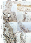
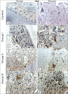
Results: The experimental side showed a higher expression of both FGF-2 and VEGF in all groups, when compared with the control side (P < 0.05). Statistically significant differences were also found between the tension and pressure areas in the experimental side.
Conclusion: Both FGF-2 and VEGF are expressed in rat periodontal tissue. Additionally, these growth factors are upregulated when orthodontic forces are applied, thereby suggesting that they play an important role in changes that occur in periodontal tissue during orthodontic movement.
Objetivo: o objetivo desse estudo foi identificar a expressão do fator de crescimento de fibroblastos 2 (FGF-2) e do fator de crescimento vascular endotelial (VEGF) nos lados de tensão e pressão do ligamento periodontal de ratos, durante movimento ortodôntico experimental, em diferentes períodos de tempo.
Métodos: uma força ortodôntica de 0,5N foi aplicada no primeiro molar superior direito de 18 ratos Wistar machos, por períodos de 3 (grupo I), 7 (grupo II) e 14 dias (grupo III). O primeiro molar do lado oposto foi utilizado como controle. Os animais foram sacrificados nos períodos de tempo mencionados, sendo a arcada superior removida e fixada. Após a desmineralização, os espécimes foram processados histologicamente e embebidos em parafina. A expressão do FGF-2 e do VEGF foram estudadas por meio de análise imuno-histoquímica.
Resultados: o ligamento periodontal dos dentes submetidos à movimentação ortodôntica mostraram maior expressão tanto de FGF-2 quanto de VEGF, em todos os grupos experimentais, quando comparados com os dentes do lado controle (p < 0,05). Diferenças estatisticamente significativas entre os lados de tensão e pressão também foram encontradas nos dentes submetidos à movimentação ortodôntica.
Conclusões: tanto o FGF-2 quanto o VEGF são expressos no tecido periodontal de ratos, e esses fatores de crescimento são aumentados quando forças ortodônticas são aplicadas, sugerindo que esses desempenham um papel importante na reorganização do periodonto durante o movimento ortodôntico.
Keywords: Fibroblast growth factor 1; Orthodontics; Periodontal ligament; Vascular endothelial growth factor A.
Figures


Similar articles
-
Evaluation of BSP expression and apoptosis in the periodontal ligament during orthodontic relapse: a preliminary study.Orthod Craniofac Res. 2014 Nov;17(4):239-48. doi: 10.1111/ocr.12049. Epub 2014 Jun 13. Orthod Craniofac Res. 2014. PMID: 24924469
-
Expression of Wnt3a, Wnt10b, β-catenin and DKK1 in periodontium during orthodontic tooth movement in rats.Acta Odontol Scand. 2016;74(3):217-23. doi: 10.3109/00016357.2015.1090011. Epub 2015 Sep 28. Acta Odontol Scand. 2016. PMID: 26414930
-
Expressions of RANKL/RANK and M-CSF/c-fms in root resorption lacunae in rat molar by heavy orthodontic force.Eur J Orthod. 2011 Aug;33(4):335-43. doi: 10.1093/ejo/cjq068. Epub 2010 Sep 10. Eur J Orthod. 2011. PMID: 20833686
-
Biological Events in Periodontal Ligament and Alveolar Bone Associated with Application of Orthodontic Forces.ScientificWorldJournal. 2015;2015:876509. doi: 10.1155/2015/876509. Epub 2015 Sep 2. ScientificWorldJournal. 2015. PMID: 26421314 Free PMC article. Review.
-
Cytokines and VEGF induction in orthodontic movement in animal models.J Biomed Biotechnol. 2012;2012:201689. doi: 10.1155/2012/201689. Epub 2012 May 14. J Biomed Biotechnol. 2012. PMID: 22665981 Free PMC article. Review.
Cited by
-
Wnt5a mediated canonical Wnt signaling pathway activation in orthodontic tooth movement: possible role in the tension force-induced bone formation.J Mol Histol. 2016 Oct;47(5):455-66. doi: 10.1007/s10735-016-9687-y. Epub 2016 Jul 25. J Mol Histol. 2016. PMID: 27456852
-
The effect of cacao bean extracts on the prevention of periodontal tissue breakdown in diabetic rats with orthodontic tooth movements.J Oral Biol Craniofac Res. 2024 Jul-Aug;14(4):384-389. doi: 10.1016/j.jobcr.2024.05.013. Epub 2024 May 24. J Oral Biol Craniofac Res. 2024. PMID: 38832299 Free PMC article.
-
Human Dental Pulp Tissue during Orthodontic Tooth Movement: An Immunofluorescence Study.J Funct Morphol Kinesiol. 2020 Aug 22;5(3):65. doi: 10.3390/jfmk5030065. J Funct Morphol Kinesiol. 2020. PMID: 33467280 Free PMC article.
-
Pulsed electromagnetic field prevents tooth relapse after orthodontic tooth movement in rat models.J Taibah Univ Med Sci. 2024 Dec 26;20(1):1-12. doi: 10.1016/j.jtumed.2024.12.009. eCollection 2025 Feb. J Taibah Univ Med Sci. 2024. PMID: 39831281 Free PMC article.
-
Effect of Caffeic Acid Phenethyl Ester Provision on Fibroblast Growth Factor-2, Matrix Metalloproteinase-9 Expression, Osteoclast and Osteoblast Numbers during Experimental Tooth Movement in Wistar Rats (Rattus norvegicus).Eur J Dent. 2021 May;15(2):295-301. doi: 10.1055/s-0040-1718640. Epub 2021 Jan 28. Eur J Dent. 2021. PMID: 33511599 Free PMC article.
References
-
- Aldridge SE, Lennard TW, Williams JR, Birch MA. Vascular endothelial growth factor receptors in osteoclast differentiation and function. Biochem Biophys Res Commun. 2005;335(3):793–798. - PubMed
-
- Anastasi G, Cordasco G, Matarese G, Rizzo G, Nucera R, Mazza M, et al. An immunohistochemical, histological, and electron-microscopic study of the human periodontal ligament during orthodontic treatment. Int J Molec Med. 2008;21(5):545–554. - PubMed
-
- Baffour R, Berman J, Garb JL, Rhee SW, Kaufman J, Friedmann P. Enhanced angiogenesis and growth of collaterals by in vivo administration of recombinant basic fibroblast growth factor in a rabbit model of acute lower limb ischemia: dose-response effect of basic fibroblast growth factor. J Vasc Surg. 1992;16(2):181–191. - PubMed
-
- Carmeliet P, Collen D. Molecular analysis of blood vessel formation and disease. Pt 2Am J Physiol. 1997;273(5):H2091–H2104. - PubMed
-
- Clarke MSF, Caldewell RW, Chiao H, Miyake K, McNeil PL. Contraction-induced cell wounding and release of fibroblast growth factor in heart. Circ Res. 1995;76(6):927–934. - PubMed
Publication types
MeSH terms
Substances
LinkOut - more resources
Full Text Sources
Other Literature Sources
