. 2014 Aug 27;9(8):e105931.
doi: 10.1371/journal.pone.0105931. eCollection 2014.

Structural and functional brain changes beyond visual system in patients with advanced glaucoma

Affiliations

Structural and functional brain changes beyond visual system in patients with advanced glaucoma

Paolo Frezzotti et al. PLoS One. .

Abstract

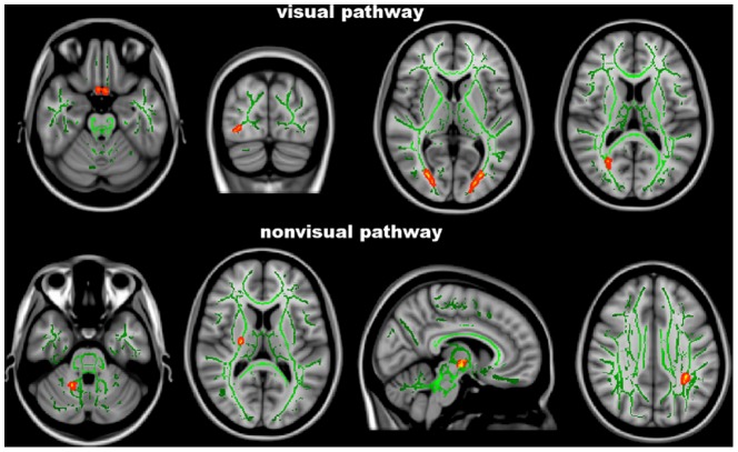
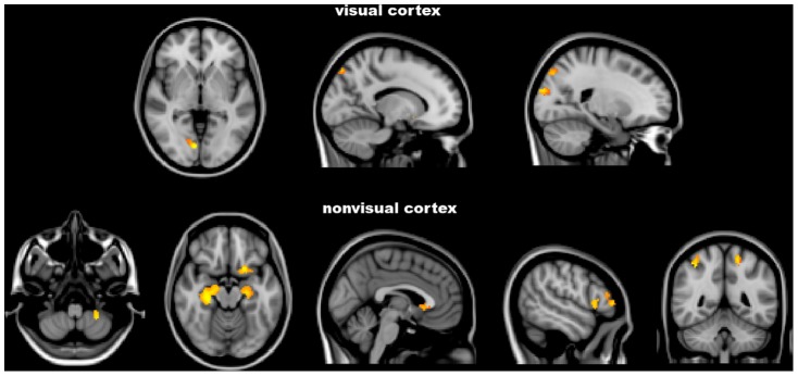
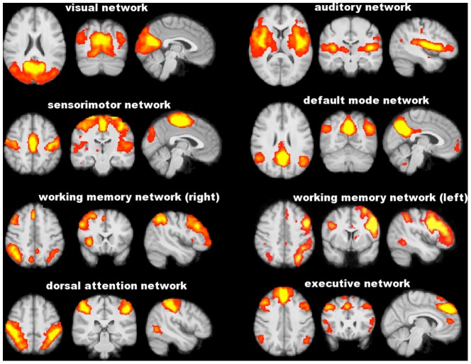
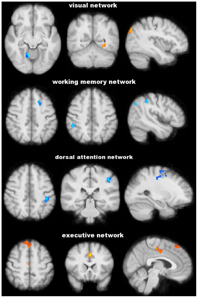
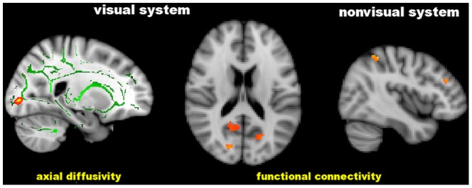
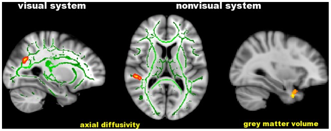
In order to test the hypothesis that in primary open angle glaucoma (POAG), an important cause of irreversible blindness, a spreading of neurodegeneration occurs through the brain, we performed multimodal MRI and subsequent whole-brain explorative voxelwise analyses in 13 advanced POAG patients and 12 age-matched normal controls (NC). Altered integrity (decreased fractional anisotropy or increased diffusivities) of white matter (WM) tracts was found not only along the visual pathway of POAG but also in nonvisual WM tracts (superior longitudinal fascicle, anterior thalamic radiation, corticospinal tract, middle cerebellar peduncle). POAG patients also showed brain atrophy in both visual cortex and other distant grey matter (GM) regions (frontoparietal cortex, hippocampi and cerebellar cortex), decreased functional connectivity (FC) in visual, working memory and dorsal attention networks and increased FC in visual and executive networks. In POAG, abnormalities in structure and FC within and outside visual system correlated with visual field parameters in the poorer performing eyes, thus emphasizing their clinical relevance. Altogether, this represents evidence that a vision disorder such as POAG can be considered a widespread neurodegenerative condition.


Conflict of interest statement

Competing Interests: Paolo Frezzotti, Antonio Giorgio, Ilaria Motolese, Alessandro De Leucio, Michele Iester, Eduardo Motolese, Antonio Federico declare no competing financial interests. Nicola De Stefano has served on scientific advisory boards, received speaker honoraria, served as a consultant or received research support from BioMS Medical, Biogen Idec, Bayer Schering Pharma, Merck Serono, NeuroRx Research, Novartis, Teva Pharmaceutical Industries Ltd and the Italian Multiple Sclerosis Society. This does not alter adherence to PLOS ONE policies on sharing data and materials.

Figures

Figure 1
Figure 1. Tract-Based Spatial Statistics analysis.
Red-yellow shows clusters where POAG patients have significant DTI abnormalities (lower fractional anisotropy or higher axial/radial diffusivity) in comparison with normal controls. Green is the white matter skeleton. Images (in standard MNI152 space) are shown in radiological convention. See Results and Table 2 for details.
Figure 2
Figure 2. FSL-Voxel Based Morphometry analysis.
Red-yellow shows clusters where POAG patients have significantly lower grey matter volume in comparison with normal controls. Images (in standard MNI152 space) are shown in radiological convention. See Results and Table 3 for details.
Figure 3
Figure 3. Functionally relevant resting state networks across study subjects (POAG patients and normal controls) identified with independent component analysis.
Images (axial, coronal, sagittal) are z-statistics overlaid on the average high-resolution scan transformed into standard (MNI152) space. Red to yellow are z values, ranging from 3 to 10. Images are shown in radiological convention.
Figure 4
Figure 4. Functional connectivity analysis at the level of resting state networks.
Blu-light blu and red-yellow show clusters where POAG patients have, respectively, significantly lower and higher functional connectivity than normal controls. Images (in standard MNI152 space) are shown in radiological convention. See Results and Table 4 for details.
Figure 5
Figure 5. Significant correlation of the visual field Mean Deviation with cerebral axial diffusivity and functional connectivity in distinct brain regions of patients with POAG.
Green is the white matter skeleton. Images (in standard MNI152 space) are shown in radiological convention. See Results and Table 5 for details.
Figure 6
Figure 6. Significant correlation of the visual field Pattern Standard Deviation with cerebral axial diffusivity and grey matter volume in in distinct brain regions of patients with POAG.
Green is the white matter skeleton. Images (in standard MNI152 space) are shown in radiological convention. See Results and Table 6 for details.

References

    1. European Glaucoma Society (2008) Terminology and guidelines for glaucoma; 3rd, editor. Savona: Dogma.
    1. Le PV, Tan O, Chopra V, Francis BA, Ragab O, et al. (2013) Regional correlation among ganglion cell complex, nerve fiber layer, and visual field loss in glaucoma. Invest Ophthalmol Vis Sci 54: 4287–4295. - PMC - PubMed
    1. Nucci C, Martucci A, Cesareo M, Mancino R, Russo R, et al. (2013) Brain involvement in glaucoma: advanced neuroimaging for understanding and monitoring a new target for therapy. Curr Opin Pharmacol 13: 128–133. - PubMed
    1. Boucard CC, Hernowo AT, Maguire RP, Jansonius NM, Roerdink JB, et al. (2009) Changes in cortical grey matter density associated with long-standing retinal visual field defects. Brain 132: 1898–1906. - PMC - PubMed
    1. Garaci FG, Bolacchi F, Cerulli A, Melis M, Spano A, et al. (2009) Optic nerve and optic radiation neurodegeneration in patients with glaucoma: in vivo analysis with 3-T diffusion-tensor MR imaging. Radiology 252: 496–501. - PubMed

MeSH terms