. 2014 Aug 27;5(4):103.
doi: 10.1186/scrt491.

Influence of insulin-like growth factor I overexpression via recombinant adeno-associated vector gene transfer upon the biological activities and differentiation potential of human bone marrow-derived mesenchymal stem cells

Influence of insulin-like growth factor I overexpression via recombinant adeno-associated vector gene transfer upon the biological activities and differentiation potential of human bone marrow-derived mesenchymal stem cells

Janina Frisch et al. Stem Cell Res Ther. .

Abstract

Introduction: The transplantation of genetically modified progenitor cells such as bone marrow-derived mesenchymal stem cells (MSCs) is an attractive strategy to improve the natural healing of articular cartilage defects. In the present study, we examined the potential benefits of sustained overexpression of the mitogenic and pro-anabolic insulin-like growth factor I (IGF-I) via gene transfer upon the biological activities of human MSCs (hMSCs).

Methods: Recombinant adeno-associated vectors (rAAV) were used to deliver a human IGF-I coding sequence in undifferentiated and chondrogenically-induced primary hMSCs in order to determine the efficacy and duration of transgene expression and the subsequent effects of the genetic modification upon the chondrogenic versus osteogenic differentiation profiles of the cells relative to control (lacZ) treatment after 21 days in vitro.

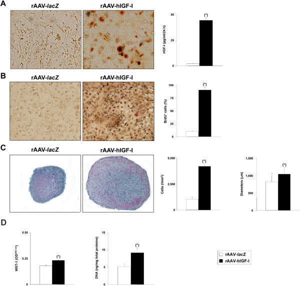
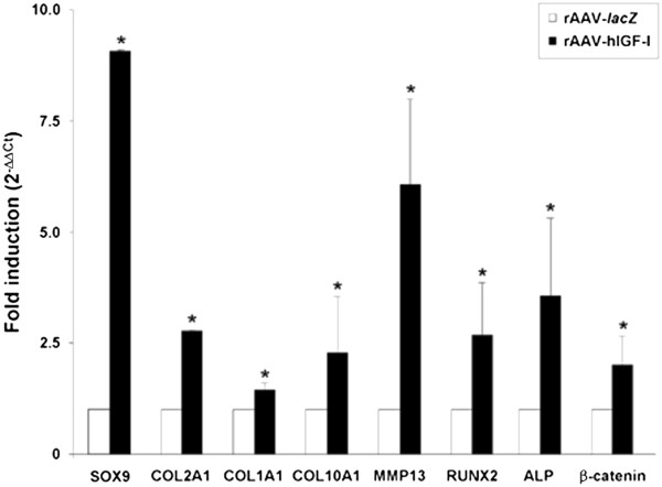
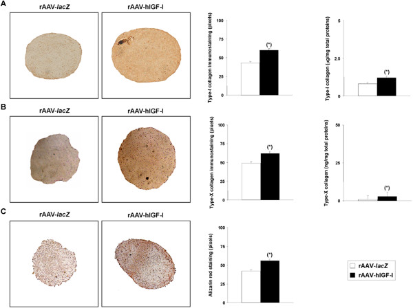
Results: Significant and prolonged expression of IGF-I was evidenced in undifferentiated and most importantly in chondrogenically-induced hMSCs transduced with the candidate rAAV-hIGF-I vector for up to 21 days, leading to enhanced proliferative, biosynthetic, and chondrogenic activities compared with rAAV-lacZ treatment. Overexpression of IGF-I as achieved in the conditions applied here also increased the expression of hypertrophic and osteogenic markers in the treated cells.

Conclusions: These results suggest that a tight regulation of rAAV expression may be necessary for further translation of the approach in clinically relevant animal models in vivo. However, the current findings support the concept of using this type of vector as an effective tool to treat articular cartilage defects via gene- and stem cell-based procedures.


Figures

Figure 1
Figure 1
Recombinant adeno-associated virus-mediated IGF-I gene transfer in undifferentiated monolayer cultures of human bone marrow-derived mesenchymal stem cells. Cells were transduced with rAAV-hIGF-I or rAAV-lacZ (20 μl each vector) as described in Methods and histologically processed after 21 days to monitor (A) transgene (insulin-like growth factor I) expression by immunocytochemical analysis and enzyme-linked immunosorbent assay, (B) levels of cell proliferation by immunocytochemical detection of bromodeoxyuridine (BrdU) incorporation and histomorphometry, by WST-1 assay, and by detection of the DNA contents, and (C) levels of proteoglycan synthesis (magnification × 4). *Statistically significant compared with rAAV-lacZ. hIGF-I, human insulin-like growth factor I; OD, optical density; rAAV, recombinant adeno-associated virus.
Figure 2
Figure 2
Recombinant adeno-associated virus-mediated IGF-I gene transfer in chondrogenically induced cultures of human bone marrow-derived mesenchymal stem cells. Human bone marrow-derived mesenchymal stem cell aggregates were transduced with rAAV-hIGF-I or rAAV-lacZ (40 μl each vector) as described in Methods and were histologically processed after 21 days to monitor (A) transgene (insulin-like growth factor I) expression by immunohistochemical analysis (magnification × 20) and enzyme-linked immunosorbent assay, and the levels of cell proliferation by immunohistochemical detection of bromodeoxyuridine (BrdU) incorporation and histomorphometry (B) (magnification × 10), by hematoxylin and eosin staining and histomorphometry (C) (magnification × 4), or by biochemical assays (WST-1 assay and detection of the DNA contents) (D). *Statistically significant compared with rAAV-lacZ. hIGF-I, human insulin-like growth factor I; OD, optical density; rAAV, recombinant adeno-associated virus.
Figure 3
Figure 3
Metabolic and differentiation activities in chondrogenically induced cultures of human bone marrow-derived mesenchymal stem cells transduced with rAAV-hIGF-I. Human bone marrow-derived mesenchymal stem cell aggregates were transduced with rAAV-hIGF-I or rAAV-lacZ as described in Figure 2 and histologically processed after 21 days to evaluate the production of matrix proteoglycans (toluidine blue staining with histomorphometry and detection of the proteoglycan contents) (A) and the expression of SOX9 with histomorphometry (B) and type II collagen (specific immunodetection with histomorphometry and detection of the type II collagen contents) (C) (all at magnification × 4). *Statistically significant compared with rAAV-lacZ. hIGF-I, human insulin-like growth factor I; rAAV, recombinant adeno-associated virus.
Figure 4
Figure 4
Expression analyses in chondrogenically induced cultures of human bone marrow-derived mesenchymal stem cells transduced with rAAV-hIGF-I. Human bone marrow-derived mesenchymal stem cell aggregates were transduced with rAAV-hIGF-I or rAAV-lacZ as described in Figure 2 and processed on day 21 for gene expression analysis by real-time reverse transcription-polymerase chain reaction amplification after total cellular RNA extraction and cDNA synthesis, as described in Methods. The genes analyzed included the transcription factor SOX9, type II, type I, and type X collagen (COL2A1, COL1A1, COL10A1), matrix metalloproteinase 13 (MMP13), the transcription factor RUNX2, alkaline phosphatase (ALP), and β-catenin, with glyceraldehyde-3-phosphate dehydrogenase (GAPDH) serving as a housekeeping gene and internal control (primers are listed in Methods). Threshold cycle (Ct) values were obtained for each target and GAPDH as a control for normalization, and fold inductions (relative to lacZ-treated aggregates) were measured using the 2–ΔΔCt method. *Statistically significant compared with rAAV-lacZ. hIGF-I, human insulin-like growth factor I; rAAV, recombinant adeno-associated virus.
Figure 5
Figure 5
Hypertrophic differentiation in chondrogenically induced cultures of human bone marrow-derived mesenchymal stem cells transduced with rAAV-hIGF-I. Human bone marrow-derived mesenchymal stem cell aggregates were transduced with rAAV-hIGF-I or rAAV-lacZ as described in Figure 2 and histologically processed after 21 days to examine the expression of type I collagen (A) and type X collagen (B) by immunohistochemistry/histomorphometry and by analysis of the type I collagen (A) and type X collagen (B) contents, and to evaluate matrix mineralization (alizarin red staining with histomorphometry) (C) (all at magnification × 4). *Statistically significant compared with rAAV-lacZ. hIGF-I, human insulin-like growth factor I; rAAV, recombinant adeno-associated virus.
Figure 6
Figure 6
Analyses in osteogenically and adipogenically differentiated cultures of human bone marrow-derived mesenchymal stem cells transduced with rAAV-hIGF-I. Cells in monolayer cultures were transduced with rAAV-hIGF-I or rAAV-lacZ (40 μl each vector) and induced toward osteogenic or adipogenic differentiation as described in Methods. Cultures were processed on day 21 for (A) alkaline phosphatase (ALP) staining with histomorphometry (osteogenesis; magnification × 4) and (B) Oil Red O staining with histomorphometry (adipogenesis; magnification × 10). *Statistically significant compared with rAAV-lacZ. hIGF-I, human insulin-like growth factor I; rAAV, recombinant adeno-associated virus.

Comment in

References

    1. Buckwalter JA. Articular cartilage: injuries and potential for healing. J Orthop Sports Phys Ther. 1998;28:192–202. doi: 10.2519/jospt.1998.28.4.192. - DOI - PubMed
    1. O'Driscoll SW. The healing and regeneration of articular cartilage. J Bone Joint Surg Am. 1998;80:1795–1812. - PubMed
    1. Bentley G, Biant LC, Carrington RW, Akmal M, Goldber A, Williams AM, Skinner JA, Pringle J. A prospective, randomised comparison of autologous chondrocyte implantation versus mosaicplasty for osteochondral defects in the knee. J Bone Joint Surg (Br) 2003;85:223–230. doi: 10.1302/0301-620X.85B2.13543. - DOI - PubMed
    1. Brittberg M, Lindahl A, Nilsson A, Ohlsson C, Isaksson O, Peterson L. Treatment of deep cartilage defects in the knee with autologous chondrocyte transplantation. N Engl J Med. 1994;331:889–895. doi: 10.1056/NEJM199410063311401. - DOI - PubMed
    1. Horas U, Pelinkovic D, Herr G, Aigner T, Schnettler R. Autologous chondrocyte implantation and osteochondral cylinder transplantation in cartilage repair of the knee joint. A prospective, comparative trial. J Bone Joint Surg Am. 2003;85-A:185–192. - PubMed

Publication types

MeSH terms

Substances

LinkOut - more resources