

Appendiceal mucocele and pseudomyxoma peritonei; the clinical boundaries of a subtle disease
- PMID: 25163976
- PMCID: PMC4156334
- DOI: 10.12659/AJCR.890837
Appendiceal mucocele and pseudomyxoma peritonei; the clinical boundaries of a subtle disease
Abstract
Patient: Male, 70 • Male, 84.
Final diagnosis: Appendiceal mucocele and pseudomyxoma peritonei.
Symptoms: -.
Medication: -.
Clinical procedure: -.
Specialty: Surgery.
Objective: Rare disease.
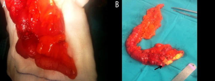
Background: Mucocele of the appendix is an uncommon cystic lesion characterized by distension of the appendiceal lumen with mucus. Most commonly, it is the result of epithelial proliferation, but it can also be caused by inflammation or obstruction of the appendix. When an underlying mucinous cystadenocarcinoma exists, spontaneous or iatrogenic rupture of the mucocele can lead to mucinous intraperitoneal ascites, a syndrome known as pseudomyxoma peritonei.
Case report: We report 2 cases that represent the clinical extremities of this heterogeneous disease; an asymptomatic mucocele of the appendix in a 70-year-old female and a case of pseudomyxoma peritonei in an 84-year-old male. Subsequently, we review the current literature focusing to the optimal management of both conditions.
Conclusions: Mucocele of the appendix is a rare disease, usually diagnosed on histopathologic examination of appendectomized specimens. Due to the existing potential for malignant transformation and pseudomyxoma peritonei caused by rupture of the mucocele, extensive preoperative evaluation and thorough intraoperative gastrointestinal and peritoneal examination is required.
Figures


References
-
- Rokitansky CF. A manual of pathological anatomy. Vol. 1855. Philadelphia (PA): Blanchard & Lea; p. 89.
-
- Ruiz-Tovar J, Teruel DG, Castiñeiras VM, et al. Mucocele of the appendix. World J Surg. 2007;31:542–48. - PubMed
-
- Aho AJ, Heinonen R, Lauren P. Benign and malignant mucocele of the appendix. Acta Chir Scand. 1973;139:392–400. - PubMed
-
- Landen S, Bertrand C, Maddern GJ, et al. Appendiceal mucoceles and pseudomyxoma peritonei. Surg Gynecol Obstet. 1992;175:401–4. - PubMed
-
- Fann JI, Vierra M, Fisher D, et al. Pseudomyxoma peritonei. Surg Gynecol Obstet. 1993;177:441–47. - PubMed
Publication types
MeSH terms
LinkOut - more resources
Full Text Sources
Other Literature Sources
Medical
