. 2014 Oct 15;74(20):5914-24.
doi: 10.1158/0008-5472.CAN-14-0834. Epub 2014 Aug 27.

Zfx facilitates tumorigenesis caused by activation of the Hedgehog pathway

Affiliations

Zfx facilitates tumorigenesis caused by activation of the Hedgehog pathway

Colin J Palmer et al. Cancer Res. .

Abstract

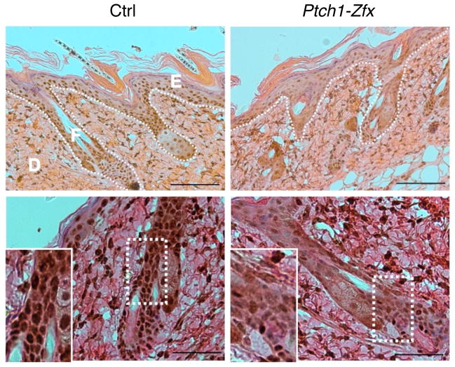
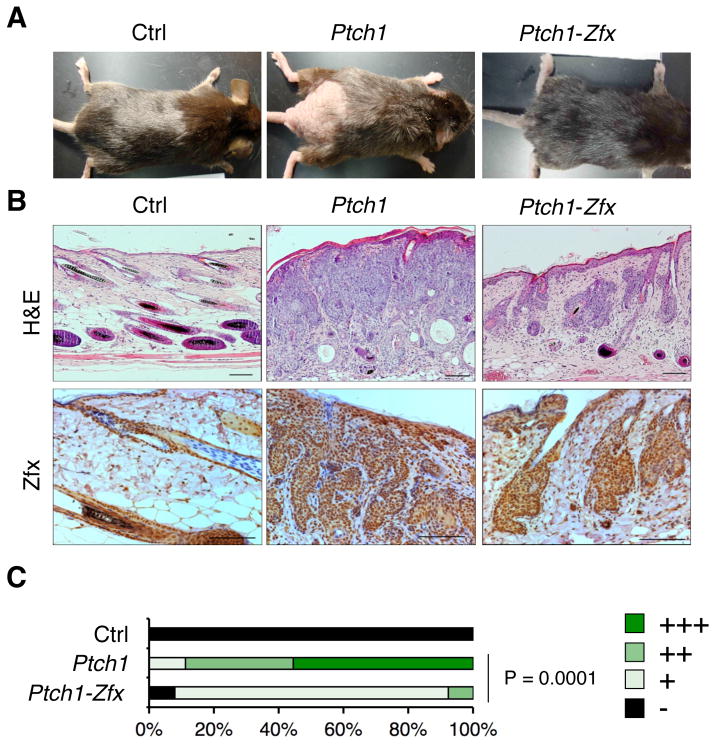
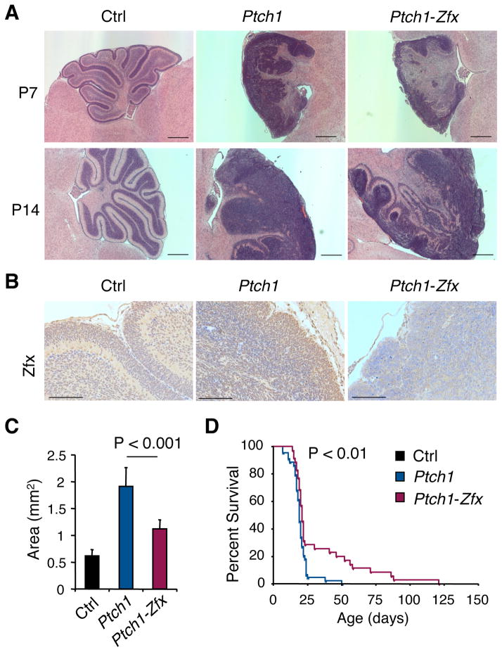
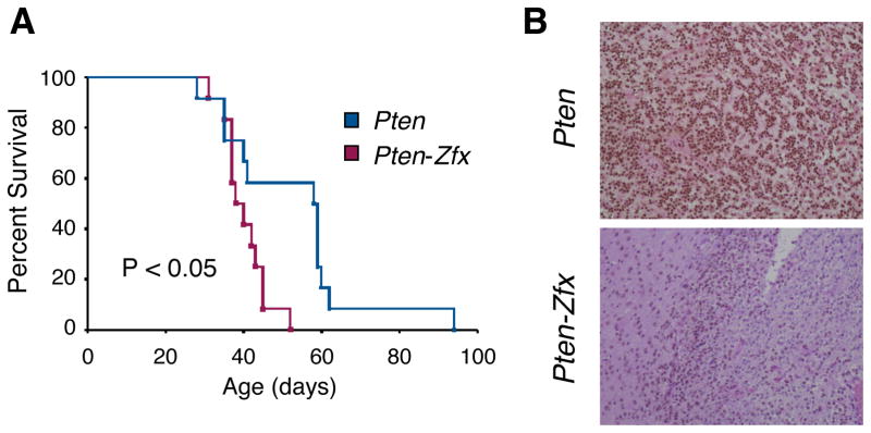
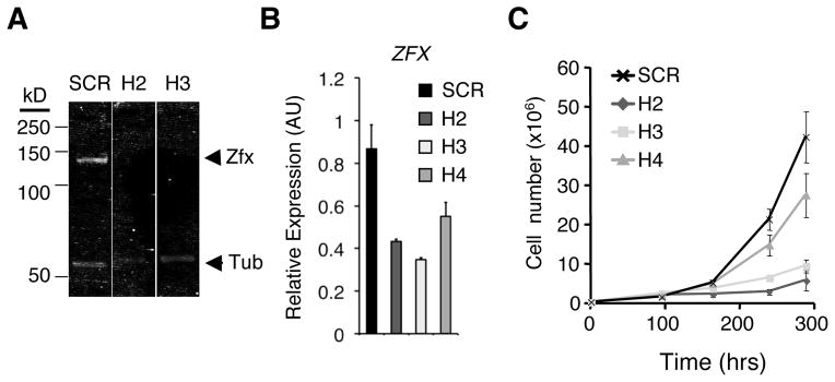
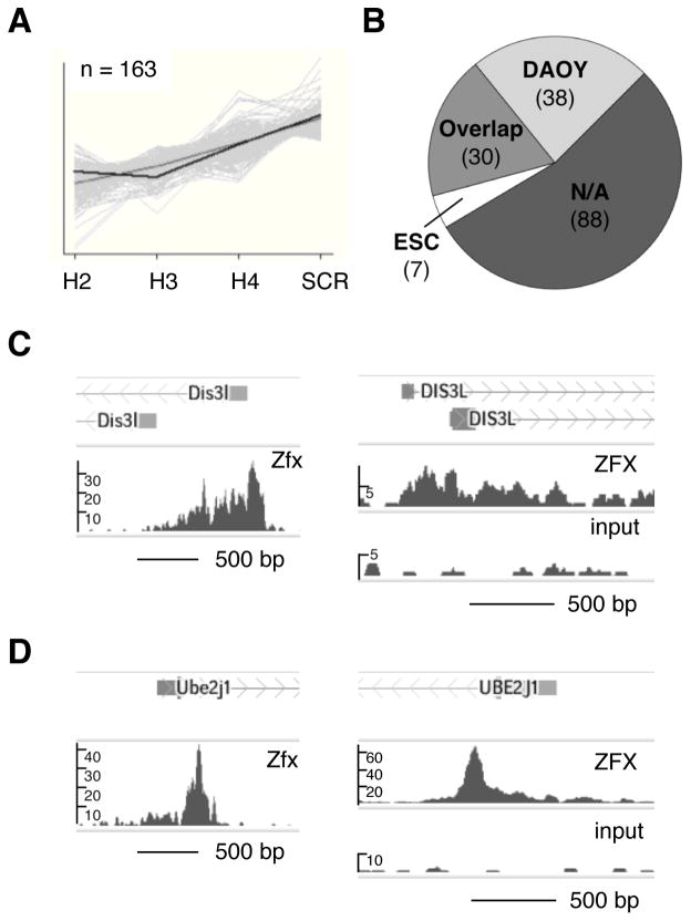
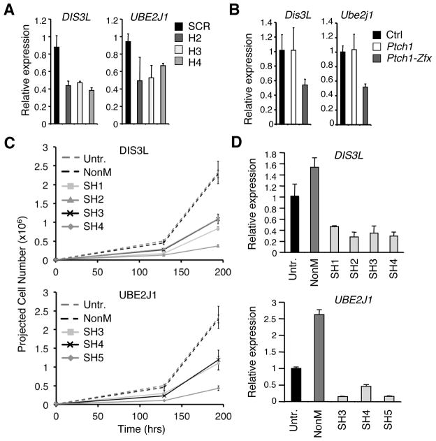
The Hedgehog (Hh) signaling pathway regulates normal development and cell proliferation in metazoan organisms, but its aberrant activation can promote tumorigenesis. Hh-induced tumors arise from various tissues and they may be indolent or aggressive, as is the case with skin basal cell carcinoma (BCC) or cerebellar medulloblastoma, respectively. Little is known about common cell-intrinsic factors that control the development of such diverse Hh-dependent tumors. Transcription factor Zfx is required for the self-renewal of hematopoietic and embryonic stem cells, as well as for the propagation of acute myeloid and T-lymphoblastic leukemias. We report here that Zfx facilitates the development of experimental BCC and medulloblastoma in mice initiated by deletion of the Hh inhibitory receptor Ptch1. Simultaneous deletion of Zfx along with Ptch1 prevented BCC formation and delayed medulloblastoma development. In contrast, Zfx was dispensable for tumorigenesis in a mouse model of glioblastoma. We used genome-wide expression and chromatin-binding analysis in a human medulloblastoma cell line to characterize direct, evolutionarily conserved targets of Zfx, identifying Dis3L and Ube2j1 as two targets required for the growth of the human medulloblastoma cells. Our results establish Zfx as a common cell-intrinsic regulator of diverse Hh-induced tumors, with implications for the definition of new therapeutic targets in these malignancies.


Conflict of interest statement

CONFLICT OF INTEREST

The authors declare no conflict of interest.

Figures

Figure 1
Figure 1. Topical tamoxifen treatment successfully ablates Zfx in the skin
Ptch1flox/flox Zfx+/y R26-CreER+ (Ptch1) and Ptch1flox/flox Zfxflox/y R26-CreER+ (Ptch1-Zfx) mice, along with Ptch1flox/flox R26-CreER controls (Ctrl), were treated topically with tamoxifen (Tmx) to induce deletion of Ptch1 alone or of both Ptch1 and Zfx in the skin. Shown is Zfx expression in the skin 3 days after Tmx treatment. Representative micrographs of IHC staining for Zfx in sections from treated dorsal skin are shown. Scale bars represent 100 μm (upper panel) and 50 μm (lower panel). Epidermis (E), dermis (D), and hair follicle (F) are labeled, and the boundary between epidermis/follicles and dermis is marked (dotted line; upper panel). Zoomed insets show decreased nuclear staining for Zfx in the hair follicle (lower panel).
Figure 2
Figure 2. Zfx loss prevents BCC formation after Ptch1 deletion in vivo
As described in Figure 1, mice carrying tamoxifen (Tmx)-inducible Cre recombinase and conditional alleles of Ptch1 alone (Ptch1) or of both Ptch1 and Zfx (Ptch1-Zfx), as well as Cre controls (Ctrl), were treated with Tmx on shaved lower dorsal skin. (A) Representative photographs showing dorsal skin of mice of the indicated genotypes following euthanasia 8 weeks after Tmx treatment. (B) BCC development in the mice of indicated genotypes. Shown are sections of treated dorsal skin isolated 8 weeks after Tmx treatment and stained with H&E or with anti-Zfx antibody. Scale bars represent 100 μm. (C) Pathology scores of BCC in treated dorsal skin 8–9 weeks after Tmx treatment. Shown are the fractions of indicated BCC severity scores in Ctrl, Ptch1 and Ptch1-Zfx mice (n = 5, 9, and 13, respectively).
Figure 3
Figure 3. Zfx loss delays MB development after Ptch1 deletion in vivo
MB development was followed in mice with CNS-specific hGFAP-Cre and conditional alleles of Ptch1 alone (Ptch1) or of both Ptch1 and Zfx (Ptch1-Zfx), as well as in Cre control mice (Ctrl). (A) H&E-stained sagittal sections of cerebella from P7 and P14 mice of the indicated genotypes. Scale bars represent 500 μm. (B) Anti-Zfx-stained sagittal sections of P7 cerebella from mice of the indicated genotypes. Scale bars represent 100 μm. (C) Cross-section area of external granular layer (EGL) in sagittal sections from P7 cerebella (mean ± SD of 6–7 mice). (D) Kaplan-Meier survival plot of Ptch1 versus Ptch1-Zfx mice.
Figure 4
Figure 4. Loss of Zfx does not impair Pten-dependent glioblastoma development
(A) Kaplan-Meier survival curve for retrovirus-injected Pten and Pten-Zfx mice. Glioblastoma was induced via stereotactic injection of retrovirus encoding PDGF and Cre recombinase into brains of young adult mice with conditional null alleles of Pten only (Pten) or of both Pten and Zfx (Pten-Zfx). (B) Immunohistochemical staining for Zfx in sections of glioblastomas from retrovirus-injected Pten and Pten-Zfx mice.
Figure 5
Figure 5. Zfx knockdown impairs cell growth in a human medulloblastoma cell line in vitro
The human MB cell line DAOY was transduced in vitro with lentiviruses encoding short hairpin RNAs (shRNAs) targeting ZFX transcript (H2, H3, H4) or a scrambled control shRNA (SCR). Transduced cells were selected for by growth in puromycin-containing medium. (A) Western blot for ZFX and tubulin control in DAOY cells transduced with control (SCR) or ZFX-targeting (H2, H3) lentivirus. (B) Expression of ZFX in DAOY cells after shRNA knockdown. Shown are ZFX expression levels 4 days after transduction, as determined by qPCR (mean ± SD of triplicate parallel cultures; representative of three independent experiments). (C) Growth curves for DAOY cells transduced with lentivirus expressing control (SCR) or ZFX-targeting (H2, H3, H4) shRNA (mean ± SD of triplicate parallel cultures, representative of three independent experiments).
Figure 6
Figure 6. Identification of conserved direct targets of ZFX in human medulloblastoma cells
(A) Downregulated genes (n= 163) identified in pattern matching analysis of Affymetrix Gene ST 1.0 array data from DAOY cells after shRNA knockdown. Full gene list is presented in Dataset S1. (B) Conserved ZFX transcriptional targets. Shown are numbers of pattern-matching genes from ZFX KD microarray in (A) that had enrichment peaks >1kb from TSS in ZFX ChIP-seq from DAOY cells, from murine embryonic stem cells (mESC), from both cell types (Overlap), or from neither cell type (N/A). DAOY, mESC, and Overlap gene lists are presented in full in Dataset S2. (C) Zfx binding to Dis3L in murine and human ChIP-seq. Shown are sequencing-read enrichment peaks near TSS of Dis3L in anti-Zfx ChIP-seq from murine ESC (Chen et al., 2008) (left), and in anti-ZFX ChIP-seq and sheared nuclear lysate control (Input) from DAOY human MB cells (right). (D) Zfx binding to Ube2j1 in murine and human ChIP-seq. Shown are sequencing-read enrichment peaks near TSS of Ube2j1 in anti-Zfx ChIP-seq from murine ESC (Chen et al., 2008) (left), and in anti-ZFX ChIP-seq and sheared nuclear lysate control (Input) from DAOY human MB cells (right).
Figure 7
Figure 7. Knockdown of ZFX targets DIS3L and UBE2J1 impairs growth of human DAOY medulloblastoma cells in vitro
(A) Expression of DIS3L and UBE2J1 in DAOY cells after ZFX knockdown. Shown are DIS3L and UBE2J1 expression levels by qPCR 4 days after transduction with lentivirus coding for ZFX-targeting (H2, H3, H4) or scrambled control (SCR) shRNA (mean ± SD of triplicate parallel cultures; representative of three independent experiments). (B) Expression levels of Dis3L and Ube2j1 in bulk epidermis and hair follicles from shaved lower dorsal skin 3 days after topical Tmx treatment in Ptch1, Ptch1-Zfx, and Cre (Ctrl) mice (as described in Figure 1). Shown are normalized expression levels relative to Ctrl skin as determined by qPCR (mean ± SD of 2–4 mice; representative of four independent experiments). (C) DAOY human MB cells were transduced in vitro with Sigma MISSION lentiviruses encoding shRNAs targeting DIS3L, UBE2J1, or a non-mammalian control gene (NonM). Shown are projected cell numbers for untreated DAOY cells versus cells transduced with NonM control virus or 3–4 shRNA lentiviruses targeting DIS3L (SH1–SH4) or UBE2J1 (SH3–SH5) (mean + SD of triplicate parallel cultures; beginning 2 days post- puromycin selection and 4 days post-transduction). (D) Expression of DIS3L and UBE2J1 in DAOY cells after shRNA knockdown. Shown are DIS3L and UBE2J1 expression levels by qPCR in DAOY cells 5 days after transduction with Sigma MISSION lentiviruses coding for DIS3L- or UBE2J1-targeting shRNA, or for NonM control shRNA (mean ± SD of triplicate reactions; normalized to expression in untreated DAOY cells).

Similar articles

Cited by

References

    1. Hooper JE, Scott MP. Communicating with Hedgehogs. Nat Rev Mol Cell Biol. 2005;6:306–17. - PubMed
    1. Ingham PW, Nakano Y, Seger C. Mechanisms and functions of Hedgehog signalling across the metazoa. Nat Rev Genet. 2011;12:393–406. - PubMed
    1. Lum L, Beachy PA. The Hedgehog Response Network: Sensors, Switches, and Routers. Science. 2004;304:1755–9. - PubMed
    1. Varjosalo M, Taipale J. Hedgehog: functions and mechanisms. Genes & Development. 2008;22:2454–72. - PubMed
    1. Kenney AM, Cole MD, Rowitch DH. Nmyc upregulation by sonic hedgehog signaling promotes proliferation in developing cerebellar granule neuron precursors. Development. 2003;130:15–28. - PubMed

Publication types

MeSH terms