. 2014 Nov 1;74(21):6036-6047.
doi: 10.1158/0008-5472.CAN-14-1084. Epub 2014 Aug 27.

Promoting thiol expression increases the durability of antitumor T-cell functions

Affiliations

Promoting thiol expression increases the durability of antitumor T-cell functions

Pravin Kesarwani et al. Cancer Res. .

Abstract

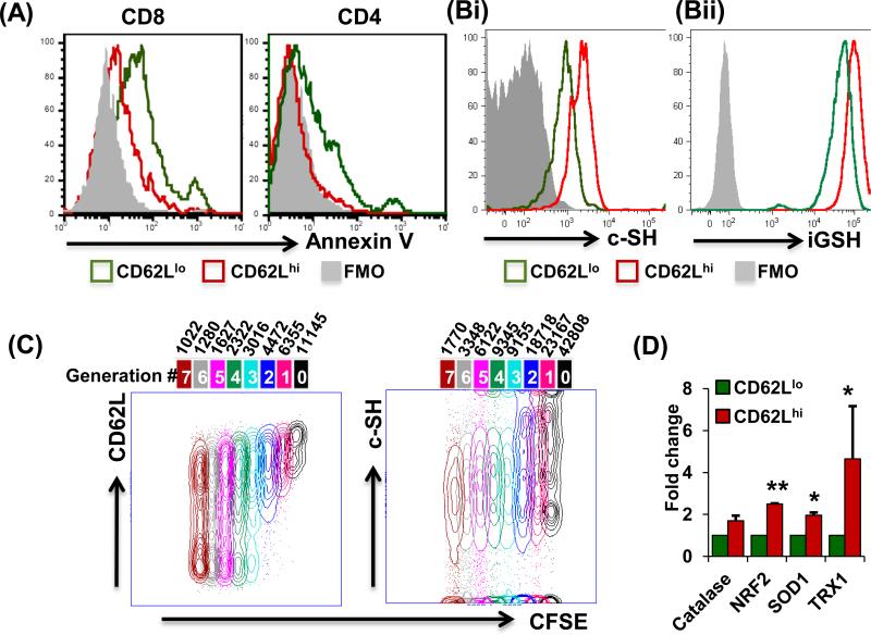
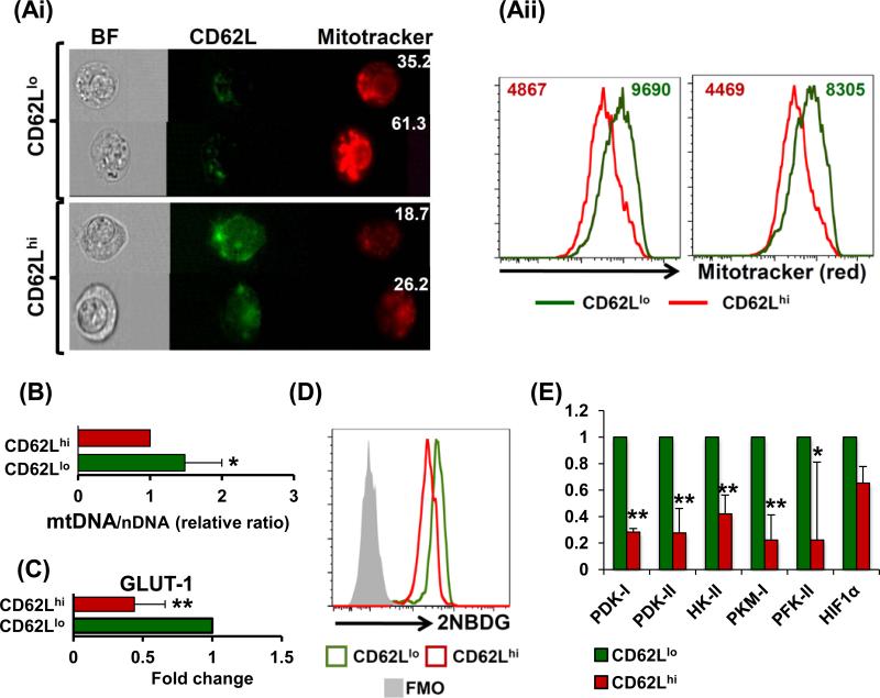
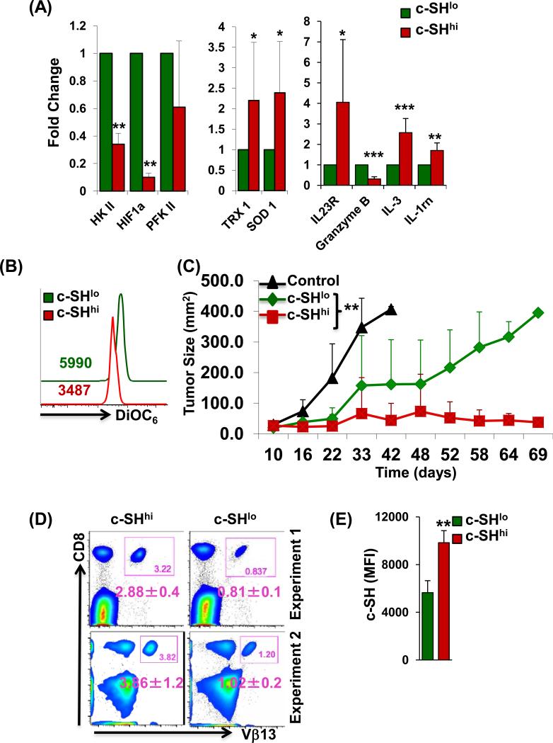
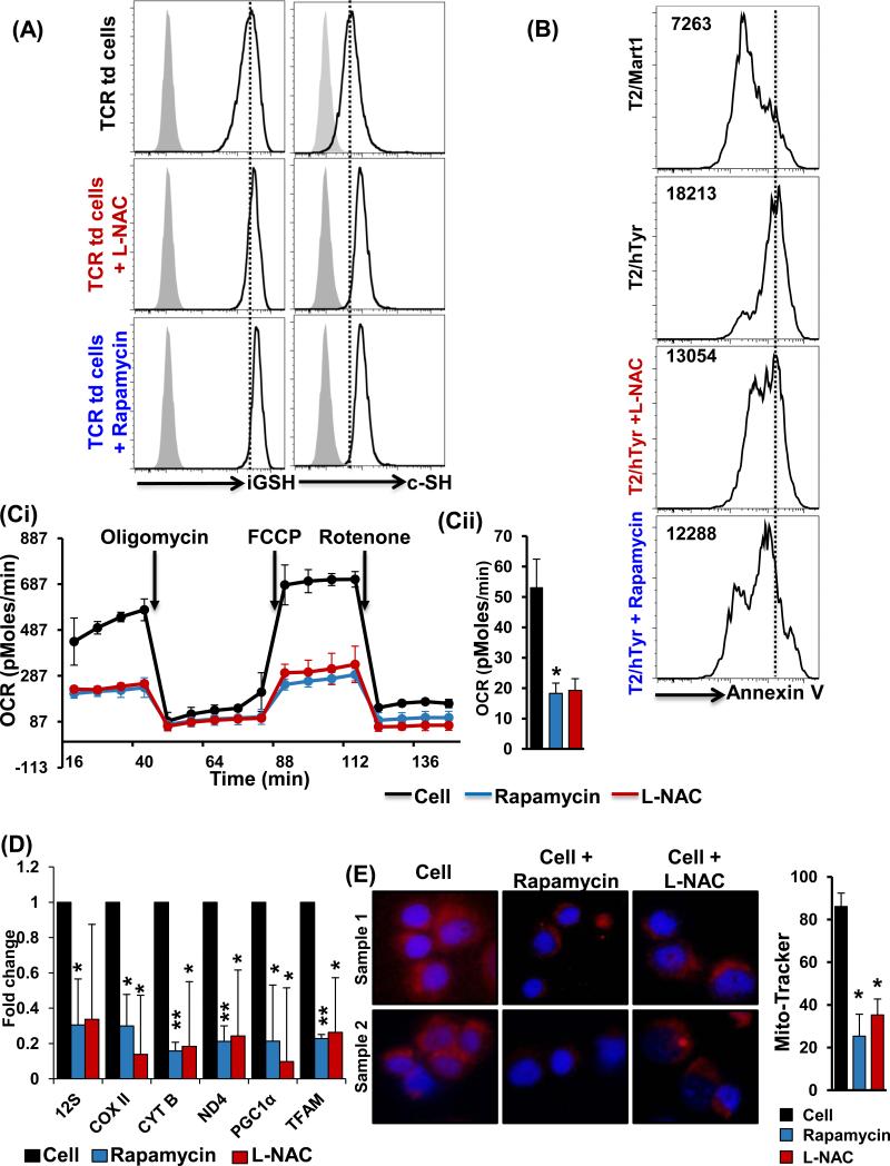
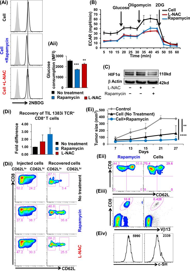
Ex vivo-expanded CD8(+) T cells used for adoptive immunotherapy generally acquire an effector memory-like phenotype (TEM cells). With regard to therapeutic applications, two undesired features of this phenotype in vivo are limited persistence and reduced antitumor efficacy, relative to CD8(+) T cells with a central memory-like phenotype (TCM cells). Furthermore, there is incomplete knowledge about all the differences between TEM and TCM cells that may influence tumor treatment outcomes. Given that TCM cells survive relatively longer in oxidative tumor microenvironments, we investigated the hypothesis that TCM cells possess relatively greater antioxidative capacity than TEM cells. Here, we report that TCM cells exhibit a relative increase compared with TEM cells in the expression of cell surface thiols, a key target of cellular redox controls, along with other antioxidant molecules. Increased expression of redox regulators in TCM cells inversely correlated with the generation of reactive oxygen and nitrogen species, proliferative capacity, and glycolytic enzyme levels. Notably, T-cell receptor-transduced T cells pretreated with thiol donors, such as N-acetyl cysteine or rapamycin, upregulated thiol levels and antioxidant genes. A comparison of antitumor CD8(+) T-cell populations on the basis of surface thiol expression showed that thiol-high cells persisted longer in vivo and exerted superior tumor control. Our results suggest that higher levels of reduced cell surface thiols are a key characteristic of T cells that can control tumor growth and that profiling this biomarker may have benefits to adoptive T-cell immunotherapy protocols.


Figures

Figure 1
Figure 1. Differential antioxidant capacity in CD62Llo and CD62Lhi T cells
(A) Activated human PBMCs cultured for seven days were restimulated overnight with anti-CD3 and stained for CD8, CD4, CD62L, and Annexin V for flow-cytometry-based analysis. (B) Human T cells were gated on CD8+CD44+CD62LhiCXCR3loCCR7hi (red) or CD8+CD44+CD62LloCXCR3hiCCR7lo (green) and analyzed for expression of: (Bi) c-SH using maleamide; p < 0.05, and (Bii) iGSH using monochlorobimane; p < 0.05. Data are representative of at-least seven experiments with similar results. (C) T cells were labeled with CFSE (1 μM) and stimulated or left unstimulated for 72 hours. Cells were then harvested and stained for CD8, Vβ13, CD62L and c-SH, and analyzed by flow cytometry. Cell cycle analysis was then performed using the unstained peak in FlowJo platform software for CD62L and c-SH on CD8+ cells. (N = 3) (D) PBMCs were cultured in IL-15 for 5 days and sorted as CD8+CD62Lhi or CD8+CD62Llo for RNA isolation. mRNA expression levels of antioxidant genes were determined by real-time PCR. All results are representative of three or more separate experiments. ** p <0.005; * p <0.05.
Figure 2
Figure 2. Differences in mitochondrial distribution and glycolysis between CD62L T cell subsets
Human tyrosinase epitope reactive TIL1383I TCR-transduced T cells were cultured in IL-2/IL-15 and used for further experimentation or sorting. (Ai) Cells were washed and labeled with fluorochrome-labeled antibodies for CD8, CD34 (TCR), CD62L and Mitotracker red. Cells were acquired on image stream (EMD Millipore Amnis) and analyzed using IDEAS v. 5.0 software. The cells were first gated on CD8+CD34+ and then on CD62Llo or CD62Lhi. The intensity of Mitotracker was compared in these cells. (Aii) In a parallel experiment, cells were acquired on BD LSR Fortessa and compared for Mitotracker staining and analyzed using FlowJo software. Experiment was repeated twice with similar results, p < 0.005. (B) Comparison between the mitochondrial DNA/nuclear DNA ratio (mDNA/nDNA) is shown, p < 0.05. (C) Real time PCR analysis for GLUT-1 expression between is shown, p < 0.005 (N = 3). (D) Glucose consumption assay was performed using 2NBGD and its florescence was compared between CD62Llo or CD62Lhi cells, p < 0.005 (N = 3). (E) RNA from sorted as CD62Llo or CD62Lhi cells and used to evaluate the differences between glycolysis pathway genes. Results shown were calculated from three separate experiments. ** p <0.005, * p <0.05.
Figure 3
Figure 3. c-SHhi anti-tumor T cells exhibit better tumor control
CD8+ transgenic T cells reactive to human gp100 were TCR-activated with cognate antigen along with IL-2 for 3 days. Cells were sorted based on c-SH expression into c-SHhi or c-SHlo fractions. (A) mRNA transcripts for key glycolysis genes, antioxidant genes, and effector molecules were analyzed. Data are representative of 3 different samples. * p <0.05, ** p <0.005, *** p <0.0005. (B) Membrane potential in c-SHhi or c-SHlo fractions as determined by DiOC6. Numerical values represent MFI, p < 0.05 (N = 3). (C) Human gp100 reactive CD8+ T cells sorted into c-SHhi or c-SHlo fraction were adoptively-transferred to C57BL/6 mice with subcutaneously established B16-F10 murine melanoma. Tumor growth was monitored as shown. Experiment was repeated twice with 3-5 mice/group/experiment. **p<0.005 Indicates a difference in tumor growth as compared by linear regression model. (D) Blood was obtained from each group of mice in C, close to the experimental end-point beyond 45 days from two different experiments, and analyzed for the presence of CD8+Vβ13+ cells. The number in each inset box represents the percentage of CD8+Vβ13+ cells. The number below each inset box represents the mean±SD percentage of CD8+Vβ13+ from 3-4 mice of similar groups. (E) Cells from blood in C were analyzed for c-SH on day 50 post-transfer, and the mean c-SH florescence is shown for mice in each group. Data were analyzed for 3-4 mice per group. ** p <0.005.
Figure 4
Figure 4. c-SH expression inversely correlates to mitochondrial metabolism in T cells
(A) TIL1383I TCR-transduced CD8+ preconditioned cells in rapamycin or L-NAC were analyzed for iGSH (p < 0.05; N = 3), and c-SH (p < 0.05; N = 3), as described in Methods. (B) TIL1383I TCR-transduced CD8+ pretreated with rapamycin or L-NAC was restimulated with either cognate peptide (human Tyrosinase) or control peptide (Mart-1) pulsed T2 cells. Cell death was determined using flow-cytometry-based Annexin V assay. Numbers represent MFI for Annexin V. (Ci) Human PBMCs were cultured in IL-15 for 3 days with rapamycin (250 nM), or L-NAC (5 mM) for 30 minutes, or kept untreated. Mitochondrial respiration was measured by determining oxygen consumption rate using a Seahorse analyzer as detailed in Methods. (Cii) Mean from three experiments with similar results is represented. (D) TIL1383I TCR-transduced CD8+ T cells preconditioned in rapamycin or L-NAC were analyzed for mitochondrial biogenesis genes and mitochondrial transcription factors using real time PCR analysis. (E) Human PBMCs treated with or without rapamycin or L-NAC were stained and analyzed using a fluorescent microscope (60X magnification). Five different fields were photographed for each slide for Mitotracker (red) and DAPI (blue) channels. ImageJ software was used to analyze the difference in Mitotracker intensity. Bar graph on the right represents an average from different experiments. Results are demonstrated as an average of two to three independent experiments with similar results. ** p <0.005, * p <0.05.
Figure 5
Figure 5. c-SH expression inversely correlates to T cell glycolysis
Human T cells transduced with TIL1383I TCR and cultured in IL-2/IL-15 were treated with/without rapamycin or L-NAC and analyzed for: (Ai) Glucose uptake using 2-NBDG on TCR-specific CD8+-gated viable cells as detailed in Methods. (Aii) Data represent MFI of 2NBDG from eight different samples. (B) ECAR levels using the Seahorse Analyzer, and (C) HIF-1α expression by Western blot. (Di) Persistence of TIL1383I CD8+ T cells in the spleen of NSG mice 24 h after adoptive transfer. Bar graph represents the number of cells recovered. Experiment was repeated twice. N=2. (Dii) Comparison of pre- and post-transfer recovered cells for CD62L expression. Experiment was repeated twice with 3-4 mice/group. (E) CD8+ transgenic T cells reactive to human gp100 were activated with cognate antigen in presence/absence of rapamycin along with IL-2 for three days. Equal number of cells were transferred into Rag−/− mice with subcutaneously established murine melanoma B16-F10. Rapamycin group received the maintenance dose of drug (i.p. dose=0.75mg/kg/day from 0-7 days). (Ei) Tumor growth was measured twice weekly as shown. Each group had 5 mice, and the experiment was repeated twice. **p <0.005, ****p <0.00005. Blood was obtained from each group of mice in Ei at the experimental end-point and donor CD8+Vβ13+ T cells were analyzed for: (Eii) CD62L expression, (Eiii) Percent of CD8+Vβ13+ cells (p < 0.05), and (Eiv) c-SH expression (p < 0.05).

Similar articles

Cited by

References

    1. Shen X, Zhou J, Hathcock KS, Robbins P, Powell DJ, Jr., Rosenberg SA, et al. Persistence of tumor infiltrating lymphocytes in adoptive immunotherapy correlates with telomere length. J Immunother. 2007;30:123–9. - PMC - PubMed
    1. Hinrichs CS, Gattinoni L, Restifo NP. Programming CD8+ T cells for effective immunotherapy. Current opinion in immunology. 2006;18:363–70. - PMC - PubMed
    1. Gattinoni L, Klebanoff CA, Palmer DC, Wrzesinski C, Kerstann K, Yu Z, et al. Acquisition of full effector function in vitro paradoxically impairs the in vivo antitumor efficacy of adoptively transferred CD8+ T cells. The Journal of clinical investigation. 2005;115:1616–26. - PMC - PubMed
    1. Klebanoff CA, Gattinoni L, Torabi-Parizi P, Kerstann K, Cardones AR, Finkelstein SE, et al. Central memory self/tumor-reactive CD8+ T cells confer superior antitumor immunity compared with effector memory T cells. Proceedings of the National Academy of Sciences of the United States of America. 2005;102:9571–6. - PMC - PubMed
    1. Berger C, Jensen MC, Lansdorp PM, Gough M, Elliott C, Riddell SR. Adoptive transfer of effector CD8+ T cells derived from central memory cells establishes persistent T cell memory in primates. The Journal of clinical investigation. 2008;118:294–305. - PMC - PubMed

Publication types

MeSH terms

Substances