. 2014 Nov 1;307(9):F1052-62.
doi: 10.1152/ajprenal.00243.2014. Epub 2014 Aug 27.

Peptidyl arginine deiminase-4 activation exacerbates kidney ischemia-reperfusion injury

Affiliations

Peptidyl arginine deiminase-4 activation exacerbates kidney ischemia-reperfusion injury

Ahrom Ham et al. Am J Physiol Renal Physiol. .

Abstract

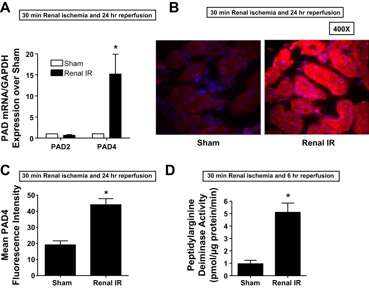
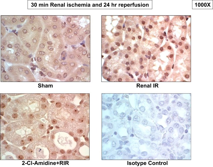
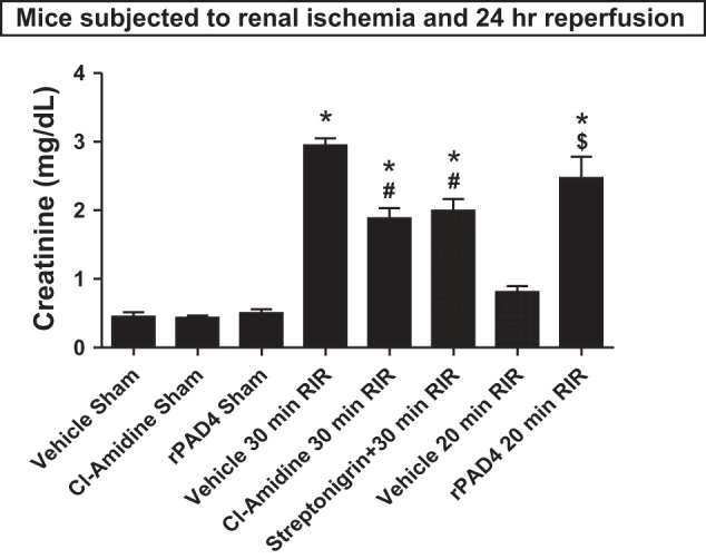
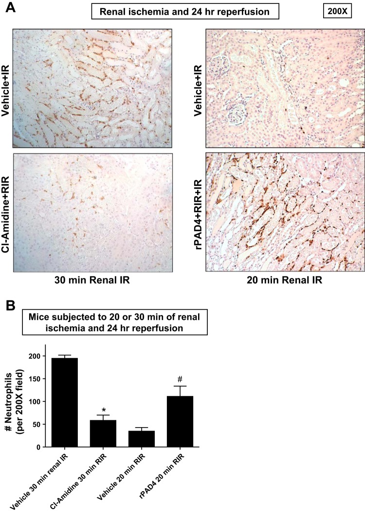
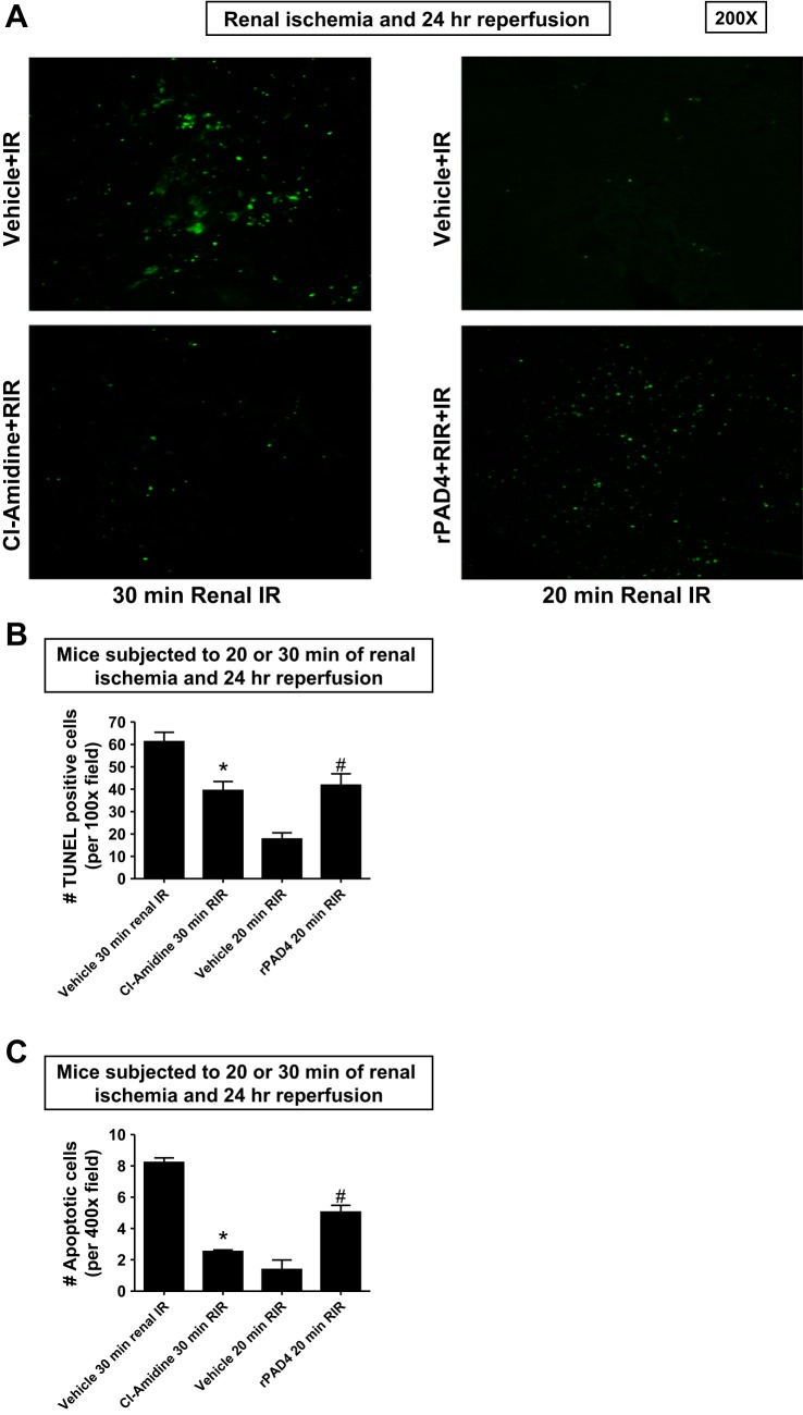
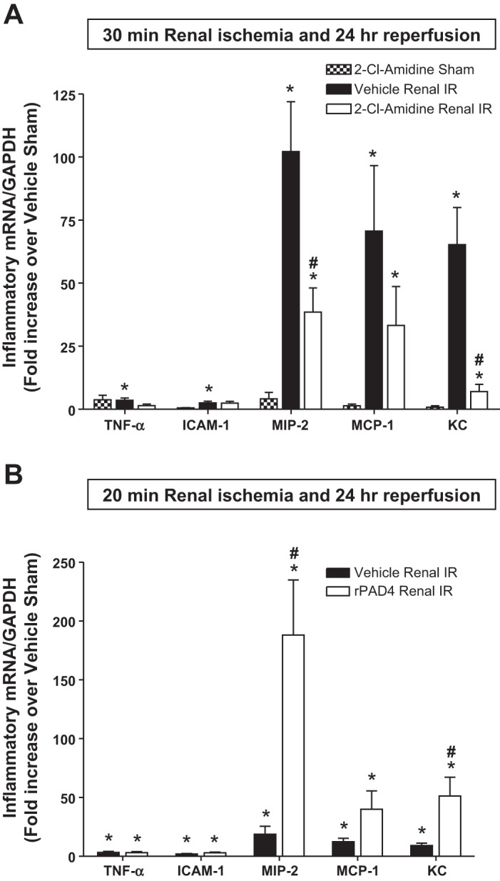
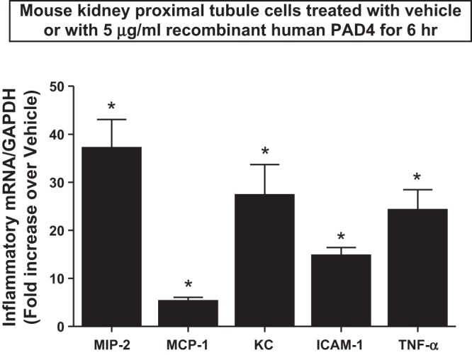
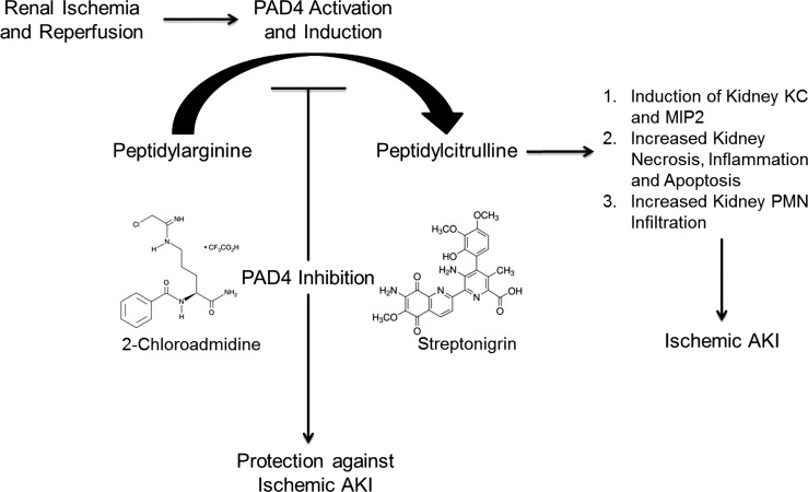
Peptidyl arginine deiminase (PAD)4 is a nuclear enzyme that catalyzes the posttranslational conversion of arginine residues to citrulline. Posttranslational protein citrullination has been implicated in several inflammatory autoimmune diseases, including rheumatoid arthritis, colitis, and multiple sclerosis. Here, we tested the hypothesis that PAD4 contributes to ischemic acute kidney injury (AKI) by exacerbating the inflammatory response after renal ischemia-reperfusion (I/R). Renal I/R injury in mice increased PAD4 activity as well as PAD4 expression in the mouse kidney. After 30 min of renal I/R, vehicle-treated mice developed severe AKI with large increases in plasma creatinine. In contrast, mice pretreated with PAD4 inhibitors (2-chloroamidine or streptonigrin) had significantly reduced renal I/R injury. Further supporting a critical role for PAD4 in generating ischemic AKI, mice pretreated with recombinant human PAD4 (rPAD4) protein and subjected to mild (20 min) renal I/R developed exacerbated ischemic AKI. Consistent with the hypothesis that PAD4 regulates renal tubular inflammation after I/R, mice treated with a PAD4 inhibitor had significantly reduced renal neutrophil chemotactic cytokine (macrophage inflammatory protein-2 and keratinocyte-derived cytokine) expression and had decreased neutrophil infiltration. Furthermore, mice treated with rPAD4 had significantly increased renal tubular macrophage inflammatory protein-2 and keratinocyte-derived cytokine expression as well as increased neutrophil infiltration and necrosis. Finally, cultured mouse kidney proximal tubules treated with rPAD4 had significantly increased proinflammatory chemokine expression compared with vehicle-treated cells. Taken together, our results suggest that PAD4 plays a critical role in renal I/R injury by increasing renal tubular inflammatory responses and neutrophil infiltration after renal I/R.

Keywords: acute kidney injury; apoptosis; citrullination; histone; inflammation; necrosis.


Figures

Fig. 1.
Fig. 1.
Induction and activation of kidney peptidyl arginine deiminase (PAD)4 after ischemia-reperfusion (I/R) injury. A: PAD4 mRNA expression measured with quantitative RT-PCR increased in the mouse kidney 24 h after renal I/R injury (n = 5–6 experiments). Note the lack of increase in PAD2 mRNA after renal I/R. B: immunohistochemistry images (n = 4 representative experiments) showing increased renal tubular PAD4 protein expression (red fluorescence, ×400) in the mouse kidney 24 h after renal I/R injury. Blue fluorescence indicates 4′,6-diamidino-2-phenylindole (DAPI) nuclear staining. C: PAD4 immunofluorescence intensities quantified in ×400 image fields were significantly higher in mice subjected to renal I/R compared with sham-operated (sham) mice (n = 4). D: increased mouse kidney PAD4 activity 24 h after renal I/R injury (n = 5 experiments). *P < 0.05 vs. the sham group. Error bars represent 1 SE.
Fig. 2.
Fig. 2.
Renal I/R causes PAD4-mediated citrullination of nuclear histone H3. Images are representative of four immunohistochemistry experiments performed to stain for citrullinated histone H3. Kidneys subjected to renal I/R showed increased nuclear citrullinated histone H3 staining (dark brown, ×1,000) compared with those from sham mice. Mice treated with the selective PAD inhibitor 2-chloroamidine (2-Cl-amidine) showed markedly attenuated nuclear citrullinated histone H3 staining after renal I/R. Kidneys incubated with negative isotype control antibody showed no staining in the nucleus (n = 4 experiments).
Fig. 3.
Fig. 3.
PAD4 plays a critical role in renal I/R (RIR) injury. C57/BL6 mice were subjected to sham operation or to 30 min of renal I/R after pretreatment with vehicle (1% DMSO) or with two distinct classes of PAD4 inhibitors (100 mg/kg 2-Cl-amidine or 0.4 mg/kg streptonigrin). Inhibition of PAD4 with 2-Cl-amidine or streptonigrin treatment protected against renal I/R injury. Separate cohorts of mice were subjected to mild (20 min) renal I/R injury after treatment with vehicle or with 10 μg recombinant human PAD4 (rPAD4). In contrast to the renal protection provided by PAD4 inhibitors, rPAD4 treatment exacerbated renal injury after I/R. *P < 0.001 vs. the vehicle-treated sham group; #P < 0.05 vs. the vehicle-treated group subjected to 30 min of renal I/R; $P < 0.001 vs. the vehicle-treated group subjected to 20 min of renal I/R. Error bars represent 1 SE; n = 5–6/group.
Fig. 4.
Fig. 4.
PAD4 inhibition reduces and rPAD4 treatment exacerbates kidney necrosis after renal I/R injury. A: representative hematoxylin and eosin-stained images (from 5 experiments) of mice subjected to 20 or 30 min of renal ischemia and 24 h of reperfusion (magnification: ×200). As expected, kidneys of vehicle-treated mice subjected to 30 min of renal I/R showed severe tubular necrosis and proteinaceous casts with increased tubular dilatation and congestion. PAD4 inhibition with 2-Cl-amidine treatment decreased renal tubular necrosis, congestion, and cast formation after 30 min of renal I/R. Mice subjected to 20 min of renal I/R had mild renal tubular necrosis and proteinaceous casts. In contrast, rPAD4-treated mice subjected to renal I/R had markedly increased renal tubular necrosis with increased tubular dilatation and congestion. B: the renal injury score (scale: 0–4, n = 5) for histology grading was used to grade renal tubular necrosis 24 h after 20 or 30 min of renal I/R. Thirty minutes of renal I/R and 24 h of reperfusion resulted in severe renal tubular injury in vehicle-treated mice. 2-Cl-treated mice subjected to 30 min of renal I/R had significantly lower renal injury scores. Furthermore, 20 min of renal ischemia and 24 h of reperfusion resulted in mild to moderate acute tubular necrosis in vehicle-treated mice. In contrast, rPAD4-treated mice had significantly higher renal injury scores after 20 min renal of I/R. *P < 0.05 vs. the vehicle-treated group subjected to 30 min of renal I/R; #P < 0.001 vs. the vehicle-treated group subjected to 20 min of renal I/R. Error bars represent 1 SE.
Fig. 5.
Fig. 5.
PAD4 inhibition decreases and rPAD4 treatment exacerbates neutrophil infiltration after renal I/R. A and B: representative images (from 5 experiments, magnification: ×200; A) and quantification of infiltrated neutrophils per ×200 field (n = 5; B) of immunohistochemistry of neutrophil infiltration (dark brown) in the kidneys (corticomedullary junction) of mice subjected to 20 or 30 min of renal ischemia and 24 h of reperfusion. Neutrophil infiltration markedly increased in vehicle-treated mice subjected to 30 min of renal I/R concentrated near the corticomedullary junction Again, 2-Cl-amidine-treated mice subjected to 30 min of renal I/R had decreased neutrophil infiltration after 24 h of reperfusion. Twenty minutes of renal ischemia and 24 h of reperfusion resulted in a scant number of neutrophils infiltrating the kidney. In contrast, rPAD4-treated mice had significantly increased numbers of neutrophils infiltrating the kidney after 20 min of renal IR (A and B). *P < 0.05 vs. the vehicle-treated group subjected to 30 min of renal I/R; #P < 0.001 vs. the vehicle-treated group subjected to 20 min of renal I/R. Error bars represent 1 SE.
Fig. 6.
Fig. 6.
PAD4 inhibition decreases and rPAD4 treatment exacerbates renal tubular apoptosis after renal IR. A and B: representative images (from 5 experiments, magnification: ×200; A) and quantification of TUNEL staining to demonstrate renal tubular apoptosis per ×100 field (n = 5; B) in the kidneys of mice subjected to 20 or 30 min of renal ischemia and 24 h of reperfusion. TUNEL staining showed that renal tubule cell apoptosis increased in vehicle-treated wild-type mice subjected to 30 min of renal I/R (representative of 5 experiments; magnification: ×100). 2-Cl-amidine-treated mice subjected to 30 min of renal I/R had significantly decreased TUNEL-positive renal tubular cells 24 h after reperfusion (B). Twenty minutes of renal ischemia and 24 h of reperfusion resulted in a few TUNEL-positive cells. In contrast, rPAD4-treated mice had significantly increased numbers of TUNEL-positive cells after 20 min of renal I/R. C: since TUNEL stains fragmented DNA rather than specifically indicating apoptotic cells, we also counted the number of apoptotic cells from 10 high-power fields in the corticomedullary junction (×400) per kidney. PAD4 inhibition significantly decreased apoptotic renal tubular cells 24 h after 30 min of renal I/R. In contrast, rPAD4-treated mice had significantly increased apoptotic renal tubule cells after 20 min of renal I/R. *P < 0.05 vs. the vehicle-treated group subjected to 30 min of renal I/R; #P < 0.001 vs. the vehicle-treated group subjected to 20 min of renal I/R. Error bars represent 1 SE.
Fig. 7.
Fig. 7.
PAD4 modulates macrophage inflammatory protein (MIP)-2 and keratinocyte-derived cytokine (KC) expression after renal I/R injury. With quantitative RT-PCR, we measured the expression of proinflammatory cytokine and chemokine mRNA in the kidney [TNF-α, ICAM-1, MIP-2, monocyte chemoattractant protein (MCP)-1, and KC] 24 h after renal I/R. Fold increases in proinflammatory mRNAs normalized to GAPDH from quantitative RT-PCRs for each indicated mRNA (n = 6) are shown. A: PAD4 inhibition with 2-Cl-amidine treatment significantly attenuated the expression of neutrophil chemotactic cytokines MIP-2 and KC after 30 min of renal I/R injury. B: mice subjected to mild (20 min) renal I/R had reduced upregulation of proinflammatory cytokines 24 h after injury compared with mice subjected to 30 min of renal I/R. Treatment of mice subjected to 20 min of renal I/R with rPAD4 selectively increased the expression of MIP-2 and KC mRNA. *P < 0.05 vs. sham mice; #P < 0.05 vs. vehicle-treated mice subjected to renal I/R. Error bars represent 1 SE.
Fig. 8.
Fig. 8.
rPAD4 induces proinflammatory mRNA expression in primary cultures of mouse proximal tubules. Primary cultures of mouse proximal tubules were treated with vehicle or 5 μg/ml rPAD4 for 6 h. With quantitative RT-PCR, we measured the expression of proinflammatory cytokine and chemokine mRNA in these cells (MIP-2, MCP-1, KC, ICAM-1, and TNF-α). Fold increases in proinflammatory mRNAs normalized to GAPDH from quantitative RT-PCR reactions for each indicated mRNA (n = 6) are shown. Quantitative RT-PCR revealed that rPAD4 treatment significantly induced the expression of all proinflammatory cytokine mRNAs measured (MIP-2, MCP-1, KC, ICAM-1, and TNF-α). *P < 0.05 vs. vehicle-treated cells. Error bars represent 1 SE.
Fig. 9.
Fig. 9.
Schematic of proposed role for PAD4 activation and induction in ischemic acute kidney injury (AKI). After renal ischemia and reperfusion, PAD4 activation and induction converts peptidyl arginine to peptidyl citrulline. Via mechanisms that are still unclear, peptidyl citrullination results in significant renal tubular inflammation, KC and MIP-2 mRNA induction, and increased kidney necrosis, inflammation, and apoptosis after renal I/R. Inhibition of PAD4 may be an effective therapeutic strategy to protect against ischemic AKI. Selective PAD4 inhibitors [2-Cl-amidine and streptonigrin (structures shown)] protected against kidney necrosis, apoptosis, and inflammation after renal I/R. PMNs, polymorphonuclear neutrophils.

Similar articles

Cited by

References

    1. Allam R, Scherbaum CR, Darisipudi MN, Mulay SR, Hagele H, Lichtnekert J, Hagemann JH, Rupanagudi KV, Ryu M, Schwarzenberger C, Hohenstein B, Hugo C, Uhl B, Reichel CA, Krombach F, Monestier M, Liapis H, Moreth K, Schaefer L, Anders HJ. Histones from dying renal cells aggravate kidney injury via TLR2 and TLR4. J Am Soc Nephrol 23: 1375–1388, 2012 - PMC - PubMed
    1. Anzilotti C, Pratesi F, Tommasi C, Migliorini P. Peptidylarginine deiminase 4 and citrullination in health and disease. Autoimmun Rev 9: 158–160, 2010 - PubMed
    1. Aronson S, Blumenthal R. Perioperative renal dysfunction and cardiovascular anesthesia: concerns and controversies. J Cardiothorac Vasc Anesth 17: 117–130, 1998 - PubMed
    1. Bicker KL, Thompson PR. The protein arginine deiminases: structure, function, inhibition, and disease. Biopolymers 99: 155–163, 2013 - PMC - PubMed
    1. Bonventre JV, Weinberg JM. Recent advances in the pathophysiology of ischemic acute renal failure. J Am Soc Nephrol 14: 2199–2210, 2003 - PubMed

Publication types