. 2014 Oct;94(2):403-17.
doi: 10.1111/mmi.12773. Epub 2014 Sep 11.

Identification of molecular mechanisms used by Finegoldia magna to penetrate and colonize human skin

Affiliations

Identification of molecular mechanisms used by Finegoldia magna to penetrate and colonize human skin

Elizabeth C Murphy et al. Mol Microbiol. 2014 Oct.

Abstract

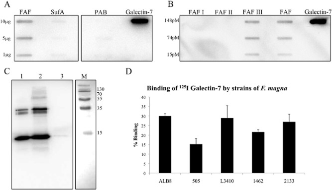
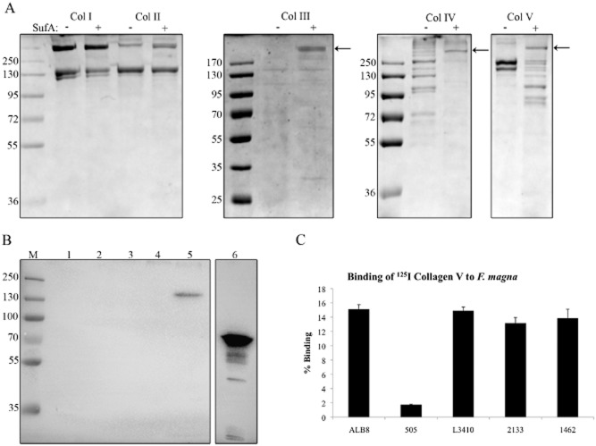
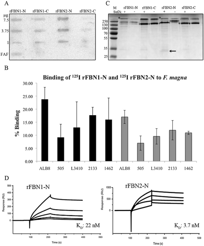
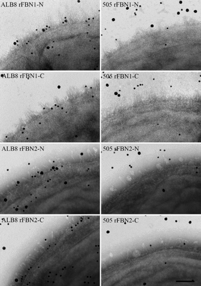
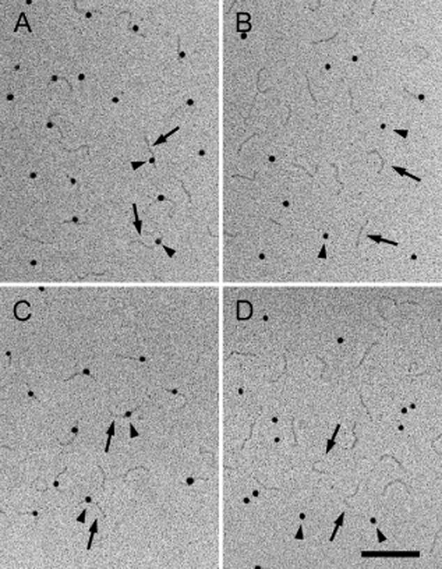
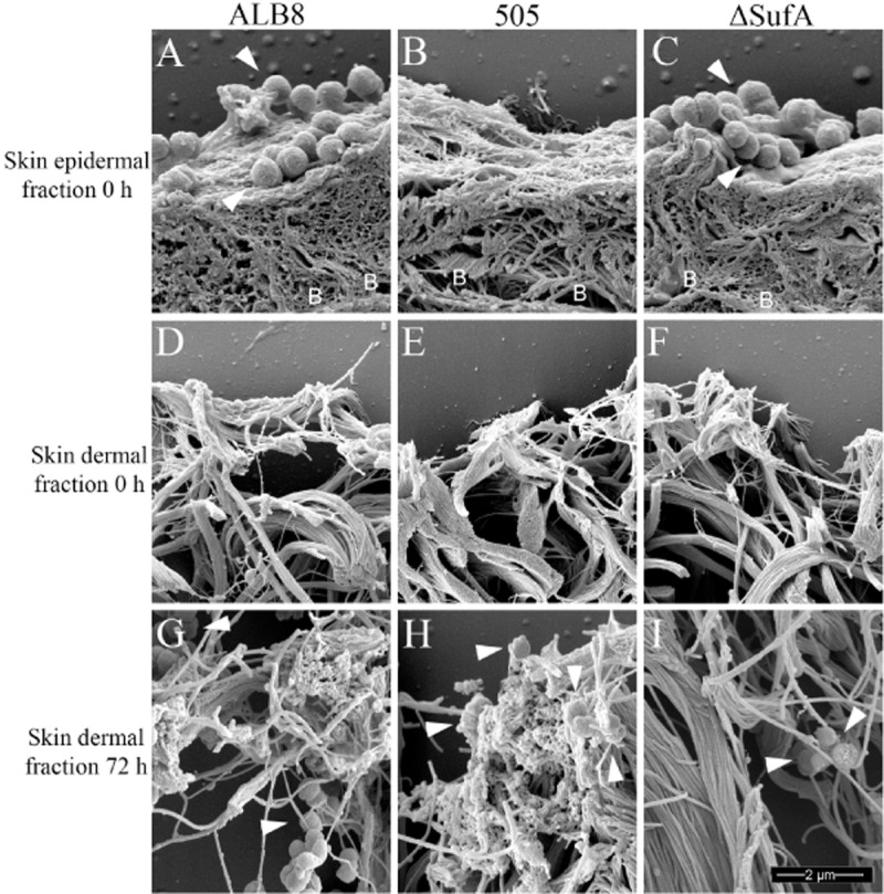
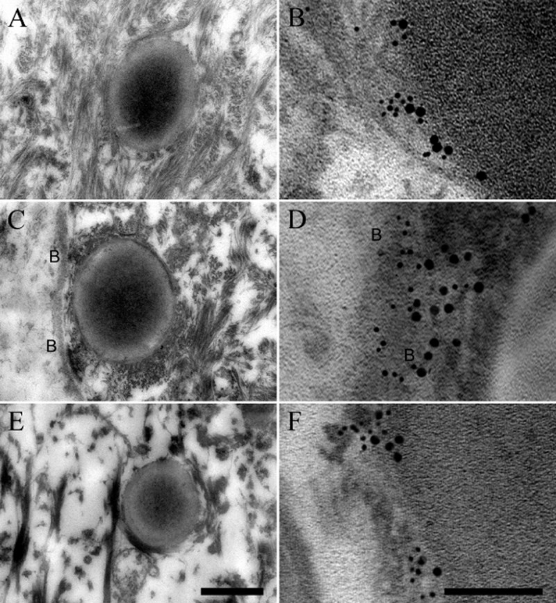
Finegoldia magna is a Gram-positive anaerobic commensal of the human skin microbiota, but also known to act as an opportunistic pathogen. Two primary virulence factors of F. magna are the subtilisin-like extracellular serine protease SufA and the adhesive protein FAF. This study examines the molecular mechanisms F. magna uses when colonizing or establishing an infection in the skin. FAF was found to be essential in the initial adherence of F. magna to human skin biopsies. In the upper layers of the epidermis FAF mediates adhesion through binding to galectin-7 - a keratinocyte cell marker. Once the bacteria moved deeper into the skin to the basement membrane layer, SufA was found to degrade collagen IV which forms the backbone structure of the basement membrane. It also degraded collagen V, whereby F. magna could reach deeper dermal tissue sites. In the dermis, FAF interacts with collagen V and fibrillin, which presumably helps the bacteria to establish infection in this area. The findings of this study paint a clear picture of how F. magna interacts with human skin and explain how it is such a successful opportunistic pathogen in chronic wounds and ulcers.


Figures

Figure 1
Figure 1
Protein FAF binds to galectin-7.A. Proteins FAF, SufA and PAB were applied in various amounts to a PVDF membrane. The membrane was incubated with a 1.33 μM solution of galectin-7 followed by probing with a rabbit anti-galectin-7 antibody. Galectin-7 was included as a positive control.B. Different recombinant length fragments of protein FAF (see Experimental procedures) were applied to a PVDF membrane and binding to galectin-7 was investigated by immunoblotting as in (A).C. A solution of F. magna strain ALB8 (2 × 109 cfu ml−1) in PBS was incubated with galectin-7 for 1 h at 37°C. Protein bound to the bacterial surface was eluted with low pH buffer and analysed by SDS-PAGE and immunoblotting using a galectin-7 antibody. M: Biorad Broad Range Molecular Weight Marker; Lane 1: Galectin-7 positive control 1 μg; Lane 2: F. magna incubated with galectin-7; Lane 3: F. magna incubated with PBS.D. Percentage binding of 125I-labelled galectin-7 to different strains of F. magna. Bars represent mean ± S.E. of at least three experiments.
Figure 2
Figure 2
SufA and FAF interact with collagens.A. SufA cleavage of collagens type I–V was determined by co-incubation for 3 h at 37°C followed by analysis on 8% SDS-PAGE. Lanes containing collagen co-incubated with SufA are indicated with a + sign. The position of SufA is indicated by a black arrow (not visible in cleavage of collagen I and II due to the position of the collagen bands). As a control, collagens were also incubated without SufA for the same length of time, indicated by a – sign.B. Binding of FAF to collagens I, II, III, IV and V was determined by electrophoresing 3.5 μg of each collagen on an 8% SDS-PAGE gel followed by incubation with FAF and immunoblotting with an anti-FAF antibody. M: Biorad Broad Range Molecular Weight Marker; Lane 1: Collagen I; Lane 2: Collagen II; Lane 3: Collagen III; Lane 4: Collagen IV; Lane 5: Collagen V; Lane 6: FAF 0.8 μg.C. Percentage binding of 125I-labelled collagen V to different strains of F. magna. Bars represent mean ± S.E. of at least three experiments.
Figure 3
Figure 3
Examination of the interaction of FAF and SufA with fibrillin.A. Indicated amounts of various recombinant fibrillin fragments and FAF (1 mg) were applied in slots to a PVDF membrane. The membrane was incubated with 125I-labelled FAF and bound FAF was determined using the Fuji Imaging system.B. Percentage binding of 125I-labelled fibrillin fragments rFBN1-N and rFBN2-N to different strains of F. magna. The black bars represent binding to rFBN1-N and the grey bars represent binding to rFBN2-N. Bars represent mean ± S.E. of at least three experiments.C. SufA cleavage of fibrillin was determined by co-incubation with the various fragments for 3 h at 37°C followed by analysis of cleavage on an 8% SDS-PAGE gel. Lanes containing fibrillin co-incubated with SufA are indicated with a + sign. As a control, fibrillin fragments were also incubated without SufA for the same length of time, indicated by a – sign. The black arrow indicates the 35 kDa fragment released by SufA from rFBN2-N. A protein band representing SufA is clearly seen above the N-terminal fibrillin fragments and is indicated by a star. The SufA band is hidden by the C-terminal fibrillin fragments due to their size.D. FAF binds rFBN1-N and rFBN2-N in surface plasmon resonance experiments. FAF was immobilized on a sensor chip, and increasing concentrations of rFBN1-N/rFBN2-N were injected over the surface. RU, response units.
Figure 4
Figure 4
Transmission electron microscopy demonstrating binding of protein FAF on the ALB8 surface to the N-terminal region of fibrillin. F. magna strains ALB8 and 505 were incubated with anti-FAF antibodies and the various fibrillin fragments labelled with colloidal gold (anti-FAF antibody, 5 nm gold and fibrillin fragments, 20 nm gold). Samples were then prepared for transmission electron microscopy. Left panels: F. magna strain ALB8. Right panels: F. magna strain 505. The scale bar represents 50 nm.
Figure 5
Figure 5
Electron micrographs after rotary shadowing showing complex formation between FAF and N-terminal fragments of fibrillin. The arrowheads point to FAF labelled with 10 nm colloidal gold and the arrows point to extended fibrillin molecules. The FAF molecules exhibit a globular appearance due to decoration with gold particles. A: Interaction of FAF with rFBN1-N; B: Interaction of FAF with rFBN1-C; C: Interaction of FAF with rFBN2-N; D: Interaction of FAF with rFBN2-C. The scale bar represents 100 nm.
Figure 6
Figure 6
Scanning electron microscopy demonstrating the effect of FAF and SufA on binding and interaction of F. magna with human skin biopsies. Different strains of F. magna were incubated with human skin biopsies anaerobically for 1 h at 37°C, followed by washing to remove unbound bacteria. Skin harbouring bound F. magna was then anaerobically incubated for 72 h at 37°C to examine the effects of FAF and SufA. The biopsies were then prepared for scanning electron microscopy. Bacteria are indicated by white arrowheads and the location of the basement membrane is shown by letters B. Images represent: Skin epidermal fraction at 0 h incubated with (A) F. magna ALB8 (B) F. magna 505 (C) F. magna ALB8 ΔSufA; Skin dermal fraction at 0 h incubated with (D) F. magna ALB8 (E) F. magna 505 (F) F. magna ALB8 ΔSufA; Skin dermal fraction at 72 h co-incubated with (G) F. magna ALB8 (H) F. magna 505 (I) F. magna ALB8 ΔSufA. The scale bar represents 2 μm. Evaluation of the data is seen in Table 1.
Figure 7
Figure 7
Transmission electron micrographs visualizing F. magna ALB8 interacting with galectin-7, BM-40 and fibrillin in human skin. Human skin biopsies were anaerobically incubated with F. magna strain ALB8 for 1 h at 37°C. Non-adherent bacteria were removed and the biopsies were anaerobically incubated for 24–72 h at 37°C and prepared for ultrathin sectioning and transmission electron microscopy. Sections were also subjected to immunolabelling with anti-FAF antibody (labelled with 5 nm colloidal gold) and anti-galectin-7, anti-BM-40 and anti-rFBN2-N antibodies (labelled with 20 nm colloidal gold) respectively. The location of the basement membrane is indicated by letters B. Trans section of F. magna ALB8 at the (A) skin epidermis after 24 h; (C) basement membrane after 48 h; (E) dermis after 72 h. The scale bar represents 0.5 μm. Gold labelled FAF on the surface of F. magna ALB8 interacting with (B) Gold labelled galectin-7 in the epidermis after 24 h; (D) Gold labelled BM-40 in the basement membrane after 48 h; (F) Gold labelled rFBN2-N in the dermis after 72 h. The scale bar represents 100 nm.

References

    1. Baschong W. Wrigley NG. Small colloidal gold conjugated to Fab fragments or to immunoglobulin G as high-resolution labels for electron microscopy: a technical overview. J Electron Microsc Tech. 1990;14:313–323. - PubMed
    1. Birk DE, Fitch JM, Babiarz JP. Linsenmayer TF. Collagen type I and type V are present in the same fibril in the avian corneal stroma. J Cell Biol. 1988;106:999–1008. - PMC - PubMed
    1. Björck L. Protein L. A novel bacterial cell wall protein with affinity for Ig L chains. J Immunol. 1988;140:1194–1197. - PubMed
    1. Björck L. Kronvall G. Purification and some properties of streptococcal protein G, a novel IgG-binding reagent. J Immunol. 1984;133:969–974. - PubMed
    1. Bober M, Enochsson C, Collin M. Mörgelin M. Collagen VI is a subepithelial adhesive target for human respiratory tract pathogens. J Innate Immun. 2010;2:160–166. - PubMed

Publication types

MeSH terms