. 2014 Oct 10;289(41):28707-18.
doi: 10.1074/jbc.M114.580696. Epub 2014 Aug 27.

Vps4 stimulatory element of the cofactor Vta1 contacts the ATPase Vps4 α7 and α9 to stimulate ATP hydrolysis

Affiliations

Vps4 stimulatory element of the cofactor Vta1 contacts the ATPase Vps4 α7 and α9 to stimulate ATP hydrolysis

Brian A Davies et al. J Biol Chem. .

Abstract

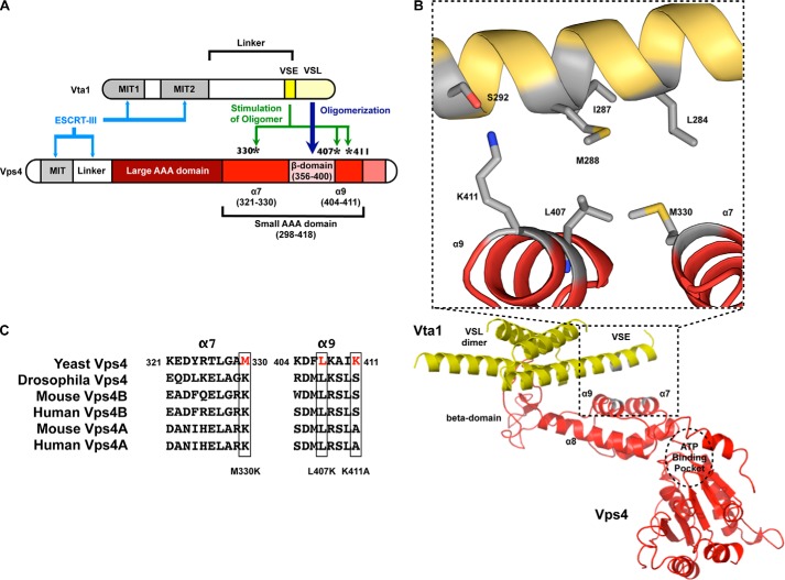
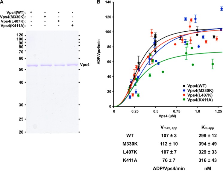
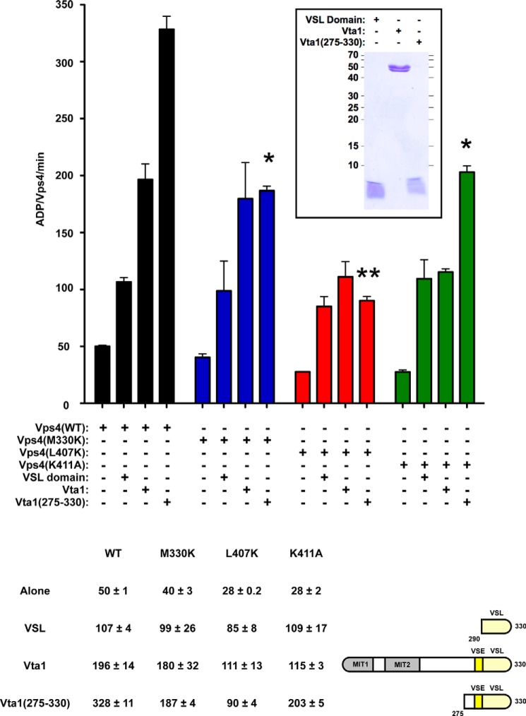
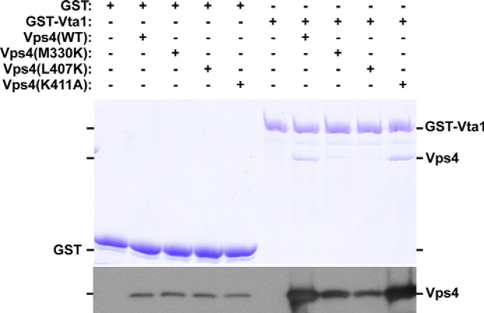
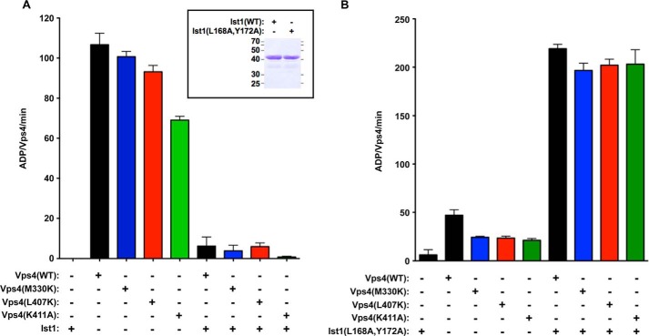
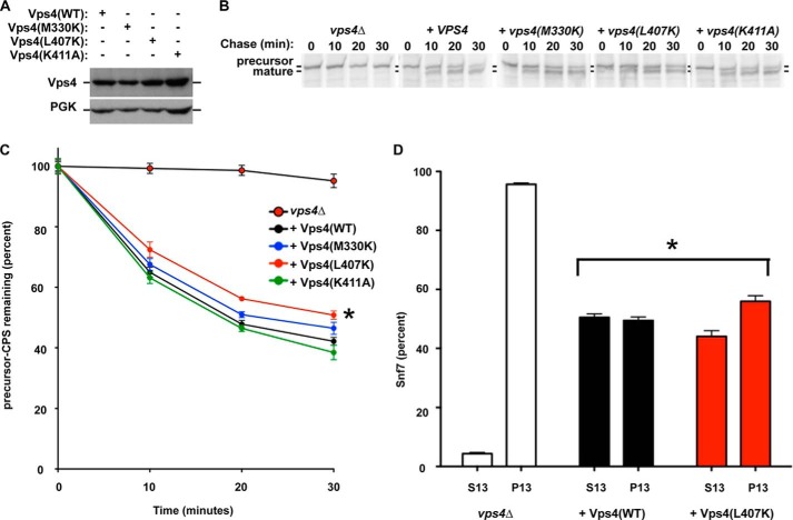
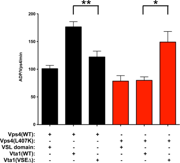
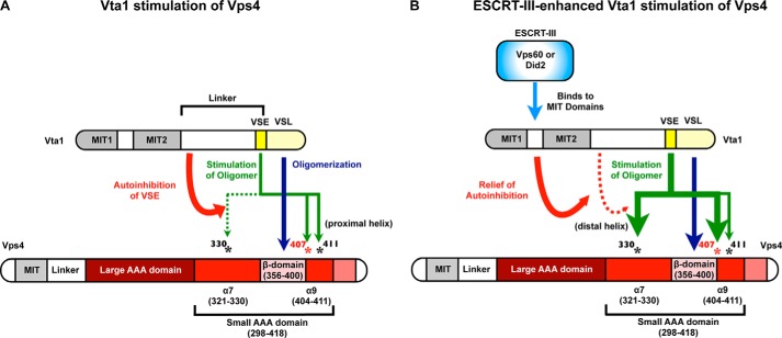
The endosomal sorting complexes required for transport (ESCRTs) function in a variety of membrane remodeling processes including multivesicular body sorting, abscission during cytokinesis, budding of enveloped viruses, and repair of the plasma membrane. Vps4 ATPase activity modulates ESCRT function and is itself modulated by its cofactor Vta1 and its substrate ESCRT-III. The carboxyl-terminal Vta1/SBP-1/Lip5 (VSL) domain of Vta1 binds to the Vps4 β-domain to promote Vps4 oligomerization-dependent ATP hydrolysis. Additionally, the Vps4 stimulatory element (VSE) of Vta1 contributes to enhancing Vps4 oligomer ATP hydrolysis. The VSE is also required for Vta1-dependent stimulation of Vps4 by ESCRT-III subunits. However, the manner by which the Vta1 VSE contributes to Vps4 activation is unknown. Existing structural data were used to generate a model of the Vta1 VSE in complex with Vps4. This model implicated residues within the small ATPase associated with various activities (AAA) domain, specifically α-helices 7 and 9, as relevant contact sites. Rational generation of Vps4 mutants defective for VSE-mediated stimulation, as well as intergenic compensatory mutations, support the validity of this model. These findings have uncovered the Vps4 surface responsible for coordinating ESCRT-III-stimulated Vta1 input during ESCRT function and identified a novel mechanism of Vps4 stimulation.

Keywords: ATPases Associated with Diverse Cellular Activities (AAA); Endosomal Sorting Complexes Required for Transport (ESCRT); Endosome; Enzyme Mechanism; Lysosome; Membrane Trafficking; Vps4; Vta1.


Figures

FIGURE 1.
FIGURE 1.
Modeling of the VSE into the structure of the VSL domain in complex with the small AAA and β-domains of Vps4. A, cartoon of Vps4 structural domains. The β-domain is an insert within the small AAA domain. Met-330 is in α7 preceding the β-domain, whereas Leu-407 and Lys-411 are in α9 immediately following the β-domain. α9 is located closer to the β-domain (proximal helix), whereas α7 is more distant (distal helix). B, the structure of the Vta1 VSL dimer bound to the Vps4 β-domain and small AAA domain (Protein Data Bank code 3MHV; MMDB 85335) was used to align structures of the Vps4 AAA domain (residues 119–437) (Protein Data Bank code 2QP9; MMDB 59522; red) and the Vta1 carboxyl terminus (residues 281–330) (Protein Data Bank code 2RKL; MMDB 62002; yellow). The Vta1 VSE falls on a flexible helix preceding the VSL domain, and alignment positioned the VSE neighboring Vps4 α7 and α9. Residues of the Vta1 VSE (Leu-284, Ile-287, and Met-288) and Vps4 small AAA domain (Met-330 of α7 and Leu-407 of α9) that appear to mediate VSE-Vps4 interaction are indicated in gray in the inset. Vta1 Ser-292 is not required for VSE stimulation but appears to interact with Vps4 Lys-411 of α9. These residues are also depicted in gray. C, sequence alignment of α7 and α9 from S. cerevisiae Vps4, D. melanogaster Vps4, M. musculus Vps4A and Vps4B, and H. sapiens Vps4A and Vps4B. Leu-407 is conserved from yeast to man.
FIGURE 2.
FIGURE 2.
Vps4 ATPase activity and Ist1 inhibition are not disrupted by mutations in Vps4 α7 and α9. A, Coomassie staining of equivalent amounts of WT and mutant Vps4 proteins. B, titration (250 nm to 1.25 μm) of WT (black) and Vps4 mutants (M330K, blue; L407K, red; K411A, green) was performed in the presence of 4 mm ATP. Rates are indicated as ADP generated/Vps4/min. The concentrations yielding half-maximal activity (Km, app) and the apparent Vmax are indicated in the accompanying table.
FIGURE 3.
FIGURE 3.
Mutations in Vps4 α7 and α9 disrupt aspects of VSE stimulation. WT (black) and mutant Vps4 (M330K, blue; L407K, red; K411A, green) ATPase activity alone (250 nm) or in the presence of 8 μm VSL domain (His6-Vta1(290–330)), WT Vta1 (His6-Vta1(1–330)), and Vta1(275–330) (His6-Vta1(275–330)). The rates are indicated as ADP generated/Vps4/min; the rates are also indicated in the table. WT and mutant Vps4 proteins exhibit comparable ATPase activity in the presence of VSL domain, and these rates are enhanced from the activities of the Vps4 proteins alone. Vps4(L407K) and Vps4(K411A) in the presence of Vta1 exhibit reduced activity compared with WT Vps4 and Vps4(M330K) in the presence of Vta1 (p value < 0.05). Vps4(M330K) and Vps4(K411A) in the presence of Vta1(275–330) (ESCRT-III-activated VSE) exhibit reduced activity compared with WT Vps4 in the presence of Vta1(275–330) (p value < 0.05; indicated by *), and activity of Vps4(L407K) with Vta1(275–330) is further reduced (p value < 0.01; indicated by **). Coomassie staining of equivalent amounts of VSL, Vta1, and Vta1(275–330) is indicated in the inset.
FIGURE 4.
FIGURE 4.
Vps4 M330K and L407K partially disrupt in vitro association with GST-Vta1. GST or GST-Vta1 pulldown experiments were performed with purified WT and Vps4 mutants. Eluted material was analyzed by Coomassie staining and Western blotting for Vps4. WT and Vps4 mutants were isolated with GST-Vta1 more effectively than with GST alone. Vps4(WT) and Vps4(K411A) exhibited similar isolation with GST-Vta1, but isolation of Vps4(M330K) and Vps4(L407K) was reduced. The data presented are representative of experiments performed more than three times.
FIGURE 5.
FIGURE 5.
Direct ESCRT-III stimulation and Ist1 inhibition are not disrupted by mutations in Vps4 α7 and α9. A, 500 nm WT (black) and mutant Vps4 (M330K, blue; L407K, red; K411A, green) ATPase activity alone or in the presence of 10 μm WT Ist1. WT Ist1 inhibits WT Vps4 and the Vps4 mutants similarly. The rates are indicated as ADP generated/Vps4/min. Coomassie staining of equivalent amounts of Ist1 and Ist1(L168A,Y172A) is indicated in the inset. B, 250 nm WT (black) and mutant Vps4 (M330K, blue; L407K, red; K411A, green) ATPase activity alone or in the presence of 1 μm Ist1(L168A,Y172A). Ist1(L168A,Y172A) stimulates WT Vps4 and the Vps4 mutants similarly.
FIGURE 6.
FIGURE 6.
Leu-407 contributes to Vps4 function in vivo. A, Western blotting analysis of Vps4 levels in vps4Δ yeast expressing WT and Vps4 mutants. Western blotting for phosphoglycerate kinase was performed as a loading control. B and C, pulse-chase immunoprecipitation was performed on endogenous CPS in vps4Δ cells transformed with vector or plasmids expressing WT Vps4 or Vps4 point mutants. CPS maturation was quantitated using a phosphoimager and plotted relative to time zero. CPS maturation kinetics in cells expressing Vps4(L407K) was significantly different (*, p values < 0.01) from cells lacking Vps4 or cells expressing WT Vps4. D, subcellular fractionation of the ESCRT-III subunit Snf7 was performed on vps4Δ cells transformed with vector or plasmids expressing WT Vps4 or Vps4(L407K). Snf7 fractionation in cells expressing Vps4(L407K) was significantly different (*, p values < 0.01) from cells lacking Vps4 or cells expressing WT Vps4.
FIGURE 7.
FIGURE 7.
Compensatory mutation of the VSE restores activation of Vps4(L407K). ATPase activity of 250 nm WT Vps4 (black) and Vps4(L407K) (red) in the presence of 8 μm VSL domain, WT Vta1, and Vta1(VSEΔ). The rates are indicated as ADP generated/Vps4/min. The activity of WT Vps4 in the presence of Vta1(VSEΔ) is reduced compared with activity with Vps4 and WT Vta1 (p value < 0.01; **). By contrast, the activity of Vps4(L407K) in the presence of Vta1(VSEΔ) is increased compared with activity with Vps4(L407K) with WT Vta1 (p value < 0.05; *).
FIGURE 8.
FIGURE 8.
Vta1 stimulates Vps4 via the β-domain and the small AAA domain α7 and α9. A, the Vta1 VSL domain stimulates Vps4 ATPase activity via the Vps4 β-domain. In the absence of ESCRT-III binding, the VSE contributes to stimulation of Vps4 ATPase activity by contacting the Vps4 α9 residues Leu-407 and Lys-411. However, Vta1 linker region autoinhibition of the VSE curtails the extent of stimulation in the absence of ESCRT-III such that contact with Vps4 α7 (Met-330) is not required. B, ESCRT-III binding Vta1 relieves autoinhibition of the VSE to facilitate contact with Vps4 α7 residue Met-330 to further enhance Vps4 ATPase activity. Contact with α9 also contributes to this ESCRT-III-enhanced stimulation but may not be required. Leu-407 of α9 is key for activation via both α7 and α9 and is conserved from yeast to humans.

References

    1. Jimenez A. J., Maiuri P., Lafaurie-Janvore J., Divoux S., Piel M., Perez F. (2014) ESCRT machinery is required for plasma membrane repair. Science 343, 1247136–1247136 - PubMed
    1. Agromayor M., Martin-Serrano J. (2013) Knowing when to cut and run: mechanisms that control cytokinetic abscission. Trends Cell Biol. 23, 433–441 - PubMed
    1. Weissenhorn W., Poudevigne E., Effantin G., Bassereau P. (2013) How to get out: ssRNA enveloped viruses and membrane fission. Curr. Opin. Virol. 3, 159–167 - PMC - PubMed
    1. Hanson P. I., Cashikar A. (2012) Multivesicular body morphogenesis. Annu. Rev. Cell Dev. Biol. 28, 337–362 - PubMed
    1. Votteler J., Sundquist W. I. (2013) Virus budding and the ESCRT pathway. Cell Host Microbe 14, 232–241 - PMC - PubMed

Publication types

MeSH terms

Substances

LinkOut - more resources