Review
. 2014 Aug 28;55(4):328-36.
doi: 10.3325/cmj.2014.55.328.

Virchow-Robin space and aquaporin-4: new insights on an old friend

Affiliations
Review

Virchow-Robin space and aquaporin-4: new insights on an old friend

Tsutomu Nakada. Croat Med J. .

Abstract

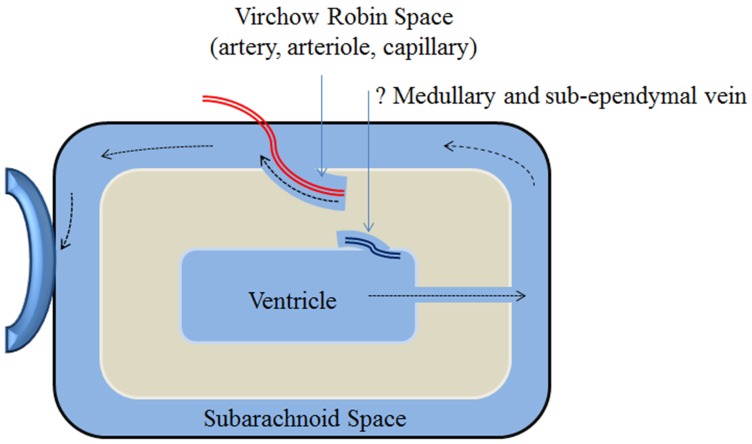
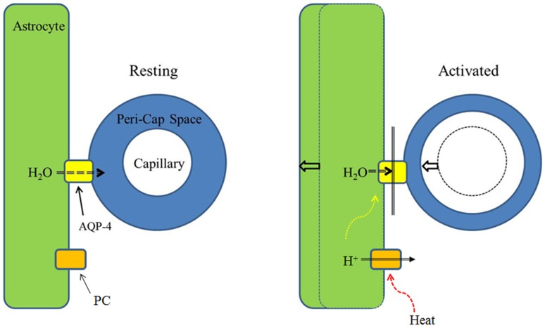
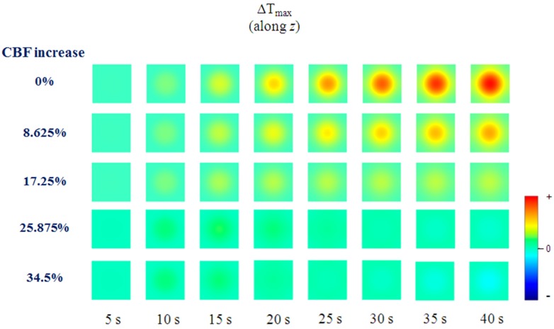
Recent studies have strongly indicated that the classic circulation model of cerebrospinal fluid (CSF) is no longer valid. The production of CSF is not only dependent on the choroid plexus but also on water flux in the peri-capillary (Virchow Robin) space. Historically, CSF flow through the Virchow Robin space is known as interstitial flow, the physiological significance of which is now fully understood. This article briefly reviews the modern concept of CSF physiology and the Virchow-Robin space, in particular its functionalities critical for central nervous system neural activities. Water influx into the Virchow Robin space and, hence, interstitial flow is regulated by aquaporin-4 (AQP-4) localized in the endfeet of astrocytes, connecting the intracellular cytosolic fluid space of astrocytes and the Virchow Robin space. Interstitial flow has a functionality equivalent to systemic lymphatics, on which clearance of β-amyloid is strongly dependent. Autoregulation of brain blood flow serves to maintain a constant inner capillary fluid pressure, allowing fluid pressure of the Virchow Robin space to regulate regional cerebral blood flow (rCBF) based on AQP-4 gating. Excess heat produced by neural activities is effectively removed from the area of activation by increased rCBF by closing AQP-4 channels. This neural flow coupling (NFC) is likely mediated by heat generated proton channels.


Figures

Figure 1
Figure 1
Schematic presentation of the Virchow Robin space and interstitial flow. The ventricles and subarachnoid space represent the cerebrospinal fluid (CSF) space in the brain. The Virchow Robin space is a continuous canal surrounding penetrating vessels. Interstitial flow runs within the Virchow Robin space and drains into the subarachnoid space. Contrary to the classical concept of CSF flow, water CSF within the subarachnoid space is now believed to be dependent on the interstitial flow in the Virchow Robin space. Although not well accepted yet, the Virchow Robin space likely exists surrounding the medullary veins and sub-ependymal veins. As shown in Figure 2, water influx from the systemic circulation into CSF is strongly dependent on the interstitial flow in the Virchow Robin space through aquaporin-4 (AQP-4). Schematic is shown in Figure 4.
Figure 2
Figure 2
In vivo dynamic study of water influx (13). Detailed description of H2O17 JJ vicinal coupling proton exchange (JJVCPE) imaging can be found in the Methods. In brief, the technique allowed for tracing water molecules in vivo non-invasively. The figure gives a summary of quantitative analysis of water influx into the region of interest (ROI). I0 (see Figure 6 for definition) represents dynamic signal intensity, the decline of which inversely correlates with H2O17 influx into the region, namely, higher water influx gives lower I0 value. Values of I0 in cortex and basal ganglia (BG) are virtually identical among the three groups. In contrast, I0 of cerebrospinal fluid (CSF) within the third ventricle is significantly higher in AQP-4 knock out (KO) mice compared to AQP-1 KO and wild type (WT) mice. I0 of CSF within the third ventricle in AQP-1 KO mice is virtually identical to WT mice. The data indicate that water influx into the CSF is regulated by AQP-4, and not by AQP-1. *P < 0.01 vs WT, §P < 0.01 vs AQP1(−/−). WT: wild type, AQP1(−/−): AQP1 knockout, AQP4(−/−): AQP4 knockout mice. Note: WT and KO mice are phenotypically indistinguishable.
Figure 3
Figure 3
Water influx study in transgenic mice. H2O17 JJ vicinal coupling proton exchange (JJVCPE) imaging dynamic study showed that only senile plaque bearing transgenic mice (5xFamilial Alzheimer Disease [FAD]) showed a decline in water influx into the cerebrospinal fluid (CSF) system similar to aquaporin-4 (AQP-4) knockout mice. β-amyloid overproducing transgenic mice without senile plaque formation (C2a-5FAD) showed a virtually identical influx with control mice (C57/BL6). The study indicates that disturbance in β-amyloid clearance through the interstitial flow play a critical, if not sole, role in the pathogenesis of Alzheimer disease.
Figure 4
Figure 4
Schematic presentation of the hypothesis. Neural activity is known to produce two distinctive phenomena, namely, astrocyte swelling and increased regional cerebral blood flow (rCBF). Since an increase in rCBF associated with neural activities occurs within an area limited to 250 µm around the site of the neural activity, it is very likely to be a phenomenon associated with capillaries. Inhibition of aquaporin-4 (AQP-4) effectively increased rCBF, supporting the hypothesis presented here. Inhibition of AQP-4 results in blockage of water flow from astrocyte into the Virchow Robin space (Peri-cap space). This in turn results in astrocyte swelling and capillary expansion due to reduction of the peri-capillary Virchow Robin space. The process associated with brain activation can be explained in the same manner. The trigger is likely to be excess heat produced by neural activities, which in turn open heat gated proton channels (PC) similar to those found in leukocyte.
Figure 5
Figure 5
Schematic presentation of temperature changes-associated activation. A detailed description of simulation study can be found in the Methods. The figure shows temperature changes of the activated area (Figure 7) with continuous neural activities in seconds and associated percentage of cerebral blood flow (CBF) increase. Note that with 34.5% increase in CBF, virtually 100% of excess heat can be removed from the cortex.
Figure 6
Figure 6
Intensities at the steady state of each area, expressed as percentage against the averaged intensity of identical pixel prior to administration of H2O17, were determined by fitting their time course by the function: I = I0+ae(-bt). I0 denotes the normalized signal intensity at infinite time (t = ∞) calculated from the fitted curve (13).
Figure 7
Figure 7
Simulation architecture.

References

    1. Oreskovic D, Klarica M. The formation of cerebrospinal fluid: Nearly a hundred years of interpretations and misinterpretations. Brain Res Rev. 2010;64:241–62. doi: 10.1016/j.brainresrev.2010.04.006. - DOI - PubMed
    1. Virchow R. Ueber die Erweiterung kleinerer Gefaesse. Arch Pathol Anat Physiol Klin Med. 1851;3:427–62.
    1. Robin C. Recherches sur quelques particularites de la structure des capillaires de l’encephale. J Physiol Homme Animaux. 1859;2:537–48.
    1. Esiri MM, Gay D. Immunological and neuropathological significance of the Virchow-Robin space. J Neurol Sci. 1990;100:3–8. doi: 10.1016/0022-510X(90)90004-7. - DOI - PubMed
    1. Weller RO. Pathology of cerebrospinal fluid and interstitial fluid of the CNS: significance for Alzheimer disease, prion disorders and multiple sclerosis. J Neuropathol Exp Neurol. 1998;57:885–94. - PubMed

Publication types