. 2014 Nov;88(21):12825-38.
doi: 10.1128/JVI.02139-14. Epub 2014 Aug 27.

Epstein-Barr virus late gene transcription depends on the assembly of a virus-specific preinitiation complex

Affiliations

Epstein-Barr virus late gene transcription depends on the assembly of a virus-specific preinitiation complex

Valentin Aubry et al. J Virol. 2014 Nov.

Abstract

During their productive cycle, herpesviruses exhibit a strictly regulated temporal cascade of gene expression that has three general stages: immediate early (IE), early (E), and late (L). Promoter complexity differs strikingly between IE/E genes and L genes. IE and E promoters contain cis-regulating sequences upstream of a TATA box, whereas L promoters comprise a unique cis element. In the case of the gammaherpesviruses, this element is usually a TATT motif found in the position where the consensus TATA box of eukaryotic promoters is typically found. Epstein-Barr virus (EBV) encodes a protein, called BcRF1, which has structural homology with the TATA-binding protein and interacts specifically with the TATT box. However, although necessary for the expression of the L genes, BcRF1 is not sufficient, suggesting that other viral proteins are also required. Here, we present the identification and characterization of a viral protein complex necessary and sufficient for the expression of the late viral genes. This viral complex is composed of five different proteins in addition to BcRF1 and interacts with cellular RNA polymerase II. During the viral productive cycle, this complex, which we call the vPIC (for viral preinitiation complex), works in concert with the viral DNA replication machinery to activate expression of the late viral genes. The EBV vPIC components have homologs in beta- and gammaherpesviruses but not in alphaherpesviruses. Our results not only reveal that beta- and gammaherpesviruses encode their own transcription preinitiation complex responsible for the expression of the late viral genes but also indicate the close evolutionary history of these viruses.

Importance: Control of late gene transcription in DNA viruses is a major unsolved question in virology. In eukaryotes, the first step in transcriptional activation is the formation of a permissive chromatin, which allows assembly of the preinitiation complex (PIC) at the core promoter. Fixation of the TATA box-binding protein (TBP) is a key rate-limiting step in this process. This study provides evidence that EBV encodes a complex composed of six proteins necessary for the expression of the late viral genes. This complex is formed around a viral TBP-like protein and interacts with cellular RNA polymerase II, suggesting that it is directly involved in the assembly of a virus-specific PIC (vPIC).


Figures

FIG 1
FIG 1
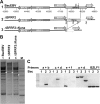
Construction of the BacEBVΔBFRF2 recombinant. (A) Schematic representation of the EBV region surrounding the BFRF2 gene (1). Homologous recombination between the wild-type EBV genome (p2089) and a PCR fragment carrying the kanamycin gene flanked by 50 bp of BcRF1 sequences was performed in the DH10B E. coli strain containing the pKD46 plasmid. Clones carrying the recombinant EBV were selected for chloramphenicol and kanamycin resistance. The resulting BAC-EBVΔBFRF2 recombinant had the kanamycin gene inserted between positions 47668 and 48700 in the BFRF2 gene (2). The kanamycin gene was then deleted by homologous recombination using the pCP20 vector in order to generate the BAC-EBVΔBFRF2ΔKana recombinant used in this study (3). (B) Restriction enzyme analysis of the BAC-EBVΔBFRF2 plasmid DNA. The BamHI restriction pattern of BAC-EBVΔBFRF2 (lane 2) and BAC-EBVΔBFRF2ΔKana (lane 3) mutant DNA was compared to that of wild-type BAC-EBV DNA (lane 3). An asterisk indicates the modified DNA pattern. Lane M, molecular markers. (C) PCR amplification analysis of the BAC-EBVΔBFRF2 (lanes 2) and BAC-EBVΔBFRF2ΔKana (lanes 3) (ΔF2) clones compared to the BAC-EBV clone (lane 1) carrying the wild-type EBV genome. PCRs were performed using primers indicated by small arrowheads in panel A (a, b, c, d, e, and f) and listed in Table 1.
FIG 2
FIG 2
BFRF2 is necessary for virion production. (A) Raji cells were incubated with supernatants from HEK293EBVΔBFRF2 cells transfected with an expression plasmid encoding the EB1 protein in order to induce the productive cycle together (or not) with an expression plasmid encoding the Flag.BFRF2 protein to complement BFRF2 deficiency. (Left side) The GFP fluorescence of Raji cells was analyzed 48 h after infection under UV light. (A, sub panels a and b) Phase contrast; (A, sub panels c and d) UV light. (Right side) Immunoblot analysis of the EB2, Flag.BFRF2, and EB1 proteins expressed in HEK293EBVΔBFRF2 cells that either had not been transfected (lane 1), had been transfected with an EB1 expression plasmid (lane 2), or had been cotransfected with expression plasmids for both EB1 and BFRF2 (lane 3). Western blots were revealed using a polyclonal anti-Flag antibody, an anti-EB2 polyclonal rabbit antibody, or an anti-EB1 MAb. * indicates an unspecified protein recognized by the anti-EB2 serum. (B) BFRF2 is not required for viral DNA replication. Viral DNA in HEK293EBV and HEK293EBVΔBFRF2 cells transfected with EB1 and Flag.BFRF2 expression plasmids, as indicated in the figure, was quantitated by qPCR. The amount of viral DNA in the induced cells (transfected with the EB1 expression plasmid) is expressed relative to the amount of viral DNA present in the uninduced cells. (C, top) BFRF2 enhances expression of the late viral genes. Total RNA from HEK293EBV (wild type [WT]) or HEK293EBVΔBFRF2 (ΔF2) cells that either had not been transfected (−), had been transfected with an EB1 expression plasmid (EB1), or had been cotransfected with expression plasmids for EB1 and Flag.BFRF2 (EB1 + BFRF2) was purified, reverse transcribed, and analyzed by qPCR using specific primers for either the BMRF1 early gene or the BDLF1 and BcLF1 late genes. (Bottom) Western blot of HEK293EBV or HEK293EBVΔBFRF2 cells either not transfected (−) or transfected with an EB1 expression plasmid or with expression plasmids for both EB1 and Flag.BFRF2. Protein extracts were analyzed by Western blotting using a polyclonal anti-Flag antibody and an anti-EB1 MAb. The asterisk indicates an unspecified protein detected by the anti-Flag antibody.
FIG 3
FIG 3
BFRF2 is required for late gene expression. (A) Schematic representation of the luciferase reporter vector used (pTATTBcLF1-Luc). (B) HEK293EBV, HEK293EBVΔBcRF1, or HEK293EBVΔBFRF2 cells were either transfected with the reporter vector alone (−) or cotransfected with an expression plasmid for EB1, Flag.BcRF1, or Flag.BFRF2, as indicated. The amount of luciferase expressed was measured 24 h after transfection. The relative luciferase fold activation was calculated relative to the value obtained with the pTATTBcLF1-Luc reporter transfected alone. The experiment was performed 3 times, and the error bars represent standard deviations. Protein extracts from the experiment whose results are shown in panel B were tested for EB1, BcRF1, and BFRF2 expression by immunoblotting using an anti-EB1 MAb, an anti-Flag MAb (BcRF1), and a polyclonal anti-Flag antibody (BFRF2).
FIG 4
FIG 4
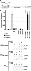
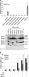
Six viral proteins are required for activation of the late viral genes. (A) The luciferase (Luc) reporter vector pTATTBcLF1-Luc was transfected into the EBV-negative cell line HEK293, together with expression vectors for the six different proteins, separately or combined. An EB1 expression vector was also used in order to show that the response of the TATTBcLF1 promoter was specific for the other six proteins. (B) Proteins expressed in HEK293 cells transfected with the different expression plasmids encoding the Flag-tagged proteins used in panel A were analyzed by Western blotting using a monoclonal anti-Flag antibody. White asterisks label the expected positions of the different proteins on the gel. − indicates the missing protein. (C) HEK293 cells were transfected, together with the pTATTBcLF1-Luc reporter, with expression vectors for the six different viral proteins combined (all) or with only five expression vectors as indicated (“all-” indicates the missing expression vector). (D) Western blot corresponding to HEK293 cells transfected with expression vectors for all 6 proteins or with different combinations of five of the proteins. All the proteins were tagged with a Flag epitope, and the blot was incubated with an anti-Flag MAb. Black asterisks label the positions of proteins destabilized in the absence of one component (the monoclonal anti-Flag antibody does not react with Flag-BDLF3.5). (E) The pBMRF1-Luc reporter was transfected into the EBV-negative cell line HEK293, together with combined expression vectors for the six different proteins (all) or an EB1 expression vector. (A, C, and E) The amount of luciferase expressed was measured 24 h after transfection. The relative luciferase fold activation was calculated relative to the value obtained with the pTATTBcLF1-Luc or the pBMRF1-Luc reporter transfected alone. The experiment was performed 3 times, and the error bars represent standard deviations. (F) BGLF3 and BVLF1 are stabilized by coexpression of BFRF2 and BDLF3.5, respectively. HEK293 cells were transfected with a Flag.BGLF3 expression vector either alone or in combination with a Flag.BFRF2 expression vector or with a Flag-BVLF1 expression vector either alone or in combination with a Flag-BDLF3.5 expression vector. The immunoblot was revealed with a polyclonal anti-Flag antibody. (G) Total RNA from the transfected cells was extracted, and RT-PCR was performed in order to show that expression of BFRF2 and BDLF3.5 did not increase the amount of BGLF3 and BVLF1 mRNA, respectively. Primers specific for the cellular GusB mRNA were used to control for the absence of contaminating DNA in the assay and to ensure that the same amount of mRNA was used for the RT.
FIG 5
FIG 5
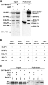
GST pulldown of the vPIC. Extracts from HEK293 cells transfected with a GST-Flag (GST) or a GST-Flag.BcRF1 (GST-BcRF1) expression plasmid together with expression vectors for components of the vPIC (all tagged with the Flag epitope) were incubated with glutathione-Sepharose beads. The purified proteins were analyzed by immunoblotting with a mix of monoclonal and polyclonal anti-Flag antibodies in order to identify the components of the vPIC associated with GST-Flag.BcRF1 (A) or with an anti-RNAP II antibody in order to analyze the interaction between BcRF1 and the RNAP II complex (C). GST-Flag and GST-Flag.BcRF1, which are very weakly expressed in the cells, are seen only after GST pulldown. Dark-gray arrows indicate their positions. Again, Flag.BDLF3.5 was not efficiently detected. II0 indicates the position of the hyperphosphorylated form of the RNAP II enzyme and IIA its unphosphorylated form. (B) Summary of the interactions found between the different components of the vPIC as determined by in vitro GST pulldown assay. In vitro-translated 35S-labeled BcRF1, BGLF3, BVLF1, BDLF4, and BFRF2 proteins were incubated with purified GST, GST-BcRF1, GST-BGLF3, GST-BDLF4, GST-BFRF2, or GST-BDLF3.5 bound to glutathione-Sepharose beads. The bound proteins were analyzed by SDS-PAGE and visualized by autoradiography. None of the in vitro-translated 35S-labeled proteins interacted with GST.
FIG 6
FIG 6
The vPIC was unable to induce late gene expression from the endogenous viral genome. (A) Total RNA from HEK293EBV or Raji cells either mock transfected (−) or transfected with the EB1 expression vector or the six expression plasmids encoding the vPIC factors was quantified by RT-qPCR using primers specific for either the BMRF1 early gene or the BDLF1 and BFRF3 late genes. The experiment was performed 3 times, and the error bars represent standard deviations. (B) EB1 and vPIC induce late gene expression in a transient-transfection assay of EBV-positive cells. HEK293EBV or Raji cells were transfected with the pTATTBcLF1-Luc reporter plasmid either alone (−) or together with the EB1 expression vector or the six expression plasmids encoding the vPIC components. The experiment was performed 3 times, and the error bars represent standard deviations. Protein extracts from the experiment whose results are shown in panel B were tested for expression of EB1, and the vPIC components were tested by immunoblotting using an anti-EB1 MAb or a mix of monoclonal and polyclonal anti-Flag antibodies (for the vPIC proteins). A star indicates an unspecified band recognized by the anti-Flag antibody.
FIG 7
FIG 7
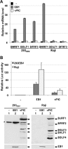
Activation of endogenous late gene expression is dependent on viral DNA replication. HEK293EBV cells were transfected with the pTATTBcLF1-Luc reporter plasmid together with an EB1 expression vector or the six expression plasmids encoding the vPIC components in the presence or absence of PAA, a specific inhibitor of viral DNA replication. (A) Both EB1 and the vPIC activate luciferase expression from the pTATTBcLF1-Luc reporter, and this expression is not highly sensitive to PAA treatment. (B) EB1 is unable to activate expression of the endogenous viral late genes in the presence of PAA (50 ng/ml), even when vPIC components are expressed. Following transfection of expression plasmids encoding EB1 or the vPIC components, as indicated in the figure, RNA expression of the endogenous early BMRF1 gene and the late BcLF1 or BDLF1 gene was quantified by RT-qPCR. (C) Control showing complete inhibition of viral DNA replication by PAA (upper panel) and expression of the early BMLF1 and late gp350 proteins analyzed by immunoblotting (bottom panel) in cells used in the experiment whose results are shown in panels A and B of the figure. The experiment was performed 2 times, and the error bars represent standard deviations.
FIG 8
FIG 8
Betaherpesviruses also encode a vPIC. (A) HEK293 cells were transfected with the pTATTBcLF1-Luc reporter plasmid together with the expression vectors encoding either the vPIC EBV components (BcRF1, BFRF2, BGLF3, BVLF1, BDLF4, and BDLF3.5), the vPIC CMV components (UL87, UL49, UL95, UL79, UL92, and UL91), or the vPIC CMV components with one component replaced by the EBV analog, as indicated. The relative luciferase fold activation was calculated relative to the value obtained with the pTATTBcLF1-Luc reporter transfected alone (−). The experiment was performed 3 times, and the error bars represent standard deviations. Protein expression from transfected cells in panel A was controlled by immunoblotting using a mix of monoclonal and polyclonal anti-Flag antibodies. Neither BDLF3.5 nor UL91 was detected. (B) The CMV vPIC can substitute for the EBV vPIC. HEK293EBVΔBFRF2 cells were transfected with the different expression plasmids indicated at the bottom of the graph. Total RNA extracted from the transfected cells was quantified by RT-qPCR using primers specific for either the BMRF1 early gene or the BDLF1 and BFRF3 late genes. The experiment was performed 3 times, and the error bars represent standard deviations.

References

    1. Rickinson AB, Kieff E. 2001. Epstein-Barr virus, p 2575–2627 In Knipe DM, Howley PM, Griffin DE, Martin MA, Lamb RA, Roizman B, Straus SE. (ed), Fields virology, 4th ed. Lippincott Williams & Wilkins, Philadelphia, PA.
    1. Babcock GJ, Decker LL, Volk M, Thorley-Lawson DA. 1998. EBV persistence in memory B cells in vivo. Immunity 9:395–404. 10.1016/S1074-7613(00)80622-6. - DOI - PubMed
    1. Laichalk LL, Thorley-Lawson DA. 2005. Terminal differentiation into plasma cells initiates the replicative cycle of Epstein-Barr virus in vivo. J. Virol. 79:1296–1307. 10.1128/JVI.79.2.1296-1307.2005. - DOI - PMC - PubMed
    1. Chevallier-Greco A, Manet E, Chavrier P, Mosnier C, Daillie J, Sergeant A. 1986. Both Epstein-Barr virus (EBV) encoded trans-acting factors, EB1 and EB2, are required to activate transcription from an EBV early promoter. EMBO J. 5:3243–3249. - PMC - PubMed
    1. Hardwick JM, Lieberman PM, Hayward SD. 1988. A new Epstein-Barr virus transactivator, R, induces expression of a cytoplasmic early antigen. J. Virol. 62:2274–2284. - PMC - PubMed

Publication types

LinkOut - more resources