. 2014 Nov;88(21):12895-906.
doi: 10.1128/JVI.02194-14. Epub 2014 Aug 27.

Structural definition of an antibody-dependent cellular cytotoxicity response implicated in reduced risk for HIV-1 infection

Affiliations

Structural definition of an antibody-dependent cellular cytotoxicity response implicated in reduced risk for HIV-1 infection

Priyamvada Acharya et al. J Virol. 2014 Nov.

Abstract

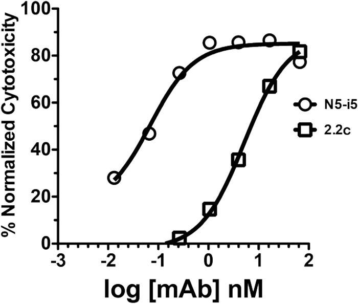
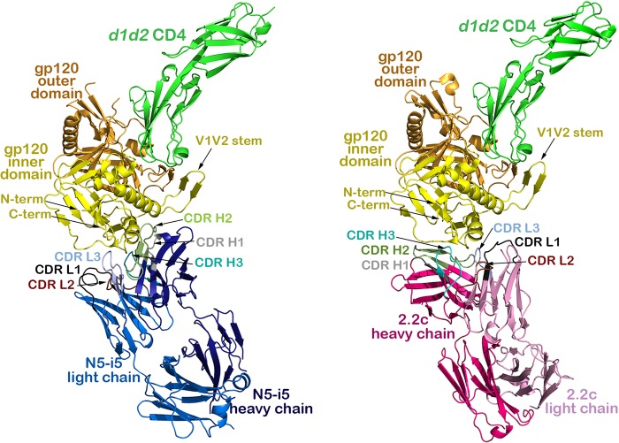
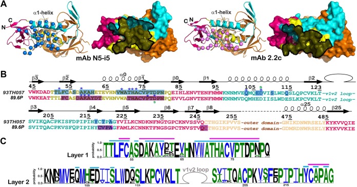
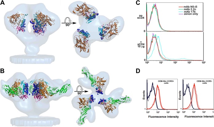
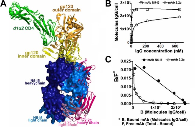
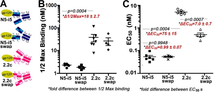
The RV144 vaccine trial implicated epitopes in the C1 region of gp120 (A32-like epitopes) as targets of potentially protective antibody-dependent cellular cytotoxicity (ADCC) responses. A32-like epitopes are highly immunogenic, as infected or vaccinated individuals frequently produce antibodies specific for these determinants. Antibody titers, as measured by enzyme-linked immunosorbent assay (ELISA) against these epitopes, however, do not consistently correlate with protection. Here, we report crystal structures of CD4-stabilized gp120 cores complexed with the Fab fragments of two nonneutralizing, A32-like monoclonal antibodies (MAbs), N5-i5 and 2.2c, that compete for antigen binding and have similar antigen-binding affinities yet exhibit a 75-fold difference in ADCC potency. We find that these MAbs recognize overlapping epitopes formed by mobile layers 1 and 2 of the gp120 inner domain, including the C1 and C2 regions, but bind gp120 at different angles via juxtaposed VH and VL contact surfaces. A comparison of structural and immunological data further showed that antibody orientation on bound antigen and the capacity to form multivalent antigen-antibody complexes on target cells were key determinants of ADCC potency, with the latter process having the greater impact. These studies provide atomic-level definition of A32-like epitopes implicated as targets of protective antibodies in RV144. Moreover, these studies establish that epitope structure and mode of antibody binding can dramatically affect the potency of Fc-mediated effector function against HIV-1. These results provide key insights for understanding, refining, and improving the outcome of HIV vaccine trials, in which relevant immune responses are facilitated by A32-like elicited responses.

Importance: HIV-1 Env is a primary target for antibodies elicited during infection. Although a small number of infected individuals elicit broadly neutralizing antibodies, the bulk of the humoral response consists of antibodies that do not neutralize or do so with limited breadth but may effect protection through Fc receptor-dependent processes, such as antibody-dependent cellular cytotoxicity (ADCC). Understanding these nonneutralizing responses is an important aspect of elucidating the complete spectrum of immune response against HIV-1 infection. With this report, we provide the first atomic-level definition of nonneutralizing CD4-induced epitopes in the N-terminal region of the HIV-1 gp120 (A32-like epitopes). Further, our studies point to the dominant role of precise epitope targeting and mode of antibody attachment in ADCC responses even when largely overlapping epitopes are involved. Such information provides key insights into the mechanisms of Fc-mediated function of antibodies to HIV-1 and will help us understand the outcome of vaccine trials based on humoral immunity.


Figures

FIG 1
FIG 1
Representative ADCC curves for MAbs N5-i5 and 2.2c. ADCC assays were performed as described in Materials and Methods using CEM-NKr-CCR5 target cells sensitized with gp120 of the HIV-1BaL isolate.
FIG 2
FIG 2
Crystal structures of the N5-i5 Fab-gp12093TH057 coree-d1d2CD4 and 2.2c Fab-gp12089.6P coree-d1d2CD4 complexes. The light/heavy chains of N5-i5 Fab and 2.2c Fab are shown in light blue/dark blue and light pink/dark pink, respectively, and the complementarity-determining regions (CDRs) are shown in black (CDR L1), brown (CDR L2), light blue (CDR L3), gray (CDR H1), green (CDR H2), and cyan (CDR H3). The gp120 inner domain is shown in yellow, and the outer domain is in orange. See also Fig. S1A in the supplemental material.
FIG 3
FIG 3
N5-i5 and 2.2c epitopes. (A) N5-i5 and 2.2c epitope footprints on monomeric gp120. The Cα atoms of the gp120 residues involved in N5-i5 and 2.2c binding are represented by blue and pink balls, respectively, and displayed over the ribbon diagram of the gp120 inner domain. Antibodies' contact surfaces displayed over the gp120 molecular surface are shown in black (right). The “layered” architecture of the gp120 inner domain is shown, with the 7-stranded β sandwich in magenta, layer 1 in yellow, layer 2 in cyan, and layer 3 in light orange. The outer domain is shown in orange. (B) Mapping of the N5-i5 and 2.2c contact residues on the gp120 primary sequence of the gp120 inner domain of the isolates used in structural studies. The topology diagrams depicting a distribution of secondary structure elements as calculated with DSSP (66) is shown above the gp120 sequences. The gp120 residues involved in N5-i5 and 2.2c binding are highlighted in blue and pink, respectively. Residues contributing to the binding through H bonds and salt bridges are indicated by blue asterisks and green lowercase letters above and below the 93TH057 and 89.6P sequences, respectively. The gp120 layers are colored as in panel A. (C) Sequence conservation of N5-i5 and 2.2c epitopes. The height of the residue at each position is proportional to its frequency of distribution among the HIV-1 isolates, as deposited in the Los Alamos database (all clades are included). Residues are colored according to hydrophobicity: black, hydrophilic; green, neutral; blue, hydrophobic. Residues forming the N5-i5 and 2.2c epitope are indicated by blue and pink lines above the sequence, respectively.
FIG 4
FIG 4
Colocalization of N5-i5 and 2.2c epitopes within the BG505 SOSIP.664 gp140 trimer. (A) N5-i5 and 2.2c epitope footprints are mapped onto the BG505 SOSIP.664 trimer structure solved by X-ray crystallography (69). The gp120 protomers are colored as in Fig. 3, and gp41 is shown in green. The spheres represent the Cα atoms of residues that contribute to formation of N5-i5 and 2.2c epitopes post-CD4 binding. The enlargements show regions of contact between the gp120 inner domain and the gp41 of the unliganded trimer on which N5-i5 Fab-gp12093TH057 coree-d1d2CD4 (top) and 2.2c Fab-gp12089.6P coree-d1d2CD4 complex (bottom) are aligned based on the gp120 outer domain. Residues contributing to both the interface with gp41 within the trimer and the interface with MAb within the complex are shown as spheres. Layers 1 and 2 of the gp120 inner domain of the gp120-gp41 and the gp120-MAb interface are in darker and lighter shades of yellow and cyan, respectively. Only variable parts of N5-i5 and 2.2c Fab are depicted, and CDRs involved in interfaces are shown. Many residues of gp120 involved in N5-i5/2.2c epitope post-CD4 binding also line the interface with the gp41 of the trimer. These include multiple residues of β2̄ and β1̄ strands and α0 helix of layer 1, all residues forming the N5-i5 epitope on the α1 helix, and multiple residues of the β4 strand and the β4 -β5 connecting loop of layer 2 (marked by black dots below the 93TH057 sequence on panel B). (B) Details of the conformational changes of layer 1 (left) and layer 2 (right) from the prefusion state of trimer (light yellow and light cyan for layers 1 and 2, respectively) to the N5-i5/2.2c-bound conformation of the CD4-triggered gp120 coree (red). A change in the distribution of secondary structure elements from unliganded to MAb-bound conformation is shown above the gp120 sequences on which residues involved in N5-i5 and 2.2c binding are highlighted in blue and pink, respectively. The CD4 triggering induces rearrangements of secondary structural elements in layer 1 of inner domain manifested primarily by formation of the α0 helix and unfolding of the α1̄ helix. The α1 helix of layer 2 is shortened and tilted.
FIG 5
FIG 5
Exposure of N5-i5 and 2.2c epitopes on a viral trimer. (A) Colocalization of N5-i5 and 2.2c epitopes within the virion-associated untriggered HIV-1 trimers. The Cα atoms of gp120 residues involved in interaction with N5-i5 (blue balls) and 2.2c (pink balls) are mapped into the trimer structure derived by cryo-electron tomography (53) and are shown as blue and pink balls, respectively. gp120 molecules are shown as ribbon diagrams and colored as in Fig. 3. The views of trimers are from the side, with the viral membrane oriented toward the bottom (left) and rotated 90° about a horizontal axis with the viral membrane at the bottom (right). (B) Colocalization of N5-i5 and 2.2c epitopes within HIV-1 trimers triggered with the soluble CD4. N5-i5 and 2.2c epitope footprints were mapped in the structure derived by cryo-electron tomography of the gp120Bal-d1d2CD4 trimer (67). The mapping of N5-i5 and 2.2c epitope footprints in the tomograms confirms that they stay largely within the interface of d1d2CD4-triggered spike and are not available for antibody recognition, due to steric hindrance. See also Fig. S4 in the supplemental material. (C) The binding curves of MAb N5-i5 (red line) and 2.2c (green line) to surface-expressed HIV-1BaL trimers in the presence or absence of soluble d1-d4CD4 (sCD4). Experiments were performed as described in Materials and Methods. CD4i antibody 17b (cyan line) was used as a positive control. The enhancement of binding to sCD4-triggered HIV-1BaL trimers was observed only for coreceptor binding site MAb 17b. (D) MAb N5-i5 binding (solid lines) and 2.2c binding (dashed lines) to CEM-Nkr-CCR5+ cells (left) or CEM-Nkr-CCR5 cells (right). The rightmost curves (red) in each histogram overlay represent the binding of N5-i5 or 2.2c to virion-sensitized cells, whereas the leftmost curves (blue) represent the binding of these MAbs on cells not sensitized with virions.
FIG 6
FIG 6
MAb N5-i5 and 2.2c binding to the gp120 antigen. (A) Superimposition of N5-i5 Fab-gp12093TH057 coree-d1d2CD4 and 2.2c Fab-gp12089.6P coree-d1d2CD4 complexes. Structures were aligned based on the gp120 molecule; a molecular surface is displayed over N5-i5 Fab, and 2.2c Fab is shown in a ribbon diagram. See also Fig. S5 in the supplemental material. (B) MAb N5-i5 and 2.2c binding kinetics to gp120-sensitized CEM-Nkr-CCR5+ target cells, as measured with an unlabeled MAb competition protocol. CEM-Nkr-CCR5+ target cells were sensitized with gp120, and a saturation curve was developed as described in Materials and Methods. (C) Scatchard plots of N5-i5 and 2.2c binding to CEM-Nkr-CCR5+ target cells were derived from the binding data using the standard equation (68) by nonlinear curve fitting (Prism; GraphPad, La Jolla, CA).
FIG 7
FIG 7
Fv-swapped versions of N5-i5 and 2.2c. (A) Design of Fv-swapped versions of N5-i5 and 2.2c. Swaps were made by moving the variable heavy (VH) domain onto the constant light (CL) domain and the variable light (VL) domain onto the constant heavy 1 (CH1) domain for each MAb to replicate the Fc domain orientations of counterpart. (B) Half-maximal binding of MAbs N5-i5 and 2.2c and their swapped versions to gp120-sensitized CEM-Nkr-CCR5+ target cells. Each binding experiment was repeated independently four to seven times as described in Materials and Methods, and half-maximal-binding and Bmax values were pooled for statistical analysis. (C) Cytotoxicity mediated by MAbs N5-i5 and 2.2c and their swapped versions on gp120-sensitized CEM-Nkr-CCR5+ target cells.

References

    1. Hessell AJ, Hangartner L, Hunter M, Havenith CE, Beurskens FJ, Bakker JM, Lanigan CM, Landucci G, Forthal DN, Parren PW, Marx PA, Burton DR. 2007. Fc receptor but not complement binding is important in antibody protection against HIV. Nature 449:101–104. 10.1038/nature06106. - DOI - PubMed
    1. Mabuka J, Nduati R, Odem-Davis K, Peterson D, Overbaugh J. 2012. HIV-specific antibodies capable of ADCC are common in breastmilk and are associated with reduced risk of transmission in women with high viral loads. PLoS Pathog. 8:e1002739. 10.1371/journal.ppat.1002739. - DOI - PMC - PubMed
    1. Haynes BF, Gilbert PB, McElrath MJ, Zolla-Pazner S, Tomaras GD, Alam SM, Evans DT, Montefiori DC, Karnasuta C, Sutthent R, Liao HX, DeVico AL, Lewis GK, Williams C, Pinter A, Fong Y, Janes H, DeCamp A, Huang Y, Rao M, Billings E, Karasavvas N, Robb ML, Ngauy V, de Souza MS, Paris R, Ferrari G, Bailer RT, Soderberg KA, Andrews C, Berman PW, Frahm N, De Rosa SC, Alpert MD, Yates NL, Shen X, Koup RA, Pitisuttithum P, Kaewkungwal J, Nitayaphan S, Rerks-Ngarm S, Michael NL, Kim JH. 2012. Immune-correlates analysis of an HIV-1 vaccine efficacy trial. N. Engl. J. Med. 366:1275–1286. 10.1056/NEJMoa1113425. - DOI - PMC - PubMed
    1. Alpert MD, Harvey JD, Lauer WA, Reeves RK, Piatak M, Jr, Carville A, Mansfield KG, Lifson JD, Li W, Desrosiers RC, Johnson RP, Evans DT. 2012. ADCC develops over time during persistent infection with live-attenuated SIV and is associated with complete protection against SIV(mac)251 challenge. PLoS Pathog. 8:e1002890. 10.1371/journal.ppat.1002890. - DOI - PMC - PubMed
    1. Lambotte O, Ferrari G, Moog C, Yates NL, Liao HX, Parks RJ, Hicks CB, Owzar K, Tomaras GD, Montefiori DC, Haynes BF, Delfraissy JF. 2009. Heterogeneous neutralizing antibody and antibody-dependent cell cytotoxicity responses in HIV-1 elite controllers. AIDS 23:897–906. 10.1097/QAD.0b013e328329f97d. - DOI - PMC - PubMed

Publication types

MeSH terms