. 2014 Nov;88(22):13015-28.
doi: 10.1128/JVI.01757-14. Epub 2014 Aug 27.

Lack of interleukin-10-mediated anti-inflammatory signals and upregulated interferon gamma production are linked to increased intestinal epithelial cell apoptosis in pathogenic simian immunodeficiency virus infection

Affiliations

Lack of interleukin-10-mediated anti-inflammatory signals and upregulated interferon gamma production are linked to increased intestinal epithelial cell apoptosis in pathogenic simian immunodeficiency virus infection

Diganta Pan et al. J Virol. 2014 Nov.

Abstract

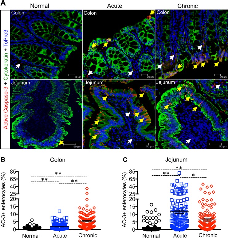
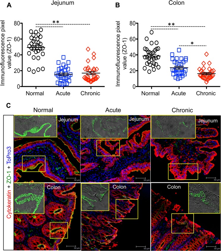
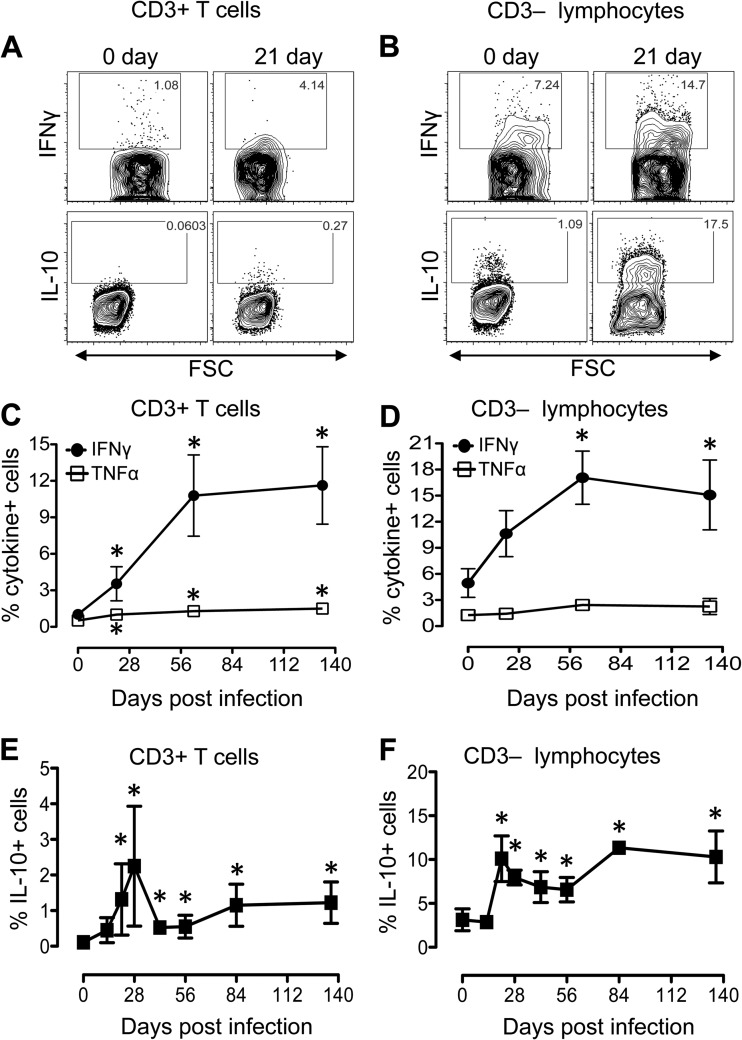
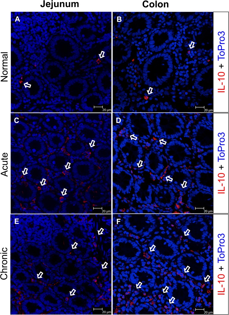
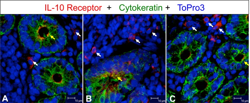
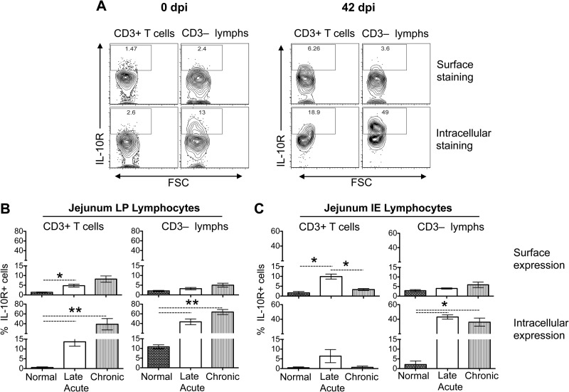
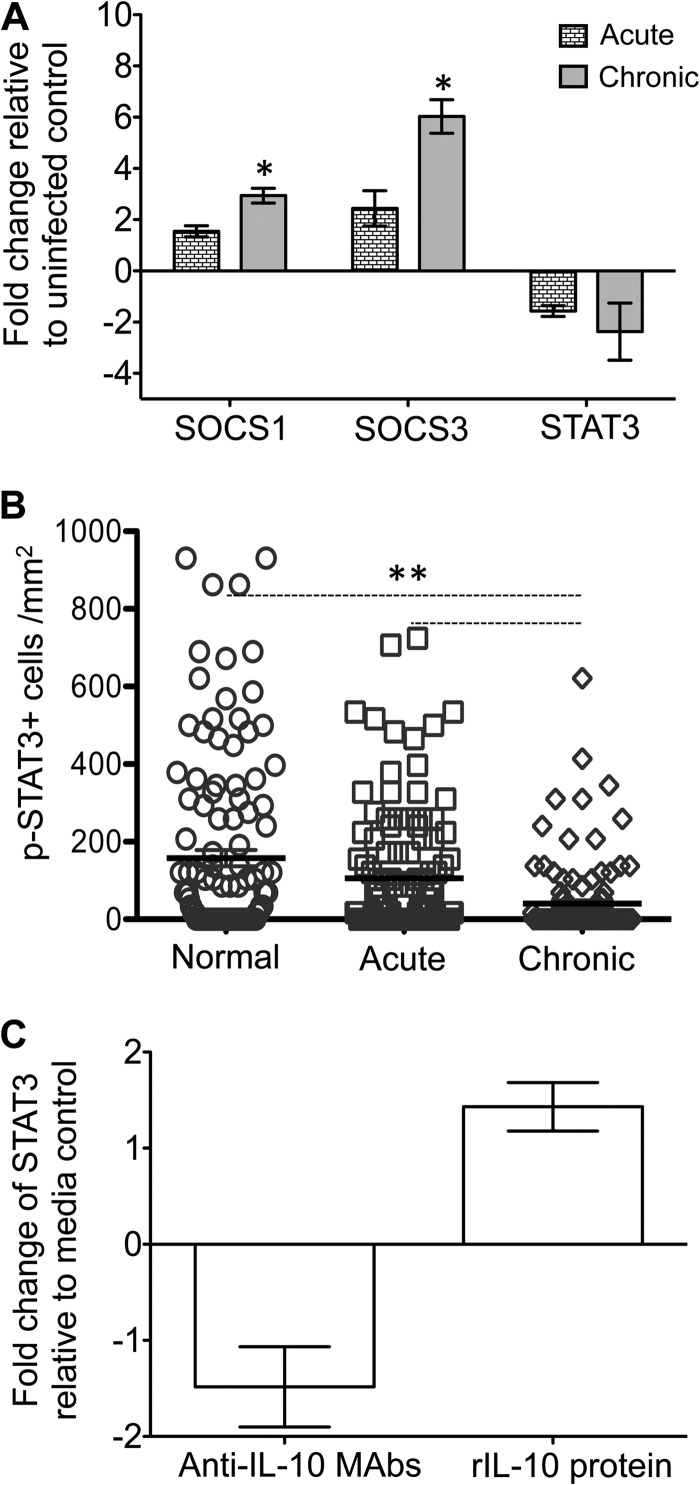
Interleukin-10 (IL-10) is an immunomodulatory cytokine that is important for maintenance of epithelial cell (EC) survival and anti-inflammatory responses (AIR). The majority of HIV infections occur through the mucosal route despite mucosal epithelium acting as a barrier to human immunodeficiency virus (HIV). Therefore, understanding the role of IL-10 in maintenance of intestinal homeostasis during HIV infection is of interest for better characterization of the pathogenesis of HIV-mediated enteropathy. We demonstrated here changes in mucosal IL-10 signaling during simian immunodeficiency virus (SIV) infection in rhesus macaques. Disruption of the epithelial barrier was manifested by EC apoptosis and loss of the tight-junction protein ZO-1. Multiple cell types, including a limited number of ECs, produced IL-10. SIV infection resulted in increased levels of IL-10; however, this was associated with increased production of mucosal gamma interferon (IFN-γ) and tumor necrosis factor alpha (TNF-α), suggesting that IL-10 was not able to regulate AIR. This observation was supported by the downregulation of STAT3, which is necessary to inhibit production of IFN-γ and TNF-α, and the upregulation of SOCS1 and SOCS3, which are important regulatory molecules in the IL-10-mediated AIR. We also observed internalization of the IL-10 receptor (IL-10R) in mucosal lymphocytes, which could limit cellular availability of IL-10 for signaling and contribute to the loss of a functional AIR. Collectively, these findings demonstrate that internalization of IL-10R with the resultant impact on IL-10 signaling and dysregulation of the IL-10-mediated AIR might play a crucial role in EC damage and subsequent SIV/HIV pathogenesis.

Importance: Interleukin-10 (IL-10), an important immunomodulatory cytokine plays a key role to control inflammatory function and homeostasis of the gastrointestinal mucosal immune system. Despite recent advancements in the study of IL-10 and its role in HIV infection, the role of mucosal IL-10 in SIV/HIV infection in inducing enteropathy is not well understood. We demonstrated changes in mucosal IL-10 signaling during SIV infection in rhesus macaques. Disruption of the intestinal epithelial barrier was evident along with the increased levels of mucosal IL-10 production. Increased production of mucosal IFN-γ and TNF-α during SIV infection suggested that the increased level of mucosal IL-10 was not able to regulate anti-inflammatory responses. Our findings demonstrate that internalization of IL-10R with the resultant impact on IL-10 signaling and dysregulation of the IL-10-mediated anti-inflammatory responses might play a crucial role in epithelial cell damage and subsequent SIV/HIV pathogenesis.


Figures

FIG 1
FIG 1
Intestinal epithelial cell (EC) apoptosis increases during SIV infection. (A) Representative immunofluorescence analysis of the colon and jejunum showing increased apoptotic ECs (active caspase-3 [AC-3]+ cytokeratin+) in colon at the chronic phase (colon, R544 at 389 dpi; jejunum, FK88 at 226 dpi) and acute phase (AV85 at 21 dpi) compared to uninfected control macaques (colon, AG71; jejunum, GN74). Apoptotic ECs and lamina propria cells are indicated by yellow and white arrows, respectively. Scatter plots (indicating means ± the standard errors) of apoptotic enterocytes are shown for both the colon (B) and jejunum (C) in normal (n = 6) and in acutely (n = 8) and chronically (n = 7) SIV-infected macaques. Asterisks indicate statistically significant differences between the respective animal groups (*, P < 0.005; **, P < 0.001).
FIG 2
FIG 2
Decreased expression of ZO-1 in SIV-infected macaques. Immunofluorescence pixel values of ZO-1 expression (indicating means ± the standard errors) in jejunum (A) and colon (B) from normal and acutely and chronically infected animals are shown (n = 6). Asterisks indicate statistically significant differences between the respective animal groups (*, P < 0.001; **, P < 0.0001). (C) Representative immunofluorescent confocal images in the jejunum and colon revealed a loss of ZO-1 expression in acutely (BA57 at 8 dpi) and chronically (AP53 at 63 dpi) SIV-infected macaques (scale bars, 50 μm) compared to normal uninfected macaques (jejunum, GN74; colon, GN70). Insets for each panel show patterns of ZO-1 expression alone. The apical mucosal surfaces of jejunum and colon exhibit ZO-1 with a typical honeycomb structure in normal uninfected animals.
FIG 3
FIG 3
Major IL-10-producing cells in intestinal tissues. (A) Expression of IL-10 by multilabel confocal microscopy in multiple cell types, including leukocytes (CD45+), T cells (CD3+), B cells/plasma cells (CD79a+), monocytes/macrophages (CD68+, Mac387+), dendritic cells (CD11c+), and epithelial cells (EC, cytokeratin+). The labels of each image are indicated at the top of each image. IL-10+ cells in lamina propria are indicated by yellow arrows. (B) Flow cytometry of cells isolated from the lower layer of a Percoll density gradient after EDTA treatment of the jejunum analyzed for the expression of cytokeratin, CD3, and IL-10. All cells were gated on singlets, followed by live cells, and then plotted based on CD3+ T cells and cytokeratin+ cells. Each gated cell population was further analyzed for IL-10 production as represented by dot plots. The data are representative of a normal uninfected InRM (i.e., macaque DE50, at the pre-SIV infection time point).
FIG 4
FIG 4
SIV infection induces increased levels of IFN-γ, TNF-α, and IL-10 cytokines in both CD3+ and CD3 lymphocytes in the jejunum lamina propria. (A) Representative contour plots showing differential expression of IFN-γ and IL-10 from one SIV-infected macaque (EB09) at days 0 and 21 dpi in CD3+ T cells (A) and CD3 lymphocytes (B). Mean percentages (± the standard errors) of CD3+ T cells (C) and CD3 lymphocytes (D) isolated from jejunum lamina propria producing IFN-γ and TNF-α cytokines are shown for SIV-infected macaques (n = 8). Increased production of IL-10 (mean ± the standard errors) was also detected in both CD3+ T cells (E) and CD3 lymphocytes (F) isolated from the jejunum lamina propria (n = 8). Asterisks indicate significant differences between day 0 and the specified time point (P < 0.05). All cells were gated on singlets, lymphocytes, followed by live cells, and then on CD3+ T cells and CD3 lymphocytes.
FIG 5
FIG 5
IL-10 expression increased in the intestinal lamina propria during SIV infection. IL-10 expression by lamina propria cells was visualized by immunofluorescence in the jejunum (A, C, and E) and colon (B, D, and F). Both jejunum and colon lamina propria cells (A and B) spontaneously express IL-10 in a normal uninfected animal (GT20). However, after SIV infection both the jejunum and colon demonstrated increased percentages of IL-10-producing cells in acute (C and D; HN29) and chronic (E and F; CL86) infections. White arrows denote the presence of IL-10-positive cells, with the majority distributed in the lamina propria region.
FIG 6
FIG 6
IL-10 receptor (IL-10R) expression increased in jejunum during SIV infection. White and yellow arrows indicate expression of IL-10R positive mononuclear cells in lamina propria and enterocytes, respectively, in jejunum from an uninfected control (A; GJ06) and from acutely (B; CF65 at 21 days postinfection) and chronically (C; AP53 at 63 days postinfection) SIV-infected macaques. Note that increased IL-10R expression was detected in both acutely and chronically SIV-infected macaques in lamina propria mononuclear cells.
FIG 7
FIG 7
SIV infection induces increased internalization of IL-10 receptors (IL-10R) in intestinal lymphoid cells compared to the expression of IL-10R on the cell surface. (A) Representative contour plots showing IL-10R expression after surface or intracellular staining in CD3+ T cells and CD3 lymphocytes in jejunum lamina propria (LP) lymphocytes from one SIV-infected macaque (EB09) at days 0 and 42 dpi. Bar charts showing the mean (± the standard errors) IL-10R surface and intracellular expression in CD3+ T cells and CD3 lymphocytes from both LP (B) and IE lymphocytes (C) derived from the jejunum tissues of normal, uninfected macaques (0 dpi) and late acutely (42 dpi) and chronically (150 to 281 dpi) SIV-infected macaques (n = 8). All cells were gated on singlets, lymphocytes, followed by live cells, and then on CD3+ T cells and CD3 lymphocytes. The percentages of IL-10R expression in cell cytoplasm were calculated by subtracting values of surface staining from values obtained from intracellular staining. Asterisks indicate statistically significant differences between the respective animal groups (*, P < 0.05; **, P < 0.005).
FIG 8
FIG 8
Expression patterns of SOCS1, SOCS3, STAT3, and pSTAT3 in SIV-infected macaques and colon explant cultures. (A) Increased fold change of SOCS1 and SOCS3 and decreased fold change of STAT3 gene expression were observed in jejunum from acute and chronically SIV-infected rhesus macaques compared to uninfected normal macaques using relative RT-PCR (mean ± the standard error; n = 6). Samples were normalized against 18S rRNA expression. (B) Scatter plots (with means ± the standard errors) of pSTAT3+ cells in uninfected normal macaques and acutely and chronically SIV-infected macaques quantified by immunohistochemistry staining are shown (n = 6; a minimum 20 fields were measured for each sample). (C) The relative fold changes (means ± the standard errors) of STAT3 gene expression are indicated for colon explants in the presence of either anti-IL-10 MAbs or recombinant IL-10 (rIL-10) protein as determined using relative RT-PCR (n = 3). Asterisks indicate significant differences between groups for the specified genes and proteins (*, P ≤ 0.005; **, P < 0.0001).

Similar articles

Cited by

References

    1. Batman PA, Kapembwa MS, Miller AR, Sedgwick PM, Lucas S, Sewankambo NK, Serwadda D, Pudney J, Moody A, Harris JR, Griffin GE. 1998. HIV enteropathy: comparative morphometry of the jejunal mucosa of HIV-infected patients resident in the United Kingdom and Uganda. Gut 43:350–355. 10.1136/gut.43.3.350. - DOI - PMC - PubMed
    1. Lim SG, Menzies IS, Lee CA, Johnson MA, Pounder RE. 1993. Intestinal permeability and function in patients infected with human immunodeficiency virus: a comparison with coeliac disease. Scand. J. Gastroenterol. 28:573–580. - PubMed
    1. Ott M, Lembcke B, Staszewski S, Helm EB, Caspary WF. 1991. Intestinal permeability in patients with acquired immunodeficiency syndrome (AIDS). Klin. Wochenschr. 69:715–721 (In German.) 10.1007/BF01649441. - DOI - PubMed
    1. Brenchley JM, Price DA, Schacker TW, Asher TE, Silvestri G, Rao S, Kazzaz Z, Bornstein E, Lambotte O, Altmann D, Blazar BR, Rodriguez B, Teixeira-Johnson L, Landay A, Martin JN, Hecht FM, Picker LJ, Lederman MM, Deeks SG, Douek DC. 2006. Microbial translocation is a cause of systemic immune activation in chronic HIV infection. Nat. Med. 12:1365–1371. - PubMed
    1. Sankaran S, George MD, Reay E, Guadalupe M, Flamm J, Prindiville T, Dandekar S. 2008. Rapid onset of intestinal epithelial barrier dysfunction in primary human immunodeficiency virus infection is driven by an imbalance between immune response and mucosal repair and regeneration. J. Virol. 82:538–545. 10.1128/JVI.01449-07. - DOI - PMC - PubMed

Publication types

MeSH terms

LinkOut - more resources