. 2014 Nov 1;25(21):3342-9.
doi: 10.1091/mbc.E14-07-1178. Epub 2014 Aug 27.

The novel component Kgd4 recruits the E3 subunit to the mitochondrial α-ketoglutarate dehydrogenase

Affiliations

The novel component Kgd4 recruits the E3 subunit to the mitochondrial α-ketoglutarate dehydrogenase

Manfred Heublein et al. Mol Biol Cell. .

Abstract

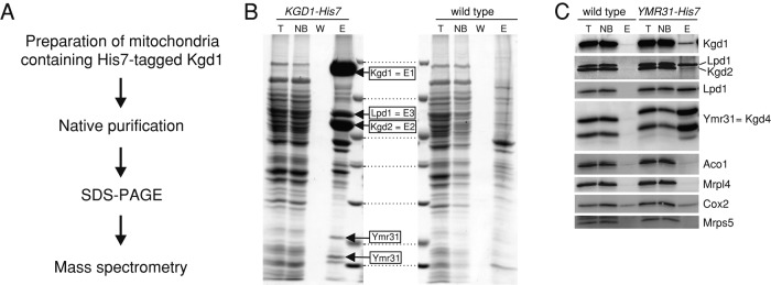
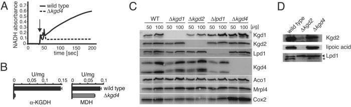
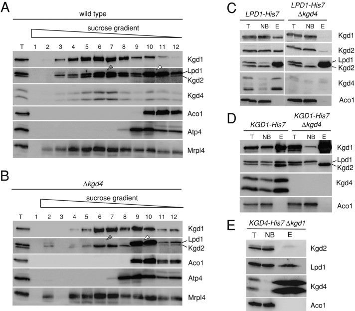
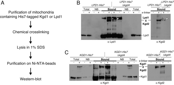
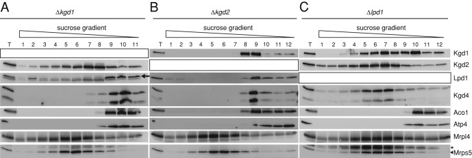
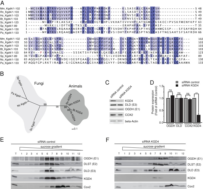
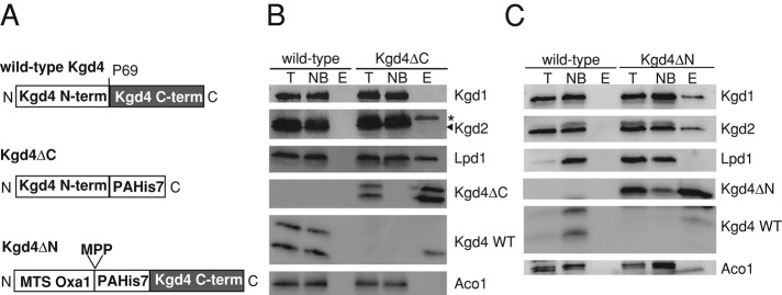
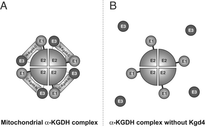
The mitochondrial citric acid cycle is a central hub of cellular metabolism, providing intermediates for biosynthetic pathways and channeling electrons to the respiratory chain complexes. In this study, we elucidated the composition and organization of the multienzyme complex α-ketoglutarate dehydrogenase (α-KGDH). In addition to the three classical E1-E3 subunits, we identified a novel component, Kgd4 (Ymr31/MRPS36), which was previously assigned to be a subunit of the mitochondrial ribosome. Biochemical analyses demonstrate that this protein plays an evolutionarily conserved role in the organization of mitochondrial α-KGDH complexes of fungi and animals. By binding to both the E1-E2 core and the E3 subunit, Kgd4 acts as a molecular adaptor that is necessary to a form a stable α-KGDH enzyme complex. Our work thus reveals a novel subunit of a key citric acid-cycle enzyme and shows how this large complex is organized.


Figures

FIGURE 1:
FIGURE 1:
Kgd4 copurifies with α-KGDH. (A) Strategy to analyze the composition of KGDH. (B) Mitochondrial KGDH was purified using a C-terminal His7-tag on Kgd1. Proteins were separated on SDS–PAGE and analyzed by Coomassie staining and mass spectrometry. Boxes indicate proteins specifically identified in the elution fraction of the sample containing His7-tagged Kgd1. (C) Complexes of Ymr31 were purified as in B, and the purifications were analyzed by Western blotting using the indicated antibodies. T, total of the lysate; NB, unbound fraction; W, wash fraction; E, elution fraction.
FIGURE 2:
FIGURE 2:
Absence of Kgd4 decreases KGDH activity in vitro without destabilizing the catalytic subunits. (A) Mitochondria from wild-type cells or cells lacking Kgd4 were lysed, and KGDH activity was photometrically determined. (B) Quantification of three independent enzyme assays for KGDH and MDH activity. (C) Total cellular protein was extracted from the indicated cells and analyzed by Western blotting. (D) Mitochondria of the indicated strains were analyzed by Western blotting using the indicated antibodies. The asterisk indicates the rest signal of the Western blotting with Kgd2 antibodies.
FIGURE 3:
FIGURE 3:
Kgd4 is necessary for a stable incorporation of the E3 subunit into the E1-E2 core of yeast α-KGDH. (A) Mitochondria from the wild-type strain were lysed in Triton X-100 and subjected to centrifugation on a linear sucrose gradient. Fractions were collected and analyzed by Western blotting. (B) Mitochondria of the Δkgd4 strain were processed and analyzed as in A. (C) Mitochondria containing a C-terminally His7-tagged Lpd1 with or without Kgd4 were lysed, and proteins were purified on Ni-NTA. The fractions of this purification were analyzed by Western blotting. (D) Mitochondria containing Kgd1-His7 with or without Kgd4 were processed and analyzed as in C. (E) Mitochondria containing Kgd4-His7 but lacking Kgd1 were processed and analyzed as in C. T, total of the lysate; NB, unbound fraction; E, elution fraction.
FIGURE 4:
FIGURE 4:
Kgd4 is required for efficient interaction of E3 with KGDH in intact organelles. (A) Cross-linking strategy to identify interactions between individual KGDH subunits. (B) Mitochondria prepared as in Figure 3C were exposed to the chemical cross-linker bismaleimidehexane, and Lpd1-His7 and its cross-linking products were purified under denaturing conditions on Ni-NTA beads. The fractions were analyzed by Western blotting. The gray arrow indicates the cross-linking product between Lpd1 and Kgd2 that decreases in the absence of Kgd4 (white arrow). (C) Mitochondria as in Figure 3D were processed and analyzed as in B. NB, unbound fraction.
FIGURE 5:
FIGURE 5:
Organization of α-KGDH in mitochondria. (A) Mitochondria lacking Kgd1 were analyzed as in Figure 3A. The black arrow indicates the signal of Lpd1. (B) Mitochondria lacking Kgd2 were analyzed as in Figure 3A. (C) Mitochondria lacking Lpd1 were lysed and analyzed as in Figure 3A. T, total of the lysate. The asterisk indicates the signal of the previous Western blotting with Mrpl4 antibodies.
FIGURE 6:
FIGURE 6:
Kgd4 function is evolutionarily conserved. (A) Alignment of amino acid sequences of Kgd4 homologues. Sequences used for comparison were extracted from the National Center for Biotechnology Information database (www.ncbi.nlm.nih.gov/protein): S. cerevisiae (Sc-gi45270840); Neurospora crassa (Nc-gi85092531), Schizosaccharomyces pombe (Sp-gi429240132), Podospora anserina (Pd-gi171678223), Homo sapiens (Hs-gi15150811), Rattus norvegicus (Rn-gi300797955), Mus musculus (Mm-gi13384742), and Bos taurus (Bt-gi78369264). (B) Phylogenetic tree of Kgd4 homologues from fungi and animals. Sequences used for comparison were extracted as in Figure 4A. (C) siRNA-mediated knockdown of KGD4 in murine glial cells. (D) Quantification of the knockdown efficiency. (E) Cells transfected with control RNA were lysed and subjected to density gradient centrifugation. The fractions were analyzed by Western blotting. (F) Cells in which KGD4 was depleted for 48 h were processed and analyzed as in D.
FIGURE 7:
FIGURE 7:
Kgd4 contains two separable domains to contact Lpd1 and the Kgd-Kgd2 core. A, Schemes of the proteins containing either the N- or the C-terminal domain of Kgd4. MPP, cleavage site of the mitochondrial processing peptidase (MPP). (B) Mitochondria containing Kgd4ΔC were lysed, and complexes containing Kgd4ΔC were purified on Ni-NTA beads. The fractions of the purifications were analyzed by Western blotting. (C) Mitochondria from wild type and Kgd4ΔN were analyzed as in 2A. T, Total of the lysate; NB, not bound fraction; E, elution fraction; * signal of Lpd1 from previous decoration.
FIGURE 8:
FIGURE 8:
Model for the organization of KGDH and the role of Kgd4. (A) Model. The novel subunit Kgd4 recruits the E3 subunit to the core of KGDH, formed by E1 and E2 subunits. The C-terminus of Kgd4 contacts Kgd1-Kgd2 core, while the N-terminus interacts with Lpd1. (B) In the absence of Kgd4, the contact of E3 to the core is dramatically reduced.

References

    1. Aevarsson A, Seger K, Turley S, Sokatch JR, Hol WG. Crystal structure of 2-oxoisovalerate and dehydrogenase and the architecture of 2-oxo acid dehydrogenase multienzyme complexes. Nat Struct Biol. 1999;6:785–792. - PubMed
    1. Behal RH, Browning KS, Hall TB, Reed LJ. Cloning and nucleotide sequence of the gene for protein X from Saccharomyces cerevisiae. Proc Natl Acad Sci USA. 1989;86:8732–8736. - PMC - PubMed
    1. Calingasan NY, Ho DJ, Wille EJ, Campagna MV, Ruan J, Dumont M, Yang L, Shi Q, Gibson GE, Beal MF. Influence of mitochondrial enzyme deficiency on adult neurogenesis in mouse models of neurodegenerative diseases. Neuroscience. 2008;153:986–996. - PMC - PubMed
    1. Cavdar Koc E, Burkhart W, Blackburn K, Moseley A, Spremulli LL. The small subunit of the mammalian mitochondrial ribosome. Identification of the full complement of ribosomal proteins present. J Biol Chem. 2001;276:19363–19374. - PubMed
    1. Edgar RC. MUSCLE: multiple sequence alignment with high accuracy and high throughput. Nucleic Acids Res. 2004;32:1792–1797. - PMC - PubMed

Publication types

MeSH terms

Substances