. 2014 Aug 28;10(8):e1004563.
doi: 10.1371/journal.pgen.1004563. eCollection 2014 Aug.

Canonical non-homologous end joining in mitosis induces genome instability and is suppressed by M-phase-specific phosphorylation of XRCC4

Affiliations

Canonical non-homologous end joining in mitosis induces genome instability and is suppressed by M-phase-specific phosphorylation of XRCC4

Masahiro Terasawa et al. PLoS Genet. .

Abstract

DNA double-strand breaks (DSBs) can be repaired by one of two major pathways-non-homologous end-joining (NHEJ) and homologous recombination (HR)-depending on whether cells are in G1 or S/G2 phase, respectively. However, the mechanisms of DSB repair during M phase remain largely unclear. In this study, we demonstrate that transient treatment of M-phase cells with the chemotherapeutic topoisomerase inhibitor etoposide induced DSBs that were often associated with anaphase bridge formation and genome instability such as dicentric chromosomes. Although most of the DSBs were carried over into the next G1 phase, some were repaired during M phase. Both NHEJ and HR, in particular NHEJ, promoted anaphase-bridge formation, suggesting that these repair pathways can induce genome instability during M phase. On the other hand, C-terminal-binding protein interacting protein (CtIP) suppressed anaphase bridge formation, implying that CtIP function prevents genome instability during mitosis. We also observed M-phase-specific phosphorylation of XRCC4, a regulatory subunit of the ligase IV complex specialized for NHEJ. This phosphorylation required cyclin-dependent kinase (CDK) activity as well as polo-like kinase 1 (Plk1). A phosphorylation-defective XRCC4 mutant showed more efficient M-phase DSB repair accompanied with an increase in anaphase bridge formation. These results suggest that phosphorylation of XRCC4 suppresses DSB repair by modulating ligase IV function to prevent genome instability during M phase. Taken together, our results indicate that XRCC4 is required not only for the promotion of NHEJ during interphase but also for its M-phase-specific suppression of DSB repair.


Conflict of interest statement

The authors have declared that no competing interests exist.

Figures

Figure 1
Figure 1. M-phase DSBs induce anaphase bridges and cause chromosome aberrations.
(A) Procedure for induction of mitotic DSBs. (B) A typical image of an M-phase cell containing an anaphase bridge after M-phase DSB introduction (upper panel). Shown in bottom are anaphase bridge formation frequencies in etoposide-treated (gray bars) or non-treated cells (white bars) cells. Anaphase bridge formation frequency was calculated from the number of cells with anaphase bridges observed in total anaphase cells (≥50 for each experiment and condition). Error bars show the standard deviation from six independent experiments. Statistical significance was determined with the Student's t-test. ** P-value<0.01. (C) Procedure for mitotic DSB introduction for chromosome aberration analysis, which excludes non-M-phase cells. (D) Effect of etoposide on genomic instability of mitotic chromosomes. Shown are representative images of chromosome spreads from etoposide-treated cells (+Noc, +Etp) and non-treated cells (+Noc, −Etp). Arrows indicate fragmented chromosomes. Arrowheads show dicentric chromosomes. Small windows show representative images of typical dicentric chromosomes, fragmented chromosomes and ring chromosomes from etoposide-treated samples. Scale bar: 10 µm.
Figure 2
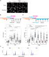
Figure 2. DSB repair occurs with low efficiency during mitosis.
(A) Asynchronous cells (Asyn.) or nocodazole-arrested (M-phase) cells were analyzed by the neutral comet assay. Typical images are shown for non-treated (−Etp) or etoposide-treated (+Etp) cells at each time point after release from etoposide treatment. (B) Comparison of DSB repair kinetics between mitotic and asynchronous cells. Shown is the comet assay procedure for detecting transiently introduced M-phase DSBs (upper figure). Shown is the distribution of tail-moment values in the non-treated cells (−Etp) or DSB-induced cells (+Etp) in the asynchronous condition (open circles) or nocodazole arrested condition (closed circles) at each time point after release from etoposide treatment. Red and blue bars show average values of tail moments for each condition. Statistical significance was analyzed using Dunnett's test. ** P-value<0.01; * P-value<0.05. (C) Cell-cycle distribution of cells with DSBs after nocodazole arrest. Cells arrested with nocodazole were treated to induce DSBs and were used for the comet assay. Cells were analyzed for DNA content by FACS at the indicated times after release from etoposide treatment. The x-axis values correspond to DNA content. (D) Procedure for mitotic DSB introduction by the comet assay during constitutive M-phase arrest (upper scheme). Shown is DSB repair kinetics in prometaphase-arrested cells by comet assay. Distribution of tail moment values under the continuous nocodazole-arrested condition is shown graphically. Statistical significance was analyzed using Dunnett's test. ** P-value<0.01.
Figure 3
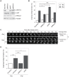
Figure 3. Anaphase bridges are formed by NHEJ and HR and are inhibited by CtIP-dependent end resection.
(A) Representative western blots showing the siRNA-mediated depletion of XRCC4, CtIP, or XRCC3 in HeLaS3 cells as well as control siRNA-treated cells. (B) Frequency of anaphase bridge formation in HeLaS3 cells transfected with an siRNA targeting XRCC4, CtIP, or XRCC3 (or control siRNA) with (+Etp) or without (−Etp) induction of mitotic DSBs by etoposide. Anaphase cells (≥50) were scored for each experiment and condition. Error bars show the standard deviation from three independent experiments. Statistical significance was analyzed with the Student's t-test. ** P-value<0.01, ns, not significant. (C) Anaphase bridge formation was observed with time-lapse live-cell imaging of a typical HeLa cell at the indicated time after release from etoposide treatment with (AB) or without (−) anaphase bridge formation. In addition, shown is an average of duration when required to transit to anaphase in non-treated (−Etp) or etoposide-treated (+Etp) cells with numbers of cells analyzed. Statistical significance was analyzed with the Student's t-test. Scale bar, 5 µm; ns, not significant. (D) Anaphase-bridge formation observed by time-lapse imaging after DSB induction in the indicated knockdown cells. Prometaphase cells (≥25) were chosen and analyzed for anaphase bridge formation for each experiment. Error bars show standard deviation from 3 to 11 independent experiments. Statistical significance was analyzed with the Student's t-test. ** P-value<0.01, * P-value<0.05.
Figure 4
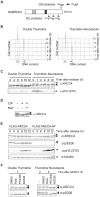
Figure 4. M-phase specific phosphorylation of XRCC4.
(A) Domain structure of XRCC4. Serine 326 near the C-terminus of human XRCC4 is a potential CDK phosphorylation site. Threonine 222, serine 256 and serine 302 are potential Plk1 phosphorylation sites. Substitution of serine 326 with alanine results in the mutant XRCC4-AP. The gray box and black box indicate XLF- and DNA ligase IV–binding sites, respectively. (B) Cell-cycle progression after release from double thymidine or thymidine-nocodazole block. Cells were arrested in G1/S via double thymidine or thymidine-nocodazole blocks and were then released to resume the cell cycle. Cells were harvested at the indicated times, lysed with Triton X-100, and analyzed for DNA content by FACS. (C) Western blot of lysates from HeLaS3 cells after release from double thymidine block or thymidine-nocodazole block. Cells were harvested at the indicated times, lysed in SDS-PAGE loading buffer, and subjected to western blotting. Each membrane was incubated with anti-XRCC4 or anti-phosphorylated histone H3 (S10), a marker for M phase. (D) Effect of phosphatase treatment on XRCC4 modification. FLAG-tagged XRCC4 was immunoprecipitated from asynchronous HeLaS3 cells expressing FLAG-tagged XRCC4. The immunoprecipitate was treated with calf intestine phosphatase (CIP) in the absence or presence of its inhibitor, NaV. (E) Western blot of lysates from FLAG-tagged XRCC4 and FLAG-tagged XRCC4-AP cells after release from double thymidine block. FLAG-tagged XRCC4 and FLAG-tagged XRCC4-AP cells were harvested at the indicated times, lysed in SDS-PAGE loading buffer, and analyzed by western blotting with anti-XRCC4, anti-phosphorylated serine 326 of XRCC4 (anti-pS326), anti-phosphorylated histone H3 (S10) (a marker for M phase), or anti-α-tubulin (internal control). (F) Effect of a CDK and Plk1 inhibitors on phosphorylation of XRCC4. Cells were arrested by double thymidine block. Cells were released into the cell cycle (double thymidine) or treated with nocodazole continuously (thymidine-nocodazole), and then treated with the CDK inhibitor roscovitine (20 µM; Rosc.) or RO3306 (20 µM) or the Plk1 inhibitor BI2536 (200 nM) for 4 h or 5 h, respectively. XRCC4 was analyzed by western blotting with anti-XRCC4 or anti-pS326.
Figure 5
Figure 5. Phosphorylation of XRCC4 at S326 is involved in suppression of DSB repair during M-phase.
(A) Staining of DNA ligase IV (green; anti-DNA ligase IV) and XRCC4 (red; anti-XRCC4) in etoposide-treated cells. Indirect immunostaining was performed as described in Materials and Methods for FLAG-XRCC4- or FLAG-XRCC4-AP-expressing cells at 1 h after release from nocodazole arrest. Shown is an image for each mitotic phase. Scale bar, 10 µm. (B) Co-immunoprecipitation of DNA ligase IV and XLF with FLAG-tagged XRCC4 or FLAG-tagged XRCC4-AP. Asynchronous cells (Asyn.) and M-phase arrested cells (M) were immunoprecipitated with anti-FLAG, and then subjected to western blotting using antibody against XRCC4, DNA ligase IV, and XLF. An overexposure image (over exp.) of the same western blot membrane using anti-XLF is shown at the bottom. (C) Frequency of anaphase bridge formation in HeLaS3 cells with control siRNA or in XRCC4-depleted, FLAG-XRCC4-expressing, and FLAG-XRCC4-AP-expressing cells. Shown are anaphase bridge formation frequencies in etoposide-treated (gray bars) or non-treated cells (white bars) of each cell line. Anaphase cells (≥50) were analyzed for each experiment and condition. Error bars show the standard deviation from seven independent experiments. Statistical significance was analyzed with the Student's t-test. * P-value<0.05; ns, not significant. (D) Detection of DSB repair kinetics in XRCC4 and XRCC4-AP cells. Shown is the distribution of tail moment (TM) values under the continuous nocodazole-arrested condition in DSB-induced FLAG-XRCC4-expressing cells (XRCC4; open circles) or FLAG-XRCC4-AP-expressing cells (closed circles) with (+Etp) or without (−Etp) etoposide at the indicated time points after release from etoposide treatment. Statistical significance was analyzed with the Student's t-test. ** P-value<0.01; ns, not significant. (E) Detection of DSB repair kinetics in XRCC4-depleted, FLAG-XRCC4-expressing (open circles), and FLAG-XRCC4-AP-expressing (closed circles) cells. Shown are kinetics of average relative TM values in etoposide-treated cells of each cell line. Relative TM values were calculated by dividing each TM value by the average peak TM value at 1 h.

References

    1. Chapman JR, Taylor MR, Boulton SJ (2012) Playing the end game: DNA double-strand break repair pathway choice. Mol Cell 47: 497–510. - PubMed
    1. Shinohara A, Ogawa H, Ogawa T (1992) Rad51 protein involved in repair and recombination in S. cerevisiae is a RecA-like protein. Cell 69: 457–470. - PubMed
    1. Bogue MA, Wang C, Zhu C, Roth DB (1997) V(D)J recombination in Ku86-deficient mice: distinct effects on coding, signal, and hybrid joint formation. Immunity 7: 37–47. - PubMed
    1. Kabotyanski EB, Gomelsky L, Han JO, Stamato TD, Roth DB (1998) Double-strand break repair in Ku86- and XRCC4-deficient cells. Nucleic Acids Res 26: 5333–5342. - PMC - PubMed
    1. Liang F, Romanienko PJ, Weaver DT, Jeggo PA, Jasin M (1996) Chromosomal double-strand break repair in Ku80-deficient cells. Proc Natl Acad Sci U S A 93: 8929–8933. - PMC - PubMed

Publication types