. 2014 Aug 27;15(9):15090-108.
doi: 10.3390/ijms150915090.

What controls DNA looping?

Affiliations

What controls DNA looping?

Pamela J Perez et al. Int J Mol Sci. .

Abstract

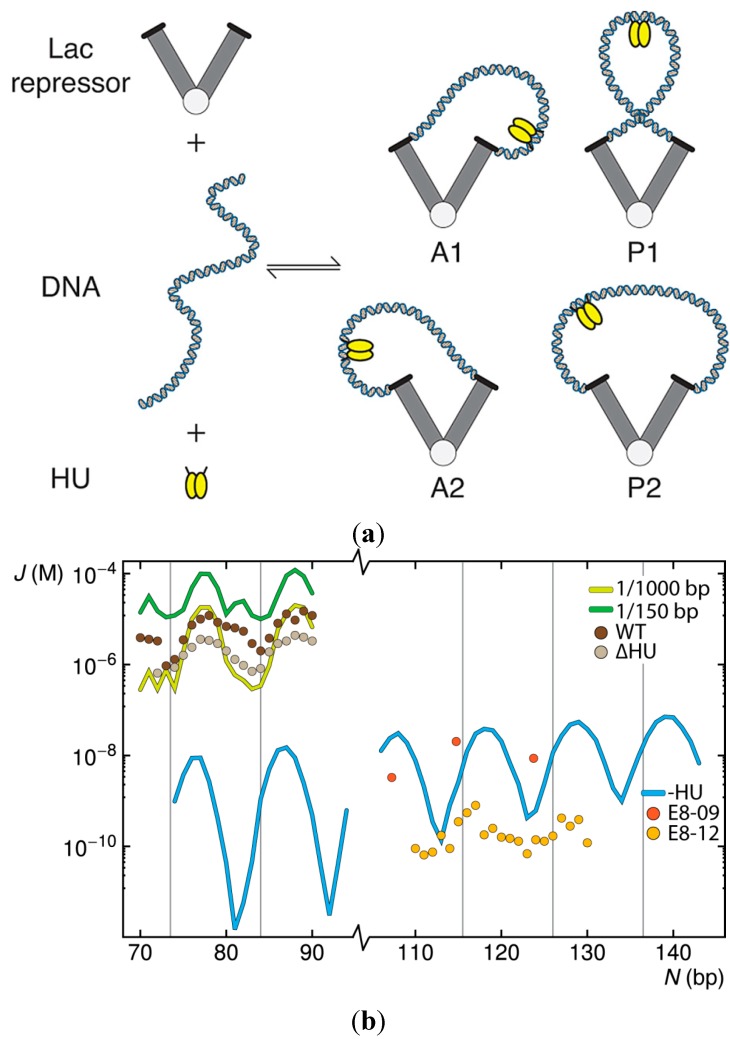
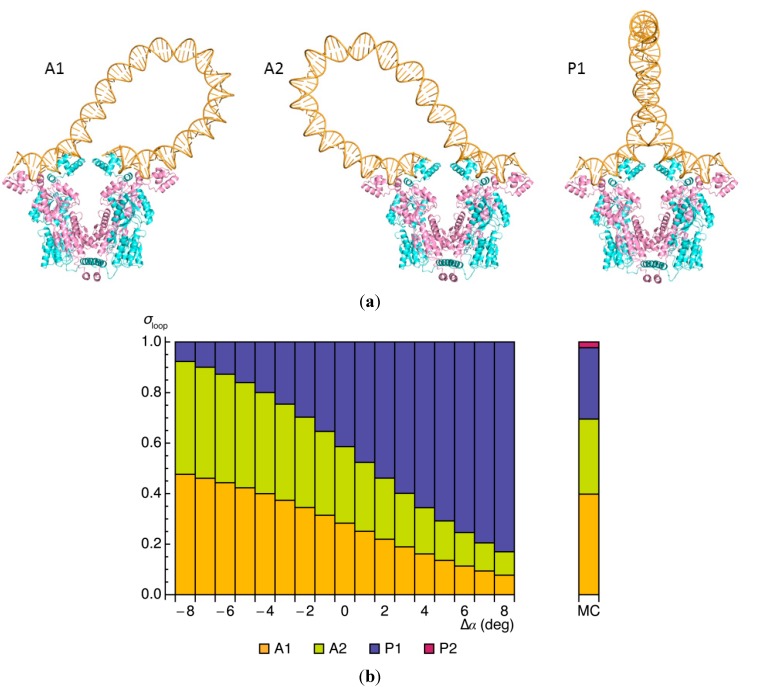
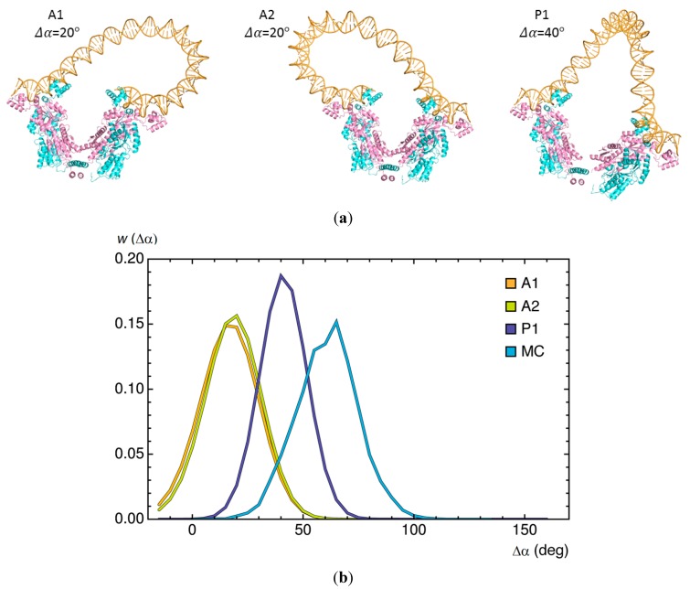
The looping of DNA provides a means of communication between sequentially distant genomic sites that operate in tandem to express, copy, and repair the information encoded in the DNA base sequence. The short loops implicated in the expression of bacterial genes suggest that molecular factors other than the naturally stiff double helix are involved in bringing the interacting sites into close spatial proximity. New computational techniques that take direct account of the three-dimensional structures and fluctuations of protein and DNA allow us to examine the likely means of enhancing such communication. Here, we describe the application of these approaches to the looping of a 92 base-pair DNA segment between the headpieces of the tetrameric Escherichia coli Lac repressor protein. The distortions of the double helix induced by a second protein--the nonspecific nucleoid protein HU--increase the computed likelihood of looping by several orders of magnitude over that of DNA alone. Large-scale deformations of the repressor, sequence-dependent features in the DNA loop, and deformability of the DNA operators also enhance looping, although to lesser degrees. The correspondence between the predicted looping propensities and the ease of looping derived from gene-expression and single-molecule measurements lends credence to the derived structural picture.


Figures

Figure 1
Figure 1
Simulated effects of HU on the ease of looping short pieces of DNA between the headpieces of a V-shaped Lac repressor protein assembly. (a) Schematic representation of the molecular components and resulting HU-decorated DNA loops anchored in antiparallel (A1, A2) and parallel (P1, P2) orientations [13] on the repressor; (b) Predicted values of the J factor, as a function of operator spacing, for double-helical molecules free of HU or binding one HU on average every 150 or 1000 bp compared, on the top left, with values deduced from the expression of lac genes in Escherichia coli cells containing the wild-type (WT) protein and a mutated strain (∆HU) that cannot express HU [5,14,15] and, on the lower right, with values deduced from tethered particle motion studies of DNA constructs (E8-09, E8-12) flanked at the 5'-end by a symmetric operator and the 3'-end by the natural O1 operator [16,17]. Computed J factors are connected by thin lines. Values deduced from experiments are shown by symbols. Vertical lines denote integral helical repeats of DNA.
Figure 2
Figure 2
Effects of DNA chain length on the orientation of DNA and the simulated uptake of HU on loops anchored to the Lac repressor. (a) Molecular images illustrating the predominant configurations of DNA and the positions of HU on 109 bp (left) and 115 bp (right) loops. Images rendered in PyMOL [22] with DNA backbones are shown as gold tubes, DNA bases as gold sticks, HU chains as blue ribbons, and repressor chains as pink and cyan ribbons. Views looking down the axis perpendicular to the long axes of the globular arms of the repressor; (b) Frequencies of occurrence σloop of the four possible looped forms. Note the marked shift in loop orientation (A1/A2→P1/P2) and HU uptake (1→2) with the increase in operator spacing, the straight segments of DNA in the models, and the potential (highly curved) site for a third HU at the center of the P2 loop.
Figure 3
Figure 3
Effect of the Lac repressor on the minimum-energy configurations of 92-bp DNA loops. (a) Molecular images of repressor-mediated loops anchored in antiparallel and parallel orientations on the rigid V-shaped protein assembly. Note the more gradually curved pathways of the dominant types of bare DNA loops compared to the corresponding forms found in the presence of HU (Figure 2a); (b) Variation in the relative weights σloop of the three looped forms with small changes Δα in the angle between the protein arms, compared with the relative populations of DNA loops found to be attached to the same Lac repressor model through Monte Carlo (MC) sampling [23] (vertical bar to the right of the data obtained by energy optimization).
Figure 4
Figure 4
Changes in DNA looping associated with large-scale opening of the Lac repressor. (a) Molecular images of 92-bp loops anchored in the dominant antiparallel and parallel orientations with the change in the opening angle Δα set to values that minimize the DNA elastic energy. Note the rearrangement of DNA on opened versus closed repressors (Figure 3a); (b) Distribution of the opening angle w (Δα) of Lac repressor proteins anchoring DNA loops subjected to energy minimization and collected in Monte Carlo (MC) sampling. The differences in repressor opening found with the two approaches reflect the different treatments of protein, i.e., precisely imposed routes of conformational change versus spatial forms indirectly captured in the course of DNA loop closure.
Figure 5
Figure 5
Changes in 92-bp DNA looping brought about by localized changes in the DNA model. (a) Superimposed images of the optimized DNA pathways found when the sites of pyrimidine-purine steps along the lac operon are softened (blue) and when the helix is overtwisted (green) compared to ideal DNA (gold). Terminal base pairs are constrained to the spatial positions imposed by the V-shaped Lac repressor assembly. The antiparallel loops are rotated ~45° and the parallel loop ~90° about the vertical axis relative to the images in Figure 3a; (b) Changes in the bending Δγ and twisting Δθ between successive base pairs along the depicted loops, with the data color-coded to match the molecular images. The yellow lines denote the locations of the pyrimidine–purine steps, where the softened DNA shows bursts of bending and untwisting. Note the relative insensitivity of the parallel loop and the responses in the antiparallel loops to imposed overtwisting.
Figure 6
Figure 6
Relative ease of forming 92-bp DNA loops anchored by various Lac repressor-bound O3···O1 and Osym···Osym operator models compared to loops terminated by identical rigid, Osym operators. (a) Cartoon images illustrating the variability in the three operator structures. All views looking down the long axis of the kinked CG·CG step (blue) at the centerof the DNA fragments; (b) Distribution of the looping propensities, expressed in terms of the logarithmic values of the J factors, for the two classes of flexible anchoring conditions and for the rigid operator pair (formula image
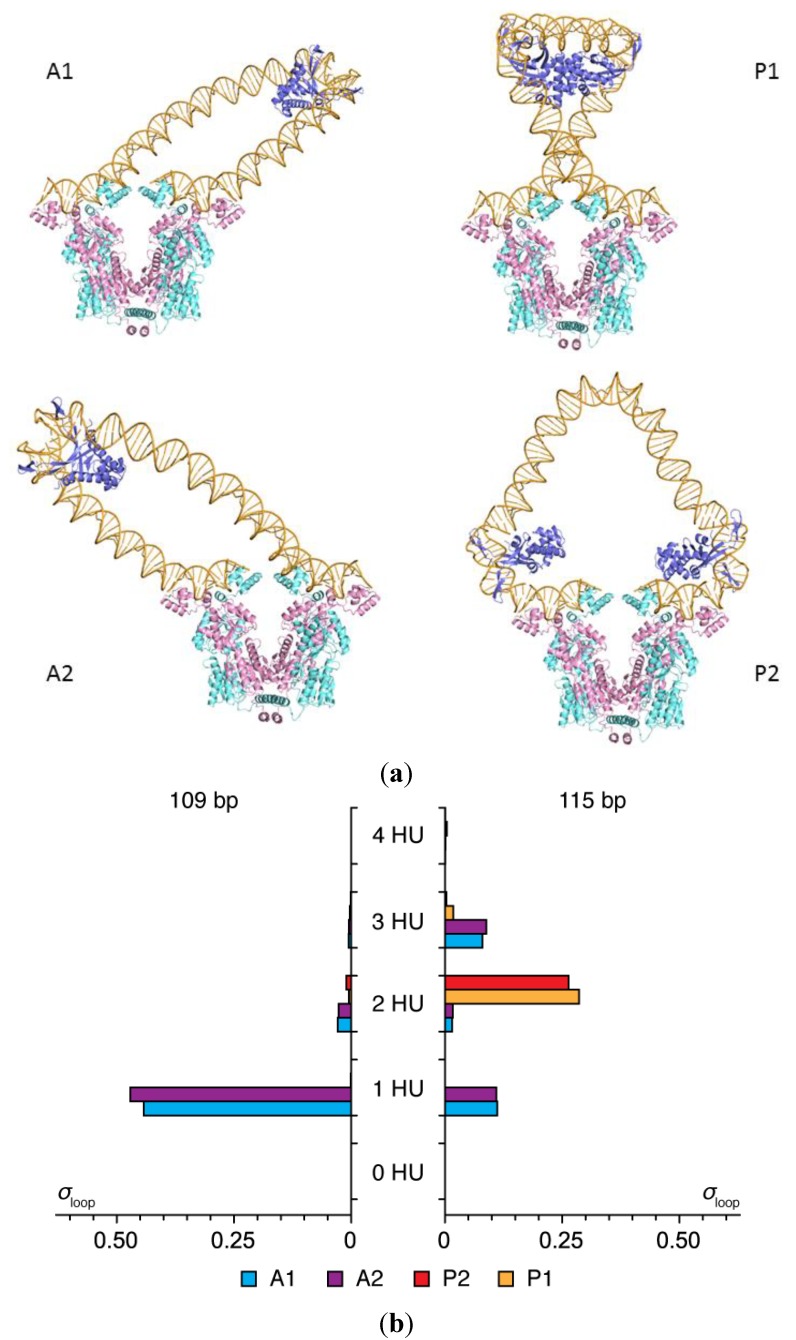
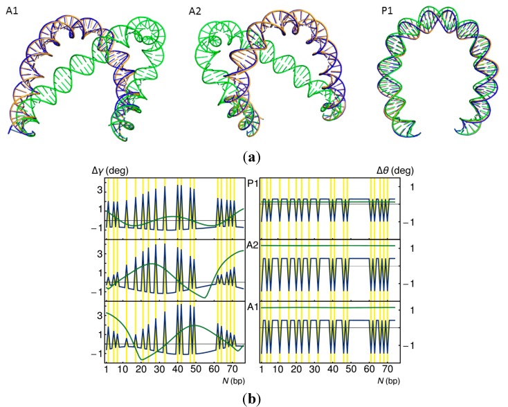
). Note the enhancement in looping with the natural operators.

References

    1. Müller-Hill B. The lac Operon. Walter de Gruyter; Berlin, Germany: 1996.
    1. Koh J., Shkel I., Saecker R.M., Record M.T., Jr. Nonspecific DNA binding and bending by HUαβ: Interfaces of the three binding modes characterized by salt-dependent thermodynamics. J. Mol. Biol. 2011;410:241–267. doi: 10.1016/j.jmb.2011.04.001. - DOI - PMC - PubMed
    1. Swinger K.K., Lemberg K.M., Zhang Y., Rice P.A. Flexible DNA bending in HU–DNA cocrystal structures. EMBO J. 2003;22:3749–3760. doi: 10.1093/emboj/cdg351. - DOI - PMC - PubMed
    1. Krämer H., Niemöller M., Amouyal M., Revêt B., von Wilcken-Bergmann B., Müller-Hill B. Lac repressor forms loops with linear DNA carrying two suitably spaced lac operators. EMBO J. 1987;6:1481–1491. - PMC - PubMed
    1. Bond L.M., Peters J.P., Becker N.A., Kahn J.D., Maher L.J., 3rd. Gene repression by minimal lac loops in vivo. Nucleic Acids Res. 2010;38:8071–8082. - PMC - PubMed

Publication types

MeSH terms

LinkOut - more resources