. 2014 Aug 28;10(8):e1004584.
doi: 10.1371/journal.pgen.1004584. eCollection 2014 Aug.

A conserved dopamine-cholecystokinin signaling pathway shapes context-dependent Caenorhabditis elegans behavior

Affiliations

A conserved dopamine-cholecystokinin signaling pathway shapes context-dependent Caenorhabditis elegans behavior

Raja Bhattacharya et al. PLoS Genet. .

Abstract

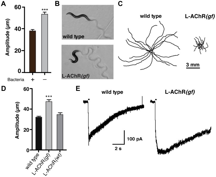
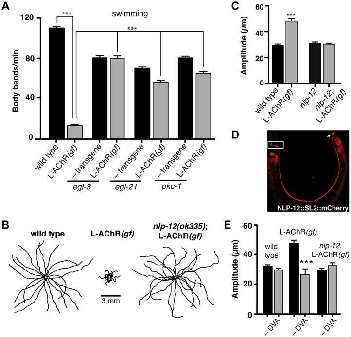
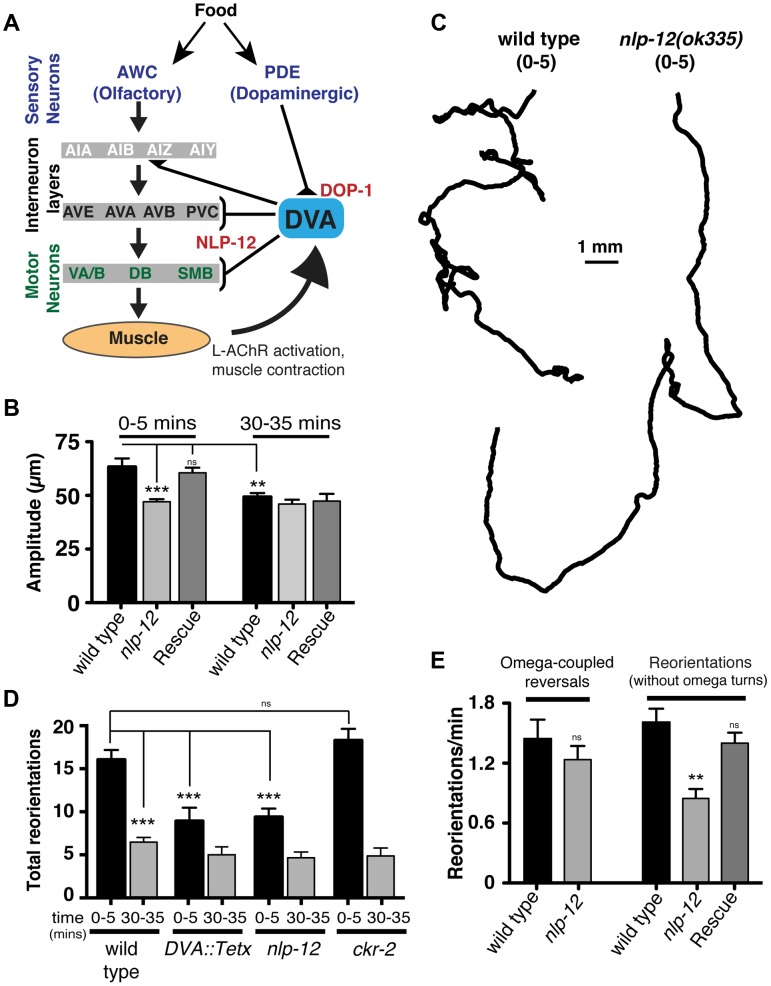
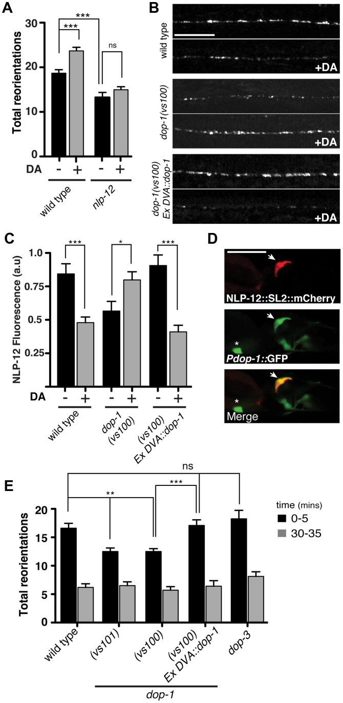
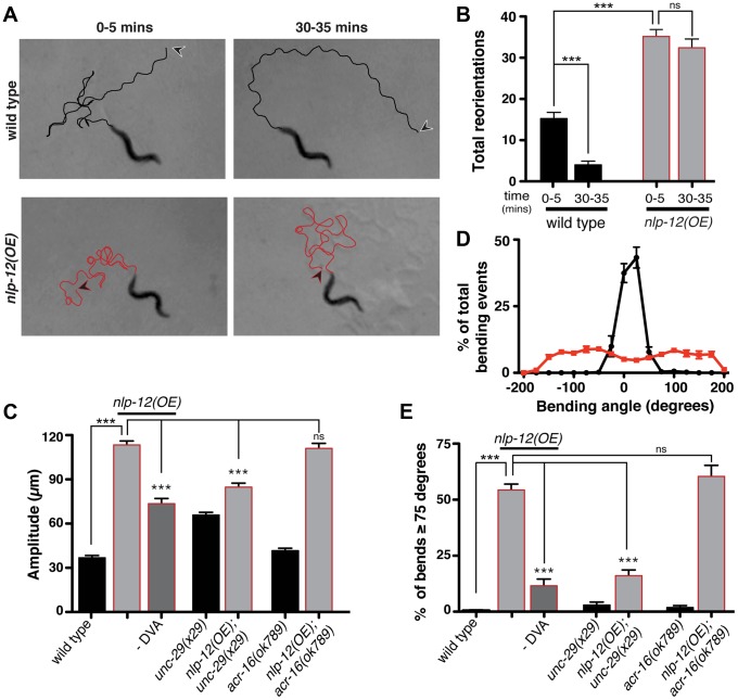
An organism's ability to thrive in changing environmental conditions requires the capacity for making flexible behavioral responses. Here we show that, in the nematode Caenorhabditis elegans, foraging responses to changes in food availability require nlp-12, a homolog of the mammalian neuropeptide cholecystokinin (CCK). nlp-12 expression is limited to a single interneuron (DVA) that is postsynaptic to dopaminergic neurons involved in food-sensing, and presynaptic to locomotory control neurons. NLP-12 release from DVA is regulated through the D1-like dopamine receptor DOP-1, and both nlp-12 and dop-1 are required for normal local food searching responses. nlp-12/CCK overexpression recapitulates characteristics of local food searching, and DVA ablation or mutations disrupting muscle acetylcholine receptor function attenuate these effects. Conversely, nlp-12 deletion reverses behavioral and functional changes associated with genetically enhanced muscle acetylcholine receptor activity. Thus, our data suggest that dopamine-mediated sensory information about food availability shapes foraging in a context-dependent manner through peptide modulation of locomotory output.


Conflict of interest statement

The authors have declared that no competing interests exist.

Figures

Figure 1
Figure 1. Enhanced L-AChR function increases neuromuscular signaling and alters the C. elegans locomotory pattern.
(A) Average body bend amplitude of wild type worms in presence (+) or absence (−) of bacterial food. Well-fed animals were transferred to assay plates with or without food and videotaped for 45 s following a recovery period of one minute. Bars represent the mean (±SEM) of values calculated from 15 animals. ***, p<0.0001 student's t-test. (B) Representative images of wild type and L-AChR(gf) animals. Note the exaggerated track amplitudes and the hypercontracted body posture of L-AChR(gf) animals. (C) Movement trajectories of wild type and L-AChR(gf) animals. Each black line shows the trajectory of one animal monitored for 45 s on food. In this and subsequent figures, tracks are aligned to a common center point (gray) for clarity. (D) Average body bend amplitude for wild type, L-AChR(gf) and L-AChR(wt) animals as indicated. Values for body bend amplitude were calculated from recordings of the tracks shown in C. Bars represent the mean (±SEM) of values calculated from at least 15 animals. ***, p<0.0001 by ANOVA and Sidak's post-hoc test. (E) Current responses to pressure application of levamisole (100 µM) recorded from body wall muscles of wild type and L-AChR(gf) animals. Holding potential was −60 mV. Black bars indicate the duration of levamisole application (200 ms). See Figure 6 for additional electrophysiological characterization of the L-AChR(gf) strain.
Figure 2
Figure 2. Locomotory phenotypes associated with L-AChR(gf) expression require neuropeptide signaling.
(A) Average body bends/min measured in liquid for the genotypes indicated. The strong locomotory defects of egl-3 and egl-21 mutants prevented analysis of L-AChR(gf) effects on agar (Fig. S3A, B). Mutation of pkc-1 normalized the locomotor effects of L-AChR(gf) in both liquid and on agar (Fig. S3C–E). (B) Movement trajectories of wild type, L-AChR(gf), nlp-12;L-AChR(gf) animals as indicated. Each black line shows the trajectory of one animal monitored for 45 s on food. (C) Average body bend amplitude for the genotypes indicated. Each bar represents the mean (±SEM) of values calculated from recordings of at least 15 animals. For (A) and (C) ***, p<0.0001 by ANOVA with Sidak's post-hoc test. (D) Wide-field epifluorescent image of an adult animal expressing nlp-12::SL2::mCherry. The image is oriented with the head to the left. White rectangle: nerve ring. Arrow: DVA cell body. (E) Average body bend amplitude for the indicated genotypes and effects of DVA ablation (–DVA). Ablations were performed on L2 stage animals. Body bend amplitude was measured from recordings of young adult animals 2 days following laser ablation. Error bars indicate mean (±SEM) of at least 8 animals. ***, p<0.0001 student's t-test.
Figure 3
Figure 3. Modulation of body bend depth through NLP-12/CCK signaling is critical for local food searching.
(A) Schematic representation of the neural circuit underlying NLP-12 modulation of local searching. Synaptic connections (triangles, brackets) are as described by . DVA receives synaptic input from the dopaminergic neuron PDE and makes connections with both motor neurons and interneurons involved in locomotory control. DVA makes synaptic contacts onto all of the motor and interneurons indicated by brackets. In addition, DVA is connected to AVB and PVC by gap junctions. Assignments of interneurons into layers are as described by . Other neuron classes are as described in the text and references therein. DOP-1 modulation of DVA activity regulates NLP-12 release from DVA, altering the motor pattern during local food searching. (B) Average body bend amplitude for the genotypes as indicated measured during an initial five minute time interval (0–5) immediately following removal from food and a second five minute time interval 30 minutes after removal from food (30–35). Bars represent mean values (±SEM) calculated from 9–14 animals. (C) Representative tracks of wild type and nlp-12(ok335) mutants during an initial five minute period (0–5) following removal from food. Note the decreased number of reorientations and increased frequency of long forward runs for nlp-12(ok335) mutant as compared to the wild type. (D) Total directional reorientations measured during the 0–5 and 30–35 minute intervals following removal from food for wild type, transgenic wild type animals expressing Tetanus toxin in DVA [DVA::Tetx], nlp-12(ok335) and ckr-2(tm3082) mutants. Bars represent mean (±SEM) for at least 12 animals. (E) Quantification of reversal coupled omega turns and reorientations in the absence of omega turns during the first 5 minutes following removal from food for the genotypes indicated. (WT and nlp-12(ok335): n = 17). Rescue refers to the nlp-12(ok335) mutant expressing an extrachromosomal array carrying Pnlp-12::nlp-12::SL2::mCherry (n = 9). Bars represent mean (±SEM). ***, p<0.0001; **, p<0.001 by ANOVA with Sidak's post-hoc test.
Figure 4
Figure 4. The dopamine receptor DOP-1 is required in DVA for NLP-12 modulation of food searching.
(A) Frequency of high angled reorientations for wild type and nlp-12(ok335) animals quantified for 5 minutes after transfer to food free plates in the presence (+) or absence (−) of dopamine (DA). Bars represent mean (±SEM) for at least 12 animals. Dopamine mechanosensory signaling is strongly enhanced at low osmotic strength . Therefore, these assays were conducted following transfer of the animals to low osmotic-strength assay plates as described previously . We observed a modest increase in basal reorientation frequency across all genotypes under these conditions. (B, C) Representative images (B) and quantification (C) of NLP-12::VenusYFP fluorescence in the ventral cord region of the DVA process of wild type, dop-1(vs100), and dop-1(vs100) Ex DVA::dop-1 animals before (−) and after (+) 10 minutes dopamine (DA) treatment (wild type: n = 12 for (−) and (+) DA; dop-1(vs100): n = 12 for (−) and 9 for (+) DA). Ex DVA::dop-1 refers to specific rescue of dop-1 expression in DVA using the nlp-12 promoter (−DA, n = 12; +DA, n = 11). Bars represent mean ±SEM. ***, p<0.0005; *, p<0.05 student's t-test. (D) Single slice confocal images of the DVA neuron in a transgenic animal expressing nlp-12::SL2::mCherry (upper panel) together with Pdop-1::GFP (middle panel). White arrow denotes the DVA interneuron in all cases. Asterix denotes a ventral cord motor neuron expressing the dop-1 reporter. Scale bars in B and D, 20 µm. (E) Total directional reorientations measured during 0–5 and 30–35 minute intervals following removal from food for the genotypes as indicated. WT: n = 10; dop-1(vs101): n = 12, dop-1(vs100): n = 14, dop-1(vs100) Ex DVA::dop-1: n = 12 and dop-3(vs106): n = 8. Bars represent mean (±SEM). For (A) and (E) ***, p<0.0005, **, p<0.005 by ANOVA with Sidak's post-hoc test.
Figure 5
Figure 5. Elevated NLP-12 signaling induces a chronic local search-like state.
(A) Representative tracks (45 s) of wild type (black lines) and nlp-12(OE) (red lines) animals during 0–5 and 30–35 minute intervals after removal from food as indicated. nlp-12(OE) refers to a transgenic strain (ufIs104) stably expressing high levels of the wild type nlp-12 genomic sequence. Note that high angled reorientations still persist during the 30–35 minute interval for nlp-12(OE) in contrast to the long forward run of the wild type animal. The starting point of each track is indicated by an arrowhead (wild type: black; nlp-12(OE): red). (B) Total directional reorientations measured during 0–5 and 30–35 minute intervals following removal from food for wild type (n = 8) and nlp-12(OE) (n = 15) animals. (C) Average body bend amplitude for the genotypes indicated. –DVA refers to DVA ablation (WT: n = 11, nlp-12(OE): n = 15, nlp-12(OE) -DVA: n = 10, unc-29: n = 30, unc-29;nlp-12(OE): n = 22, acr-16: n = 16, acr-16;nlp-12(OE): n = 15). (D) Frequency distribution of bending angles for wild type (black) and nlp-12(OE) (red) animals monitored on bacteria seeded plates for 30 s (WT: n = 12, nlp-12(OE): n = 15). (E) Bar graph depicting total percentage of bending angles ≥75° for the genotypes indicated (WT: n = 12, nlp-12(OE): n = 14, nlp-12(OE) -DVA: n = 9, unc-29: n = 10, unc-29;nlp-12(OE): n = 12, acr-16: n = 10, acr-16;nlp-12(OE): n = 12). Note that wild type worms rarely execute bends ≥75°. Bars represent mean (±SEM). For (B), (C) and (E) ***, p<0.0001 by ANOVA with Sidak's post-hoc test.
Figure 6
Figure 6. Evoked synaptic responses are prolonged in L-AChR(gf) animals and require nlp-12.
(A) Time course of paralysis in the presence of the L-AChR agonist levamisole (200 µM) for wild type, L-AChR(gf), nlp-12 mutants and nlp-12;L-AChR(gf) animals as indicated. Each data point represents the mean (± SEM) for at least 10 trials. (B) Current responses to pressure application (1 s) of levamisole (500 µM) recorded from body wall muscles of control (− transgene) (n = 5), L-AChR(gf) (n = 6) and nlp-12;L-AChR(gf) (n = 4) animals as indicated. Holding potential was −60 mV. (C) The ratio of the current amplitude 1 s after the start of levamisole application (steady state) to the peak current for control, L-AChR(gf), and nlp-12;L-AChR(gf) animals as indicated. (D) Current responses to photostimulation of motor neurons (10 ms, upper or 800 ms, lower) recorded from body wall muscles of control, L-AChR(gf), and nlp-12;L-AChR(gf) animals as indicated. Holding potential was −80 mV. Recordings were made in the presence of the N-AChR antagonist dHβE (10 µM) to isolate the L-AChR mediated current. (E, F) Average amplitude (E) decay time constant (F) for synaptic responses to 10 ms motor neuron photostimulation for the genotypes indicated. The decay phase of L-AChR mediated evoked current responses were fit with a single exponential. Bars represent mean (± SEM) of amplitude and decay time constant values calculated for responses from control (n = 19), L-AChR(gf) (n = 11), and nlp-12;L-AChR(gf) (n = 11) animals. ***, p<0.0001 by ANOVA with Sidak's post-hoc test. Each strain stably expressed the Pacr-2::ChR2-GFP transgene (ufIs23) for photostimulation of motor neurons.

References

    1. Marder E (2012) Neuromodulation of neuronal circuits: back to the future. Neuron 76: 1–11. - PMC - PubMed
    1. Nusbaum MP, Blitz DM (2012) Neuropeptide modulation of microcircuits. Curr Opin Neurobiol 22: 592–601. - PMC - PubMed
    1. DeLong ND, Nusbaum MP (2010) Hormonal modulation of sensorimotor integration. J Neurosci 30: 2418–2427. - PMC - PubMed
    1. Doi A, Ramirez JM (2010) State-dependent interactions between excitatory neuromodulators in the neuronal control of breathing. J Neurosci 30: 8251–8262. - PMC - PubMed
    1. Nusbaum MP, Marder E (1989) A modulatory proctolin-containing neuron (MPN). II. State-dependent modulation of rhythmic motor activity. J Neurosci 9: 1600–1607. - PMC - PubMed

Publication types

LinkOut - more resources