Review
. 2014 Aug 28;4(11):a018481.
doi: 10.1101/cshperspect.a018481.

Autophagy in tuberculosis

Affiliations
Review

Autophagy in tuberculosis

Vojo Deretic. Cold Spring Harb Perspect Med. .

Abstract

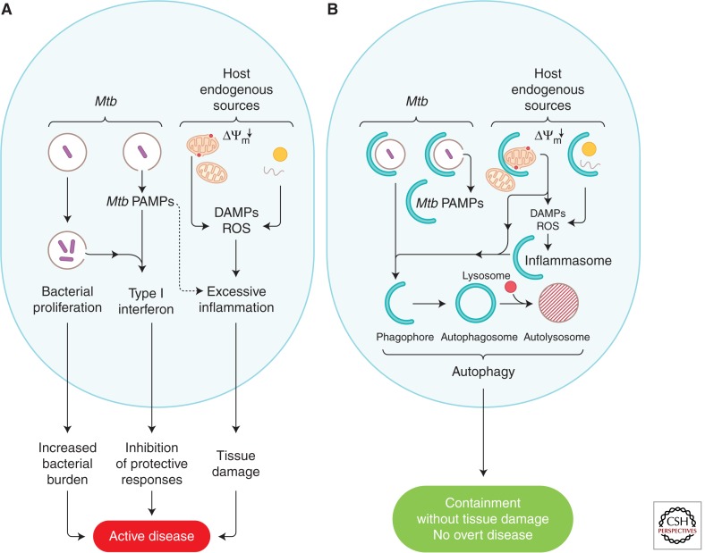
Autophagy as an immune mechanism controls inflammation and acts as a cell-autonomous defense against intracellular microbes including Mycobacterium tuberculosis. An equally significant role of autophagy is its anti-inflammatory and tissue-sparing function. This combination of antimicrobial and anti-inflammatory actions prevents active disease in animal models. In human populations, genetic links between autophagy, inflammatory bowel disease, and susceptibility to tuberculosis provide further support to these combined roles of autophagy. The autophagic control of M. tuberculosis and prevention of progressive disease provide novel insights into physiological and immune control of tuberculosis. It also offers host-based therapeutic opportunities because autophagy can be pharmacologically modulated.


Figures

Figure 1.
Figure 1.
Autophagy pathway. Shown are a simplified macroautophagy pathway, protein factors, and membrane sources for the formation of autophagosomes in mammalian cells. (Top) Different sources of membranes contributing to the formation of autophagosomes. ER, endoplasmic reticulum; PM, plasma membrane. Within the cartoon: Crescent, autophagic phagophore or isolation membrane; double-membrane, closed autophagosome; red circle, lysosome; hatched circle delimited by a single membrane, autolysosome; green dots, LC3B (one of six mammalian Atg8s listed on the right side; typically used as a marker for autophagosomes). Other key factors are shown using their mammalian nomenclature.
Figure 2.
Figure 2.
Autophagy protects against M. tuberculosis infection and pathogenesis. (A) Autophagy induction by starvation or rapamycin kills virulent M. tuberculosis in macrophages. (B) Increased gross lung pathology in mice defective for autophagy in the myeloid lineage (Cre+) relative to autophagy-competent (Cre) mice infected with M. tuberculosis H37Rv. (C) Increased lung tissue necrosis and bacillary load in the lungs of mice defective for autophagy in the myeloid lineage (Cre+) relative to their autophagy-competent (Cre) littermates. (Bottom panels) Acid-fast stain. Arrows, M. tuberculosis H37Rv bacilli. (A, Reprinted, with permission, from Gutierrez et al. 2004; B,C, reprinted, with permission, from Castillo et al. 2012.)
Figure 3.
Figure 3.
Sequestosome 1/p62–like receptors (SLRs), a new class of pattern-recognition receptors involved in autophagic elimination of intracellular microbes. LIR, LC3-interacting region (consensus sequence shown and key positions in red); aromatic pocket-filling W (or F/Y) and aliphatic pocket-filling L (or I/V) form an intermolecular parallel β sheet with LC3s or GABARAPs, at the interface between the amino-terminal α-helical domain and ubiquitin-like fold of LC3s/GABARAPs. CLIR, a LIR specific for LC3C, whereby aromatic residue is not present (X) to fill in the aromatic pocket and instead the interactions are stabilized by compensatory hydrophobic contacts provided by additional aliphatic residues located between the W and L position anchors. KIR, KAEP1-interacting region; NES, nuclear export signal; NLS, nuclear localization signal; PB1, protein-binding domain (homopolymerization of p62 hetero-oligomerization between p62 and NBR1, or interactions with other partners); TR, TRAF6-interacting region (also a multipartner binding region); ZZ, ZZ-type zinc finger (ZnF) domain; FW, four-Trp domain, also known as the NBR1 box; UBAN, a parallel coiled-coiled dimer UBD with specificity for linear ubiquitin chains; CC, coiled coil; UBA, a three-helix bundle UBD (ubiquitin-binding domain) with affinity for monoubiquitin and the more open conformation of K63 ubiquitin linkages; UBZ, a Zn finger ββα fold UBD binding mono- and polyubiquitin; SKICH, skeletal muscle and kidney enriched inositol phosphatase carboxyl homology domain; GIR, galectin-interacting region. Sequestosome 1/p62 has been shown to affect M. tuberculosis clearance (Ponpuak et al. 2010). NDP52 has been implicated in elimination of M. tuberculosis in murine macrophages (Watson et al. 2012); note, however, that NDP52 is severely truncated in this species. (Image modified from Deretic et al. 2013.)
Figure 4.
Figure 4.
LC3-associated phagocytosis (LAP) as an intersection between autophagy and phagocytosis. Autophagy is frequently morphologically described as formation of double-membrane autophagosomes in the cytoplasm. This is requisite when an internal phagosome is derived from intracellular membranes such as the endoplasmic reticulum (ER). One important exception in the context of the role of autophagy during infection is the formation of conventional phagosomes that are also decorated with LC3 (green dots). Note that LC3 is only on the cytofacial side of the phagosomal membrane, and that this is a single membrane as in the case of conventional phagosomes. Depicted also is a Toll-like receptor molecule recognizing pathogen-associated molecular patterns (asterisks), involved in the induction of LAP and capable of concomitantly inducing conventional autophagy in the cell. The presence of LC3 on these phagosomes promotes maturation of the standard phagosome into autolysosomes. LAP depends on Beclin 1-hVPS34, LC3-conjugation systems, and other parts of autophagy pathway but is independent of ULK1, which is needed to generate double-membrane autophagosomes during starvation from internal ER membranes. The role of LC3 may be a manifestation of the tethering and fusogenic properties of LC3; furthermore, concomitantly generated autolysosomes are enriched in their bactericidal properties, whereas other immunologically active compartments such as those involved in antigen presentation and TLR signaling (e.g., TLR9) can enhance or exacerbate a variety of immune responses. (Image modified from Deretic et al. 2013.)
Figure 5.
Figure 5.
Proposed model of how autophagy interferes with progression to active disease during M. tuberculosis infection. (A) Proposed processes contributing to progression into active disease pertinent to the role of autophagy: uncontrolled M. tuberculosis growth; endogenous sources of excessive inflammation (e.g., damaged organelles such as depolarized mitochondria, which are the source of reactive oxygen species [ROS] and mitochondrial DNA released into the cytosol) acting as damage-associated molecular patterns (DAMPs) that amplify inflammatory responses to the point of causing excessive tissue damage; M. tuberculosis pathogen-associated molecular patterns (PAMPs; e.g., mycobacterial N-glycolyl muramyl dipeptide or bacterial DNA released from or associated with the bacilli) inducing type I IFN, which is a biomarker of active disease and suppresses measured host responses that inhibit M. tuberculosis proliferation and thus curtails their protective effectiveness. (B) Autophagy (represented by crescents) eliminates the above promoters of active disease and thus acts as an antibacterial and tissue-sparing process.

References

    1. Ablasser A, Goldeck M, Cavlar T, Deimling T, Witte G, Rohl I, Hopfner KP, Ludwig J, Hornung V. 2013. cGAS produces a 2′-5′-linked cyclic dinucleotide second messenger that activates STING. Nature 498: 380–384 - PMC - PubMed
    1. Alonso S, Pethe K, Russell DG, Purdy GE. 2007. Lysosomal killing of Mycobacterium mediated by ubiquitin-derived peptides is enhanced by autophagy. Proc Natl Acad Sci 104: 6031–6036 - PMC - PubMed
    1. Anandaiah A, Sinha S, Bole M, Sharma SK, Kumar N, Luthra K, Li X, Zhou X, Nelson B, Han X, et al. 2013. Vitamin D rescues impaired Mycobacterium tuberculosis–mediated tumor necrosis factor release in macrophages of HIV-seropositive individuals through an enhanced Toll-like receptor signaling pathway in vitro. Infect Immun 81: 2–10 - PMC - PubMed
    1. Axe EL, Walker SA, Manifava M, Chandra P, Roderick HL, Habermann A, Griffiths G, Ktistakis NT. 2008. Autophagosome formation from membrane compartments enriched in phosphatidylinositol 3-phosphate and dynamically connected to the endoplasmic reticulum. J Cell Biol 182: 685–701 - PMC - PubMed
    1. Bahari G, Hashemi M, Taheri M, Naderi M, Eskandari-Nasab E, Atabaki M. 2012. Association of IRGM polymorphisms and susceptibility to pulmonary tuberculosis in Zahedan, Southeast Iran. Scientific World Journal 2012: 950801. - PMC - PubMed

Publication types

MeSH terms