. 2015 Feb;22(2):258-73.
doi: 10.1038/cdd.2014.128. Epub 2014 Aug 29.

Stress-induced ceramide generation and apoptosis via the phosphorylation and activation of nSMase1 by JNK signaling

Affiliations

Stress-induced ceramide generation and apoptosis via the phosphorylation and activation of nSMase1 by JNK signaling

T Yabu et al. Cell Death Differ. 2015 Feb.

Abstract

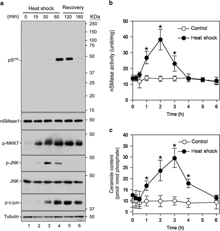
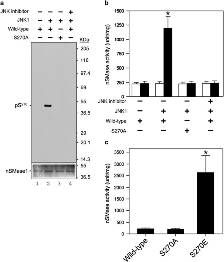
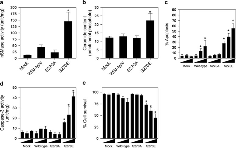
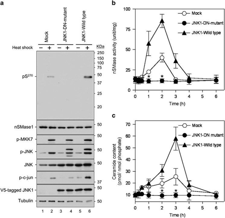
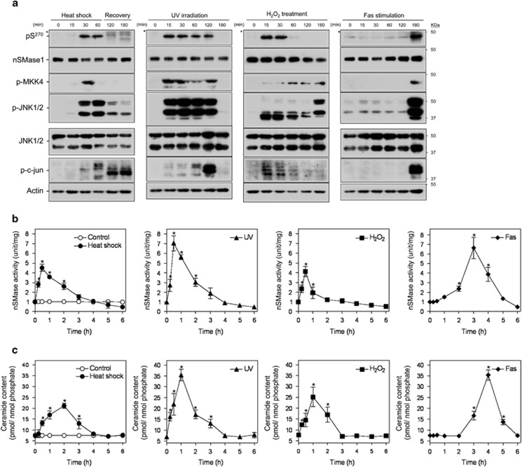
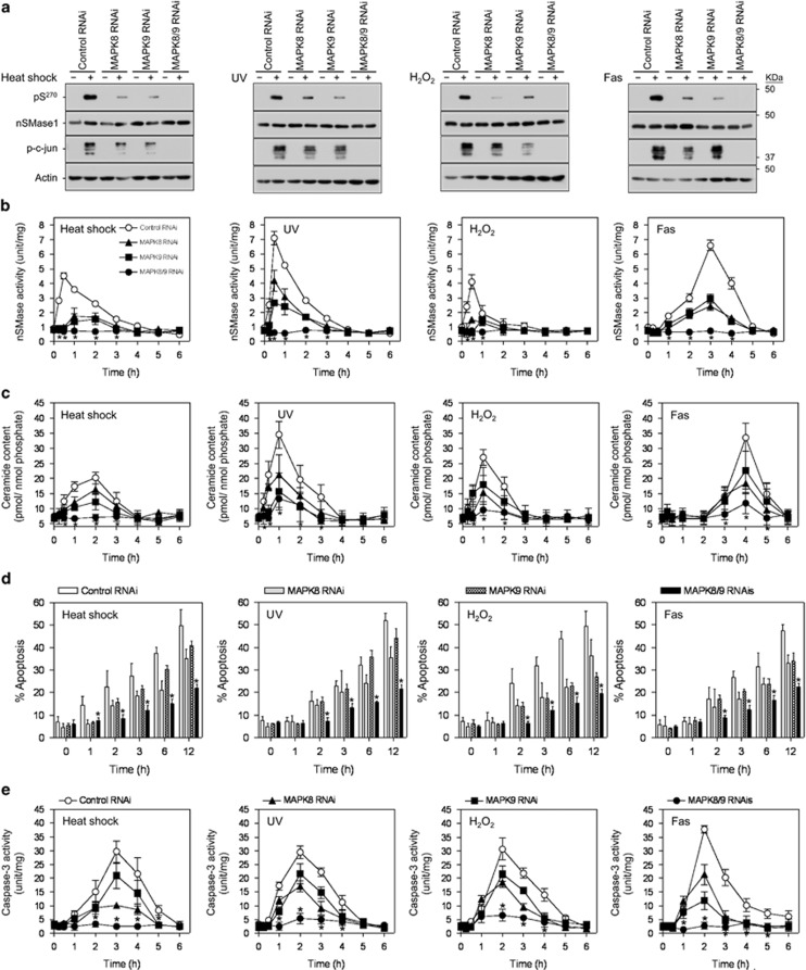
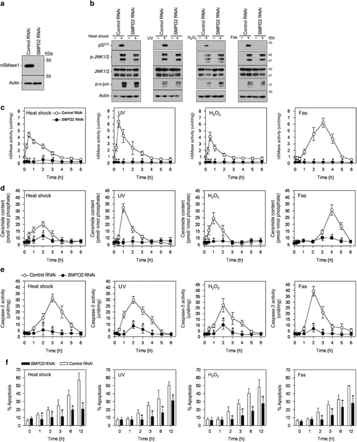
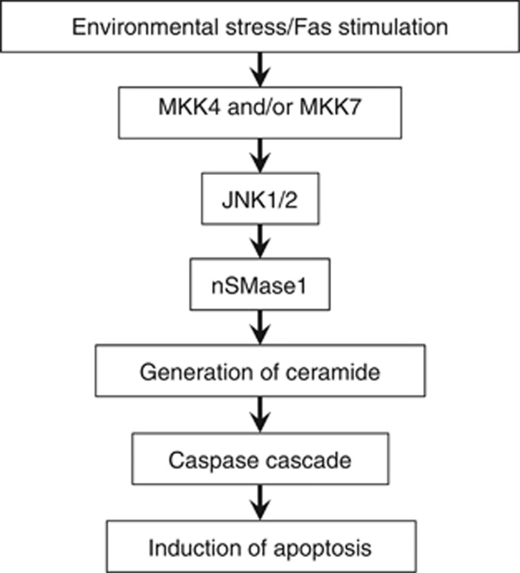
Neutral sphingomyelinase (nSMase) activation in response to environmental stress or inflammatory cytokine stimuli generates the second messenger ceramide, which mediates the stress-induced apoptosis. However, the signaling pathways and activation mechanism underlying this process have yet to be elucidated. Here we show that the phosphorylation of nSMase1 (sphingomyelin phosphodiesterase 2, SMPD2) by c-Jun N-terminal kinase (JNK) signaling stimulates ceramide generation and apoptosis and provide evidence for a signaling mechanism that integrates stress- and cytokine-activated apoptosis in vertebrate cells. An nSMase1 was identified as a JNK substrate, and the phosphorylation site responsible for its effects on stress and cytokine induction was Ser-270. In zebrafish cells, the substitution of Ser-270 for alanine blocked the phosphorylation and activation of nSMase1, whereas the substitution of Ser-270 for negatively charged glutamic acid mimicked the effect of phosphorylation. The JNK inhibitor SP600125 blocked the phosphorylation and activation of nSMase1, which in turn blocked ceramide signaling and apoptosis. A variety of stress conditions, including heat shock, UV exposure, hydrogen peroxide treatment, and anti-Fas antibody stimulation, led to the phosphorylation of nSMase1, activated nSMase1, and induced ceramide generation and apoptosis in zebrafish embryonic ZE and human Jurkat T cells. In addition, the depletion of MAPK8/9 or SMPD2 by RNAi knockdown decreased ceramide generation and stress- and cytokine-induced apoptosis in Jurkat cells. Therefore the phosphorylation of nSMase1 is a pivotal step in JNK signaling, which leads to ceramide generation and apoptosis under stress conditions and in response to cytokine stimulation. nSMase1 has a common central role in ceramide signaling during the stress and cytokine responses and apoptosis.


Figures

Figure 1
Figure 1
nSMase1 Ser-270 phosphorylation is induced by heat shock. (a) The Ser-270 phosphorylation of nSMase1 was induced in heat-shocked zebrafish cells. ZE cells were heat-shocked at 38 °C for 0, 15, 30, or 60 min and were then incubated at 25 °C for up to 2 h. The cell lysates were analyzed by western blotting with anti-phospho-Ser-270 nSMase1, anti-nSMase1, anti-phospho-MKK7, anti-phospho-JNK, anti-JNK, anti-phospho-c-jun, or anti-tubulin antibodies as indicated. Molecular weight markers are shown in kDa on the right. (b) Changes in the nSMase activity in response to heat-shock treatment in zebrafish cells. (c) Changes in ceramide generation in response to heat-shock treatment in zebrafish cells. The cells were heat-shocked at 38 °C for 1 h, and were then allowed to recover at 25 °C for the indicated times. The values represent the means of three independent experiments, and the error bars represent the S.D.s. *P<0.01 versus the control
Figure 2
Figure 2
Phosphorylation and activation of nSMase1 by JNK in vitro. (a) The serine-270 phosphorylation of nSMase1 by JNK in vitro. Mouse JNK1 was used for an in vitro kinase assay using [γ-32 P]-ATP and either nSMase1 (wild-type) or mutant protein (S270A) as the substrate. The reactions were incubated at 30 °C for 30 min with or without 2 ng of mouse JNK1 and in the presence (+) or absence (−) of 10 μM SP600125. Recombinant proteins from each reaction were separated on 10% SDS-polyacrylamide gels and transferred to PVDF membranes. Phosphorylated nSMase1 was detected by autoradiography (upper panel). The levels of substrate protein present in each reaction was determined using Coomassie Brilliant Blue R-250 staining (lower panel). (b) The effect of phosphorylation on the activation of nSMase1. The nSMase activity in each column is shown. After the in vitro kinase assay, the postreaction mixture was analyzed in an nSMase enzymatic assay using C6-NBD-sphingomyelin (black columns). White columns indicate the basal enzyme activity before the kinase assay. The basal enzyme activities of the recombinant nSMase1 (wild-type) and nSMase1 mutant (S270A) were 22.4±0.48 and 22.6±0.35 μmol/mg/h, respectively. Column 1, nSMase1 wild-type; column 2, nSMase1 treated with JNK; column 3, nSMase1 mutant (S270A) treated with JNK; column 4, nSMase1 treated with JNK in the presence of a JNK inhibitor. The enzyme activity after JNK treatment (black column 2) was 120.1±21.4 μmol/mg/h. Each value represents the mean of three independent experiments, and the error bars represent the S.D.s. *P<0.01 versus the wild-type. (c) Activities of the wild-type and S270A and S270E nSMase1 mutants. The nSMase activities of the recombinant proteins were determined using C6-NBD-sphingomyelin. Column 1, recombinant nSMase1 (wild-type); column 2, S270A mutant (S270A); column 3, S270E mutant (S270E). The activity of the S270E mutant (column 3) was 237.8±81.7 μmol/mg/h. Each value represents the mean of three independent experiments, and the error bars represent the S.D.s. *P<0.01 versus basal enzyme activity (white column 2)
Figure 3
Figure 3
Induction of ceramide generation and apoptosis by the overexpression of mutant S270E in zebrafish ZE cells. (a) The nSMase activities of nSMase1 transfectants. ZE cells were transiently transfected with three different doses (1, 2, or 3 μg) of mock, FLAG-tagged wild-type, S270A, or S270E SMase1 vector DNA constructs and cultured for 24 h. nSMase activity was measured using C6-NBD-sphingomyelin as a substrate. Column 1, mock; column 2, wild-type; column 3, S270A; column 4, S270E. (b) The ceramide content in nSMase1 transfectants. Cellular lipids were extracted from the nSMase1 transfectants, and the levels of ceramide were quantified using a diacylglycerol kinase assay after thin layer chromatography separation. Column 1, mock; column 2, wild-type; column 3, S270A; column 4, S270E. (c) Apoptosis in nSMase1-transfected cells. ZE cells were transfected with three different doses (1, 2, or 3 μg) of mock, FLAG-tagged wild-type, S270A, or S270E SMase1 vector DNA constructs, cultured for 24 h, and stained with DAPI to quantify apoptosis. (d) Caspase-3 activity in nSMase1 transfectants. Caspase-3 activation was assessed by measuring Ac-DEVD-MCA hydrolysis. (e) The viability of nSMase1-transfected cells, as determined using Trypan Blue dye exclusion. (f) Apoptotic cells in the nSMase1 transfectants after heat shock. ZE cells were transfected with three different doses (1, 2, or 3 μg) of nSMase1 constructs per dish and cultured for 24 h. After heat shock at 38 °C for 1 h, the cells were incubated at 25 °C for up to 23 h, and all apoptotic cells were identified using DAPI staining. Each value represents the mean of three independent experiments, and the error bars represent the S.D.s. *P<0.01 versus mock
Figure 4
Figure 4
Activation and phosphorylation of nSMase1 after the expression of a JNK DN mutant in heat-stressed zebrafish cells. (a) Detection of nSMase1 phosphorylation by the overexpression of JNK and its DN mutant (K55R). Mock cells (lanes 1 and 2), JNK1-DN (DN) mutant cells (lanes 3 and 4), and JNK1 wild-type cells (lanes 5 and 6) were either unstressed (normal growth, −) at 25 °C for 60 min, or heat-shocked (+) at 38 °C for 60 min. They were then analyzed by western blotting with antibodies against phosphorylated nSMase1, nSMase1, phosphorylated MKK7, phosphorylated JNK, JNK, phosphorylated c-jun, V5-tagged JNK1 wild-type, V5-tagged JNK1-DN mutant, and tubulin. Molecular weight markers are shown in kDa on the right. (b) Changes in nSMase activity. Mock, JNK1-DN, and JNK1 wild-type cells were heat-shocked at 38 °C for 0, 15, 30, or 60 min, allowed to recover at 25 °C for up to 5 h, and then harvested at the indicated times. nSMase activity was measured using C6-NBD-sphingomyelin as a substrate. Values represent the means of three independent experiments, and the error bars represent the S.D.s. *P<0.01 versus the mock control. (c) Changes in ceramide content. The ceramide content was measured using the diacylglycerol kinase assay. Values represent the means of three independent experiments, and error bars represent S.D.s. *P<0.01 versus the mock control
Figure 5
Figure 5
Stress-induced nSMase1 Ser-270 phosphorylation, nSMase activation, and ceramide generation in human Jurkat T cells. (a) The phosphorylation of Ser-270 nSMase1 was detected in Jurkat T cells. Cells were heat-shocked at 43 °C for 0, 15, 30, or 60 min and were then recovered at 37 °C for up to 2 h. For UV irradiation, the cells were irradiated by 254 nm UV at 5 mJ/cm2. For H2O2 treatments, cells were incubated in the presence of 1 mM H2O2. For Fas stimulation, cells were treated with 50 ng/ml of anti-Fas antibody and incubated at 37 °C for 0, 15, 30, 60, 120, or 180 min. The cell lysates were analyzed by western blotting with anti-phospho-Ser-270 nSMase1, anti-nSMase1, anti-phospho-MKK7, anti-phospho-MKK4, anti-phospho-JNK, anti-JNK, anti-phospho-c-Jun, or anti-actin antibodies, as indicated. Molecular weight markers are shown in kDa on the right. Asterisks indicate nonspecific signals. (b) The effect of heat shock, ultraviolet light (UV), hydrogen peroxide (H2O2), and anti-Fas antibody (Fas) on nSMase activity in Jurkat T cells. (c) The effect of heat shock, UV, H2O2, and Fas on ceramide generation in Jurkat T cells. Values represent the means of three independent experiments, and the error bars represent the S.D.s. *P<0.01 versus the control
Figure 6
Figure 6
An essential role for MAPK8 and MAPK9 in nSMase1 activation and ceramide generation in human Jurkat T cells in a loss-of-function study. (a) Detection of phosphorylated-nSMase1 by western blotting after the RNAi-mediated knockdown of MAPK8 and/or MAPK9 in Jurkat T cells. Jurkat T cells were transfected with control, MAPK8, and/or MAPK9 RNAi and then heat-shocked at 43 °C for 30 min, UV-irradiated at 254 nm and 5 mJ/cm2 for 30 min, treated with 1 mM H2O2 for 30 min, or stimulated with 50 ng/ml of anti-Fas antibody for 3 h. Cell lysates were harvested and analyzed by western blotting with anti-phospho-nSMase1, anti-nSMase1, anti-JNK, anti-phospho-c-jun, or anti-actin antibodies. Molecular weight markers are shown in kDa on the right. MAPK8 and MAPK9 are the human genes encoding JNK1 and JNK2, respectively. (b) nSMase activation. RNAi-transfected cell lines were stressed as in panel (a) and then incubated at 37 °C for up to 5 h. (c) Ceramide generation. (d) Effect of loss of function of MAPK8 and MAPK9 on the induction of apoptosis. Twenty-four hours after RNAi transfection, apoptosis was quantified in the MAPK8- and/or MAPK9-knockdown cells using DAPI staining after stress treatment as described in panel (a). (e) Effect of loss of function of MAPK8 and MAPK9 on apoptosis induction assessed using a caspase-3 assay. Each value represents the mean of three independent experiments, and the error bars represent the S.D.s. *P<0.01 versus control RNAi
Figure 7
Figure 7
Essential role of nSMase1 in stress-induced ceramide generation in human Jurkat T cells in a loss-of-function study. (a) Knockdown of SMPD2 expression in human Jurkat T cells. (b) Detection of phosphorylated-nSMase1 by western blotting. The cells were transfected with control or SMPD2 RNAi and cultured for 24 h. The transfected cells were stressed using heat shock at 43 °C for 30 min, UV-irradiated at 5 mJ/cm2 for 30 min, 1 mM H2O2 treatment for 30 min, and stimulation with 50 ng/ml anti-Fas antibody for 3 h. Cell lysates were harvested and analyzed by western blotting with anti-phospho-nSMase1, anti-nSMase1, anti-phospho-JNK, anti-JNK, anti-phospho-c-jun, or anti-actin antibodies. Molecular weight markers are shown in kDa on the right. (c) nSMase1 activation by SMPD2 knockdown. The cell lines were heat-shocked at 43 °C for 0, 15, 30, or 60 min, irradiated at 5 mJ/cm2 UV for 0, 15, 30, or 60 min, treated with 1 mM H2O2 for 0, 15, 30, or 60 min, and stimulated with 50 ng/ml of anti-Fas antibody for 0, 15, 30, or 60 min. The cells were then incubated at 37 °C for up to 5 h. (d) Ceramide generation after SMPD2 knockdown. (e and f) Effect of SMPD2 knockdown on the induction of apoptosis in human Jurkat T cells. Each value represents the mean of three independent experiments, and the error bars represent the S.D.s. *P<0.01 versus the control
Figure 8
Figure 8
Diagram connecting nSMase1 and the JNK signaling pathway

References

    1. Verheij M, Bose R, Lin XH, Yao B, Jarvis WD, Grant S, et al. Requirement for ceramide-initiated SAPK/JNK signalling in stress-induced apoptosis. Nature. 1996;380:75–79. - PubMed
    1. Chang Y, Abe A, Shayman JA. Ceramide formation during heat shock: a potential mediator of alpha B-crystallin transcription. Proc Natl Acad Sci USA. 1995;92:12275–12279. - PMC - PubMed
    1. Hannun YA, Obeid LM. Principles of bioactive lipid signalling: lessons from sphingolipids. Nat Rev Mol Cell Biol. 2008;9:139–150. - PubMed
    1. Jaffrézou JP, Levade T, Bettaïeb A, Andrieu N, Bezombes C, Maestre N, et al. Daunorubicin-induced apoptosis: triggering of ceramide generation through sphingomyelin hydrolysis. EMBO J. 1996;15:2417–2424. - PMC - PubMed
    1. Jana A, Pahan K. Fibrillar amyloid-beta peptides kill human primary neurons via NADPH oxidase-mediated activation of neutral sphingomyelinase. Implications for Alzheimer's disease. J Biol Chem. 2004;279:51451–51459. - PMC - PubMed

Publication types

Substances