. 2015 Jan 1;24(1):230-42.
doi: 10.1093/hmg/ddu441. Epub 2014 Aug 28.

Mutations in IFT172 cause isolated retinal degeneration and Bardet-Biedl syndrome

Affiliations

Mutations in IFT172 cause isolated retinal degeneration and Bardet-Biedl syndrome

Kinga M Bujakowska et al. Hum Mol Genet. .

Abstract

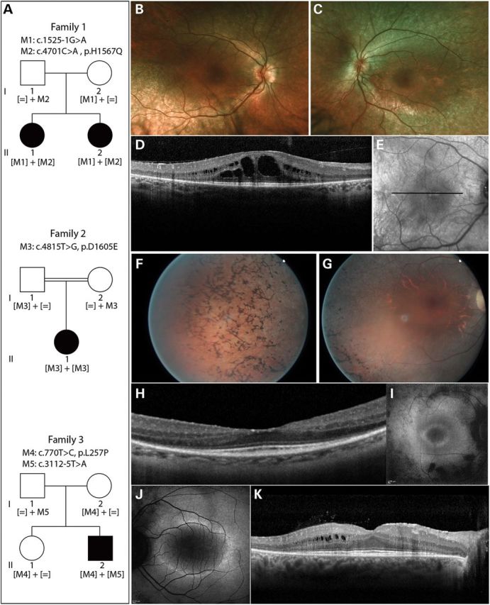
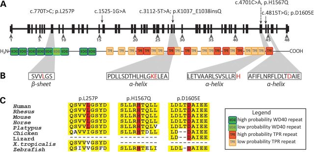
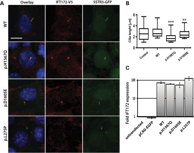
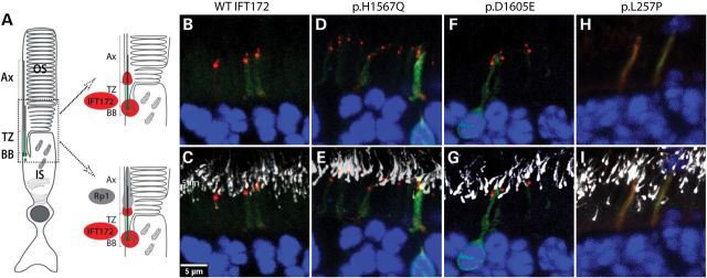
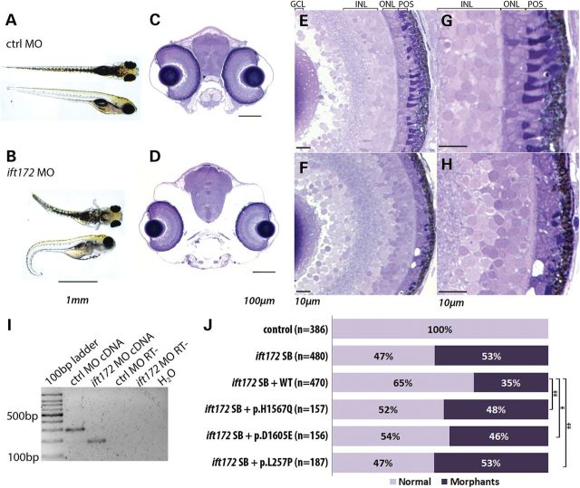
Primary cilia are sensory organelles present on most mammalian cells. The assembly and maintenance of primary cilia are facilitated by intraflagellar transport (IFT), a bidirectional protein trafficking along the cilium. Mutations in genes coding for IFT components have been associated with a group of diseases called ciliopathies. These genetic disorders can affect a variety of organs including the retina. Using whole exome sequencing in three families, we identified mutations in Intraflagellar Transport 172 Homolog [IFT172 (Chlamydomonas)] that underlie an isolated retinal degeneration and Bardet-Biedl syndrome. Extensive functional analyses of the identified mutations in cell culture, rat retina and in zebrafish demonstrated their hypomorphic or null nature. It has recently been reported that mutations in IFT172 cause a severe ciliopathy syndrome involving skeletal, renal, hepatic and retinal abnormalities (Jeune and Mainzer-Saldino syndromes). Here, we report for the first time that mutations in this gene can also lead to an isolated form of retinal degeneration. The functional data for the mutations can partially explain milder phenotypes; however, the involvement of modifying alleles in the IFT172-associated phenotypes cannot be excluded. These findings expand the spectrum of disease associated with mutations in IFT172 and suggest that mutations in genes originally reported to be associated with syndromic ciliopathies should also be considered in subjects with non-syndromic retinal dystrophy.


Figures

Figure 1.
Figure 1.
Pedigrees of subjects with IFT172 mutations and their phenotypes. (A) Pedigree information and segregation of IFT172 variants in the three families. (B and C) Fundus photographs of subject II.2 from family 1, indicating attenuation of blood vessels and RPE atrophy. (D) Horizontal optical coherence tomography (OCT) scan image through the center of the macula of subject II.2 (Family 1), showing thinning of the outer retina and macular cystic edema. (E) Infrared image of the fundus depicting the area where the OCT image was taken in E. (F and G) Fundus photographs of the right eye of subject II.1 from family 2, showing bone-spicule pigmentation in the periphery of the retina (F), attenuated blood vessels and optic disc pallor (G). (H) Horizontal OCT scan image through the center of the macula of the right eye of subject II.1 (family 2), showing thinning of the outer retina. (I and J) Fundus autofluorescence image of subject II.1 from family 2 (I) and subject II.2 from family 3 (J), depicting a perifoveal ring of increased autofluorescence and loss of autofluorescence outside the vascular arcades. (K) Horizontal OCT scan photograph of subject II.2 from family 3, showing an absent photoreceptor layer outside the macular region and cysts in the inner nuclear layer.
Figure 2.
Figure 2.
IFT172 mutations in BBS and rod-cone dystrophy subjects. (A) New mutations indicated on the gene and protein structures. Protein domains were taken from Taschner et al. (13) (WD40: WD40 repeat domain, TPR: tetratricopeptide repeats). (B) IFT172 protein secondary structures predicted by REPPER program (PSIPRED and COILS algorithms) in the Bioinformatics toolkit at the Max-Plank Institute for Developmental Biology (http://toolkit.tuebingen.mpg.de/repper). The mutated residues are indicated in red. Histidine (H) lies just outside of a predicted α-helix. (C) Conservation of missense changes found in this study, the sequences were taken from UCSC genome browser (http://genome.ucsc.edu/).
Figure 3.
Figure 3.
Expression of WT and mutant IFT172 constructs in mIMCD3 cells. (A) Anti-V5 staining of IFT172 WT and mutant proteins (red) in mIMCD3 cells expressing somatostatin receptor 3 (SSTR3)—GFP fusion protein (the size bar represents 10 µm). Cell nuclei were visualized by Hoechst staining. (B) Quantification of cilia length in mIMCD3 cells transfected with a control pCAG-enhanced green fluorescent protein (EGFP) vector, WT or mutant constructs. The boxes represent 25–75% quintiles and the whiskers represent the 5–95% quintiles. The p.L275P mutant did not locate to the cilium and therefore this condition was excluded from the cilia length quantification (**P< 0.01, ***P < 0.0001). (C) Real-time quantitative PCR of mouse and human IFT172 transcripts in the transfected and untransfected mIMCD3 cells. The untransfected mIMCD3 cells condition was used as a calibrator sample and mIMCD3 cells transfected with the empty pCAG-EGFP served as a control.
Figure 4.
Figure 4.
IFT172 localization in rat retinae. (A) A structure of the rod photoreceptor with cilia compartments indicated (BB: basal body, TZ: transition zone, Ax: axoneme, OS: outer segment, IS: inner segment). A magnified version of photoreceptors indicates localization of Rp1 (gray) and Ift172 (red). (B and C) Localization of WT-IFT172 without (B) and with (C) Rp1 staining. (D and E) Localization of p.H1567Q IFT172 protein without (D) and with (E) Rp1 staining. (F and G) Localization of p.D1605E IFT172 protein without (F) and with (G) Rp1 staining. (H and I) Localization of p.L257P IFT172 protein without (H) and with (I) Rp1 staining.
Figure 5.
Figure 5.
Phenotype of ift172 zebrafish morphants. (A and B) Zebrafish injected with a standard control (ctrl) MO and ift172 splice block MO, showing ventral body curvature, body swelling and hydrocephaly at day post fertilization 5 (dpf5). (C and D) Coronal sections of control (C) and ift172 morphant (D) zebrafish at dpf5, depicting smaller eye size and altered cranial bone structure. (E–H) Retinae of control (E and G) and ift172 morphant (F and H) fish showing thinning of photoreceptor cell layer and shortening of the photoreceptor outer segments in the ift172 morphant fish. GCL, ganglion cell layer; INL, inner- nuclear layer; ONL; outer-nuclear layer; POS, photoreceptor outer segments. (I) RT-PCR on RNAs obtained from whole control and ift172 morphant larvae at dpf5, indicating exon 3 skipping due to the donor site blocking by the MO. The primers annealed to ift172 exon 2 and 4. RT- indicates RT-PCR negative control. (J) Zebrafish ift172 morphant phenotype rescue with total quantification of fish phenotypes induced by ift172 MO and rescue by the IFT172 WT and mutant mRNAs. Normalizing total number of morphants in the IFT172 mutant conditions by the number of WT total morphants in the same experiment shows a statistically significant difference between the complementation of the WT IFT172 and the mutants (p.H1567Q, t-test, P = 0.007; p.D1605E, P = 0.039; p.L257P, P = 0.001).

References

    1. Mockel A., Perdomo Y., Stutzmann F., Letsch J., Marion V., Dollfus H. Retinal dystrophy in Bardet–Biedl syndrome and related syndromic ciliopathies. Prog. Retin. Eye Res. 2011;30:258–274. - PubMed
    1. Waters A.M., Beales P.L. Ciliopathies: an expanding disease spectrum. Pediatr. Nephrol. 2011;26:1039–1056. - PMC - PubMed
    1. Beales P.L., Elcioglu N., Woolf A.S., Parker D., Flinter F.A. New criteria for improved diagnosis of Bardet–Biedl syndrome: results of a population survey. J. Med. Genet. 1999;36:437–446. - PMC - PubMed
    1. Pierce E.A., Quinn T., Meehan T., McGee T.L., Berson E.L., Dryja T.P. Mutations in a gene encoding a new oxygen-regulated photoreceptor protein cause dominant retinitis pigmentosa. Nat. Genet. 1999;22:248–254. - PubMed
    1. Guillonneau X., Piriev N.I., Danciger M., Kozak C.A., Cideciyan A.V., Jacobson S.G., Farber D.B. A nonsense mutation in a novel gene is associated with retinitis pigmentosa in a family linked to the RP1 locus. Hum. Mol. Genet. 1999;8:1541–1546. - PubMed

Publication types

MeSH terms