. 2014 Nov;35(11):1342-53.
doi: 10.1002/humu.22680.

Comprehensive analysis of pathogenic deletion variants in Fanconi anemia genes

Affiliations

Comprehensive analysis of pathogenic deletion variants in Fanconi anemia genes

Elizabeth K Flynn et al. Hum Mutat. 2014 Nov.

Abstract

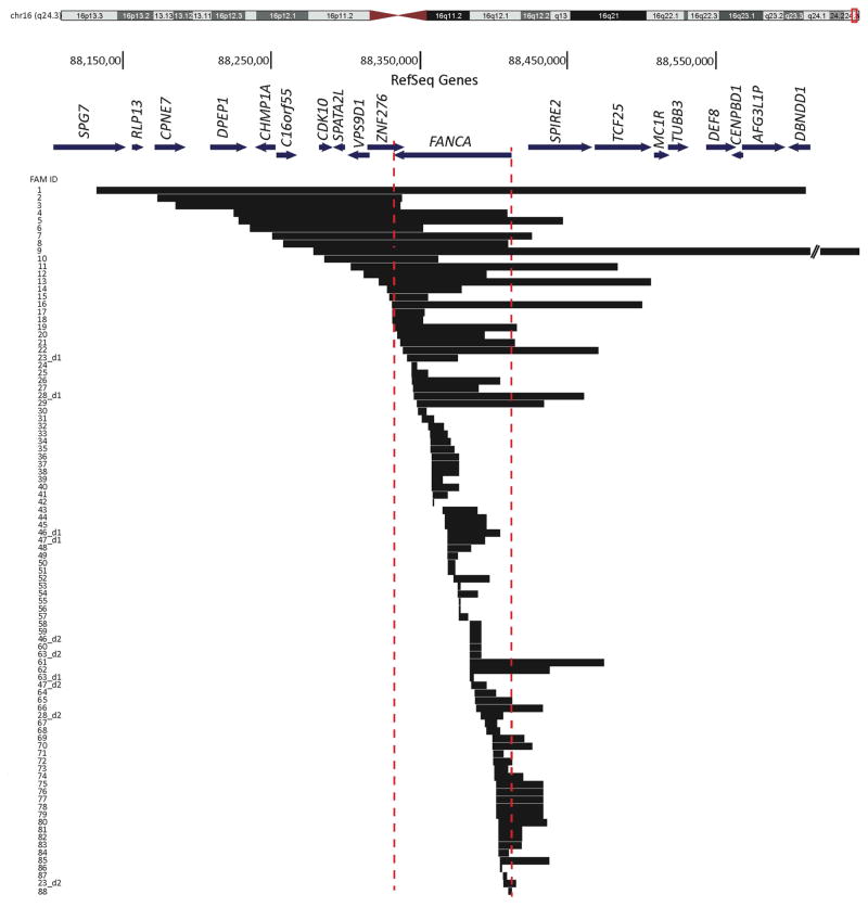
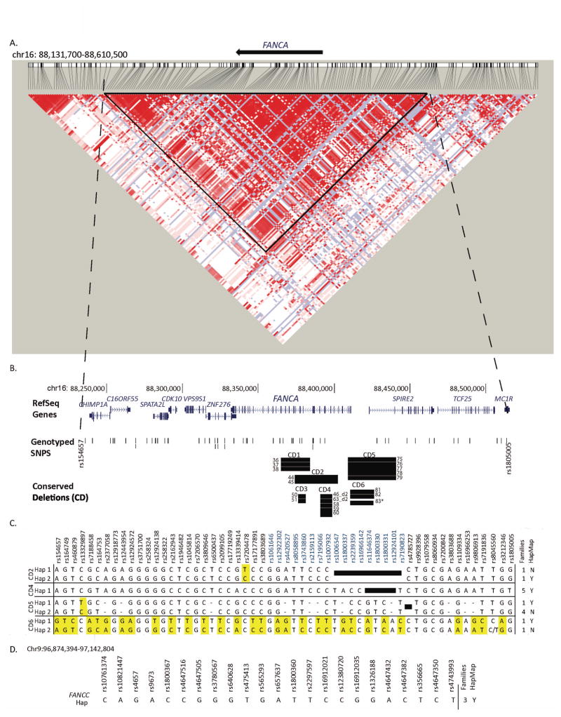
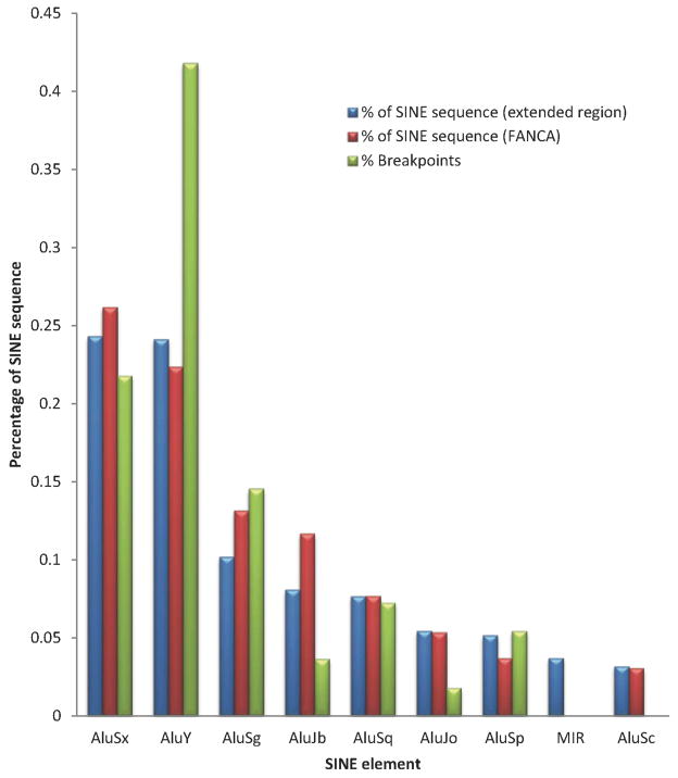
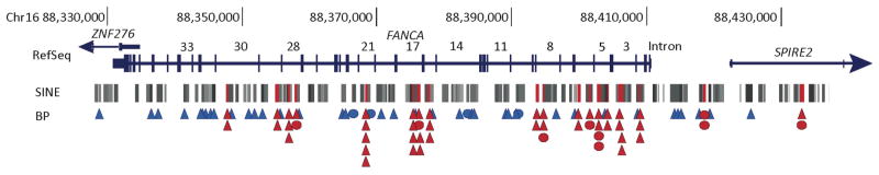
Fanconi anemia (FA) is a rare recessive disease resulting from mutations in one of at least 16 different genes. Mutation types and phenotypic manifestations of FA are highly heterogeneous and influence the clinical management of the disease. We analyzed 202 FA families for large deletions, using high-resolution comparative genome hybridization arrays, single-nucleotide polymorphism arrays, and DNA sequencing. We found pathogenic deletions in 88 FANCA, seven FANCC, two FANCD2, and one FANCB families. We find 35% of FA families carry large deletions, accounting for 18% of all FA pathogenic variants. Cloning and sequencing across the deletion breakpoints revealed that 52 FANCA deletion ends, and one FANCC deletion end extended beyond the gene boundaries, potentially affecting neighboring genes with phenotypic consequences. Seventy-five percent of the FANCA deletions are Alu-Alu mediated, predominantly by AluY elements, and appear to be caused by nonallelic homologous recombination. Individual Alu hotspots were identified. Defining the haplotypes of four FANCA deletions shared by multiple families revealed that three share a common ancestry. Knowing the exact molecular changes that lead to the disease may be critical for a better understanding of the FA phenotype, and to gain insight into the mechanisms driving these pathogenic deletion variants.

Keywords: FANCA; FANCB; FANCC; FANCD2; Fanconi anemia; arrayCGH.


Conflict of interest statement

Disclosure Statement: The authors declare no conflict of interest.

Figures

Figure 1
Figure 1
Extent of FANCA deletions in 88 FA families. BLAT alignment of deletion mutations involving FANCA and other surrounding genes identified in 88 FA families on UCSC genome browser (http://genome.ucsc.edu) (NCBI36/hg18). Chromosome 16 ideogram is shown at the top with the region of interest at q24.3 boxed in red. FANCA and neighboring genes are drawn to scale, and their transcription orientation is indicated by an arrow. Each horizontal block represents one distinct deletion and the family ID is indicated to the left. Families with two deletions are distinguished by “ _d1” (for deletion 1) and “_d2” (for deletion 2). Dotted vertical lines define the boundaries of FANCA. Twenty deletions originate centromeric and 32 terminate telomeric to FANCA, resulting in 47% of the deletions affecting genomic regions beyond the FANCA limits. Details of these deletions are in Table 2, Table 3, and Supp. Table S2.
Figure 2
Figure 2
Conserved deletions in FANCA and FANCC. A. HaploView generated LD plot of human chr16:88,130,00–88,610,500 (FANCA region). SNP data downloaded from HapMap Version 3 Release 2, CEU and TSI panel (http://broadinstitute.org/haploview)(Barrett, et al., 2005) Triangle indicates the one large LD block used for haplotype analysis. B. Location of RefSeq genes, genotyped SNPs and conserved deletions (CD) identified in FANCA families on UCSC genome browser NCBI36/hg18. RefSeq genes are drawn to scale, and vertical lines are exons and the arrows indicate the direction of transcription. Black bar represents the deletion from one family and the Family IDs sharing the deletion are indicated. * indicates that the deletion in FAM83 is similar but not identical to the other members of CD6. C. Haplotypes on the specific allele carrying the conserved deletions. Haplotypes for four of the six conserved deletions are displayed. The SNP names are indicated by rs number, and those within the FANCA are in blue. Black boxes indicate SNPs that fall within the respective deleted regions. Number of families carrying the specific background haplotype and its presence (Y) or absence (N) in the HapMap Phase 2 phased haplotypes is indicated on the right. Differences between haplotypes for a given conserved deletion are highlighted in yellow. Dashed line indicates no SNP data. D. Haplotype of the FANCC allele carrying the conserved deletion in three FA families tested.
Figure 3
Figure 3
FANCA Breakpoints preferentially occur in AluY elements. The distribution of the sequences (percent) from specific SINE elements in relation to the total SINE sequence within the FANCA (chr16.hg18:g.88331460_88410446) and the extended genomic region (chr16.hg18:g.88171112_88549994) are represented by blue and red bars respectively. The distribution (percent) of breakpoints in specific SINE elements is shown by green bars.
Figure 4
Figure 4
Alu elements with multiple breakpoints indicate hotspots for FANCA deletions. Breakpoints in and around FANCA identified by cloning and sequencing are displayed along with the distribution of Alu elements. Exons are vertical lines, and the introns are numbered for FANCA. The SINE track from the UCSC Genome Browser NCBI36/hg18 (Repeat Masker track) is displayed. Alu elements with multiple breakpoints are highlighted in red. A triangle shows each unique breakpoint in a given Alu element. Circles show the Alu elements with breakpoints from conserved deletions, a circle for each conserved deletion (CD1–CD5) except two for CD6. Blue and red triangles and circles indicate a single and multiple breakpoints within a given Alu element respectively. Details of the Alu elements with multiple hits are in Supp. Table S6.

References

    1. Ameziane N, Errami A, Leveille F, Fontaine C, de Vries Y, van Spaendonk RM, de Winter JP, Pals G, Joenje H. Genetic subtyping of Fanconi anemia by comprehensive mutation screening. Hum Mutat. 2008;29:159–66. - PubMed
    1. Amouri A, Talmoudi F, Messaoud O, d’Enghien CD, Rekaya MB, Allegui I, Azaiez H, Kefi R, Abdelhak A, Meseddi SH, et al. High frequency of exon 15 deletion in the FANCA gene in Tunisian patients affected with Fanconi anemia disease: implication for diagnosis. Mol Genet Genomic Med. 2014;2:160–5. - PMC - PubMed
    1. Auerbach AD. Fanconi anemia and its diagnosis. Mutation research. 2009;668:4–10. - PMC - PubMed
    1. Auerbach AD, Greenbaum J, Pujara K, Batish SD, Bitencourt MA, Kokemohr I, Schneider H, Lobitzc S, Pasquini R, Giampietro PF, et al. Spectrum of sequence variation in the FANCG gene: an International Fanconi Anemia Registry (IFAR) study. Hum Mutat. 2003;21:158–68. - PubMed
    1. Barrett JC, Fry B, Maller J, Daly MJ. Haploview: analysis and visualization of LD and haplotype maps. Bioinformatics. 2005;21:263–5. - PubMed

Publication types

Substances