

Investing in the future: stimulation of the medial prefrontal cortex reduces discounting of delayed rewards
- PMID: 25168685
- PMCID: PMC4289950
- DOI: 10.1038/npp.2014.211
Investing in the future: stimulation of the medial prefrontal cortex reduces discounting of delayed rewards
Abstract
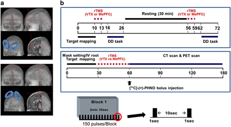
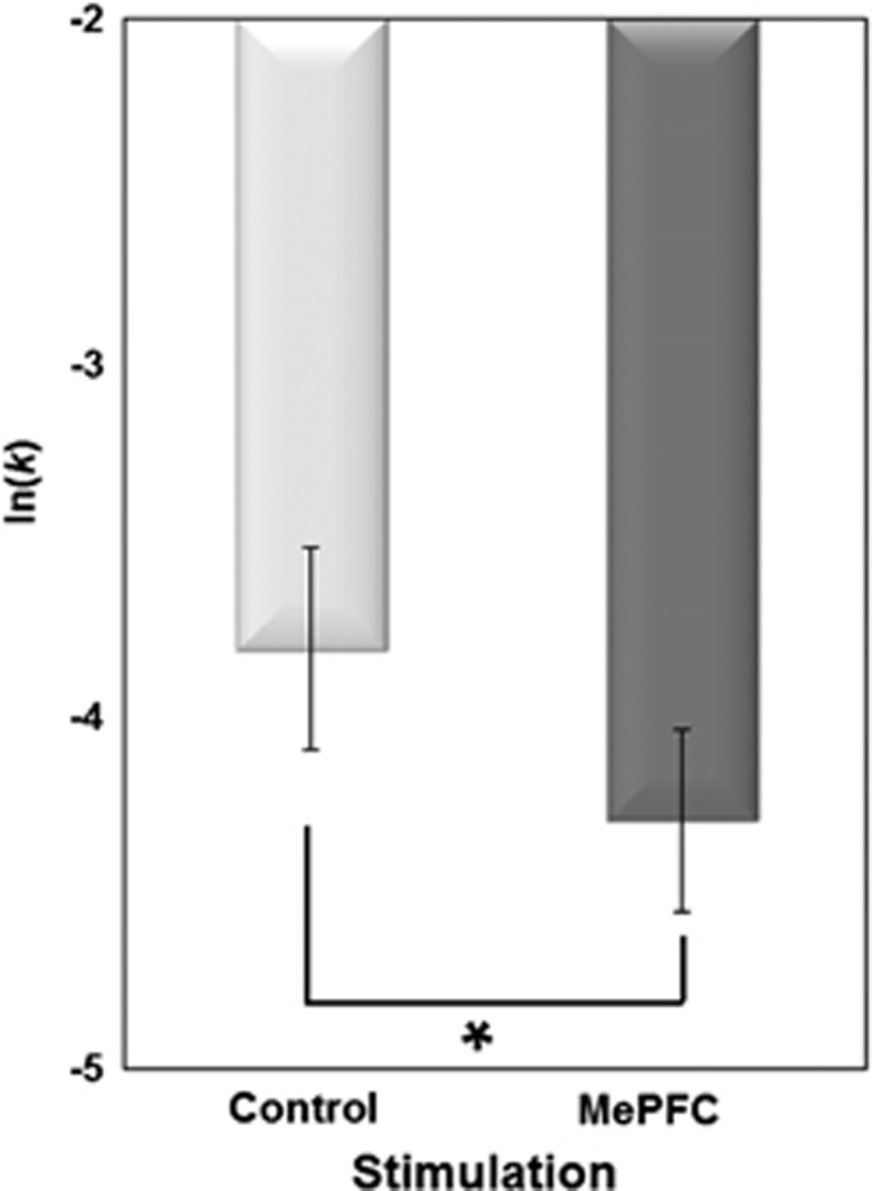
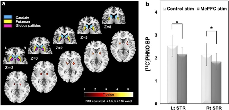
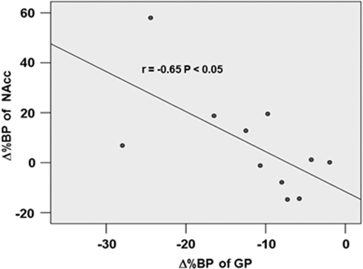
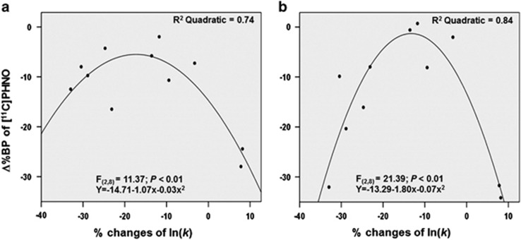
Generally, rewards that are received sooner are often preferred over future rewards, and the time between the choice and the reception of the reward is an important factor that influences our decisions, a phenomenon called delay discounting (DD). In DD, the medial prefrontal cortex (MePFC) and striatal dopamine neurotransmission both play an important role. We used repetitive transcranial magnetic stimulation (rTMS) to transiently activate the MePFC to evaluate its behavioral effect on the DD paradigm, and subsequently to measure its effect on striatal dopamine. Twenty-four right-handed young healthy subjects (11 females; age: 22.1±2.9 years) underwent DD following 10 Hz-rTMS of the MePFC and vertex stimulation (control condition). Thereafter, 11 subjects (5 females; age: 22.2±2.87 years) completed the PET study at rest using [(11)C]-(+)-PHNO following 10 Hz-rTMS of the MePFC and vertex. Modulation of the MePFC excitability influenced the subjective level of DD for delayed rewards and interfered with synaptic dopamine level in the striatum. The present study yielded findings that might reconcile the role of these areas in inter-temporal decision making and dopamine modulation, suggesting that the subjective sense of time and value of reward are critically controlled by these important regions.
Figures
References
-
- Allison T, McCarthy G, Luby M, Puce A, Spencer DD. Localization of functional regions of human mesial cortex by somatosensory evoked potential recording and by cortical stimulation. Electroencephalogr Clin Neurophysiol. 1996;100:126–140. - PubMed
-
- Baker F, Johnson MW, Bickel WK. Delay discounting in current and never-before cigarette smokers: similarities and differences across commodity, sign, and magnitude. J Abnorm Psychol. 2003;112:382–392. - PubMed
-
- Barkley RA, Edwards G, Laneri M, Fletcher K, Metevia L. Executive functioning, temporal discounting, and sense of time in adolescents with attention deficit hyperactivity disorder (ADHD) and oppositional defiant disorder (ODD) J Abnorm Child Psychol. 2001;29:541–556. - PubMed
-
- Beckstead RM. An autoradiographic examination of corticocortical and subcortical projections of the mediodorsal-projection (prefrontal) cortex in the rat. J Comp Neurol. 1979;184:43–62. - PubMed
Publication types
MeSH terms
Substances
Grants and funding
LinkOut - more resources
Full Text Sources
Other Literature Sources
