Comparative Study
. 2014 Dec;31(12):3194-205.
doi: 10.1093/molbev/msu251. Epub 2014 Aug 27.

Evolutionary origin and diversification of epidermal barrier proteins in amniotes

Affiliations
Comparative Study

Evolutionary origin and diversification of epidermal barrier proteins in amniotes

Bettina Strasser et al. Mol Biol Evol. 2014 Dec.

Abstract

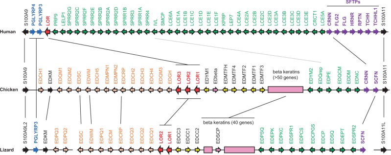
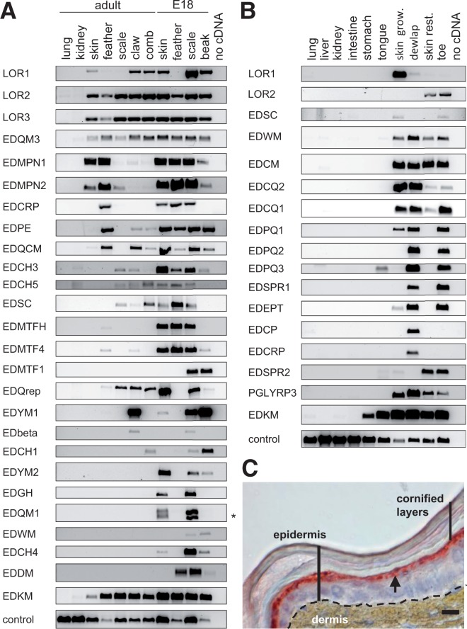
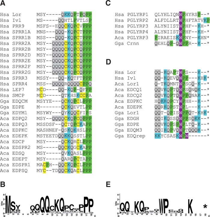
The evolution of amniotes has involved major molecular innovations in the epidermis. In particular, distinct structural proteins that undergo covalent cross-linking during cornification of keratinocytes facilitate the formation of mechanically resilient superficial cell layers and help to limit water loss to the environment. Special modes of cornification generate amniote-specific skin appendages such as claws, feathers, and hair. In mammals, many protein substrates of cornification are encoded by a cluster of genes, termed the epidermal differentiation complex (EDC). To provide a basis for hypotheses about the evolution of cornification proteins, we screened for homologs of the EDC in non-mammalian vertebrates. By comparative genomics, de novo gene prediction and gene expression analyses, we show that, in contrast to fish and amphibians, the chicken and the green anole lizard have EDC homologs comprising genes that are specifically expressed in the epidermis and in skin appendages. Our data suggest that an important component of the cornified protein envelope of mammalian keratinocytes, that is, loricrin, has originated in a common ancestor of modern amniotes, perhaps during the acquisition of a fully terrestrial lifestyle. Moreover, we provide evidence that the sauropsid-specific beta-keratins have evolved as a subclass of EDC genes. Based on the comprehensive characterization of the arrangement, exon-intron structures and conserved sequence elements of EDC genes, we propose new scenarios for the evolutionary origin of epidermal barrier proteins via fusion of neighboring S100A and peptidoglycan recognition protein genes, subsequent loss of exons and highly divergent sequence evolution.

Keywords: birds; epidermis; gene family; gene fusion; reptiles.


Figures

F<sc>ig</sc>. 1.
Fig. 1.
Organization of the EDC in sauropsids. Genes of the EDC in human (chromosome 1q21), chicken (chromosome 25), and green anole lizard (locus not yet assigned to a chromosome) are schematically depicted. Arrows indicate the orientation of the genes. SEDC genes with two exons are represented by colored arrows with a black frame whereas other genes are shown as filled arrows. Clusters of beta-keratin genes are shown as boxes. Colors indicate groups of genes as defined in the text. Black vertical lines connect orthologs; a gray line connects putative orthologs. Note that the schemes are not drawn to scale.
F<sc>ig</sc>. 2.
Fig. 2.
EDC genes of sauropsids are differentially expressed in the skin of different body sites and in other tissues that contain keratinocytes. The expression of EDC genes was determined by RT-PCR in tissues of the chicken (A) and of the green anole lizard (B). Images of RT-PCR products are ordered to highlight similarities of expression patterns of individual genes. Amplification of EDQM1 cDNA yielded two PCR products (asterisk) that result from two alleles of this gene. Note that the RT-PCR screening of these tissue panels (A and B) was performed on a subset of the predicted EDC genes. The skin of the green anole lizard was immunostained (red) with an antibody against lizard loricrin (C). The arrow points to the suprabasal epidermal layer in which loricrin is expressed. Skin grow., skin growing; skin rest., skin resting. Scale bar, 10 µm.
F<sc>ig</sc>. 3.
Fig. 3.
SEDC proteins have evolved highly diverse contents of amino acid residues in mammals and sauropsids. The diagrams show the amino acid compositions of SEDC proteins of human (A), chicken (B), and the green anole lizard (C). The protein data are shown in the order of the corresponding genes in the EDC (fig. 1). For better overview, the homologous loricrin proteins of the three species are highlighted with red letters.
F<sc>ig</sc>. 4.
Fig. 4.
EDC proteins contain conserved amino acid sequence motifs at their amino-terminus and carboxy-terminus. (A) Amino acid sequence alignment showing the conserved sequence motif at the amino-terminus of SEDC proteins. (B) Sequence logo of the amino-terminal motif. (C) Chicken cornulin (Crnn) and PGLYRPs contain sequences similar to the conserved carboxy-terminal sequence motif of SEDCs (D). Note that the genes encoding PGLYRP1 and 2 are not located in the EDC. (E) Sequence logo of the carboxy-terminal motif of SEDCs. Amino acid residues involved in covalent molecular cross-linking (C, Q, K) as well as P and W are highlighted by color shading. Asterisks mark the end of the protein. Aca, Anolis carolinensis; Gga, Gallus gallus; Hsa, Homo sapiens.
F<sc>ig</sc>. 5.
Fig. 5.
A scenario for the origin and diversification of EDC genes. (A) Data on the presence of conserved sequence elements as well as on the arrangement and orientation of genes in the EDC and their exon–intron structures were integrated into a hypothesis about the evolution of EDC genes. On the left, a phylogenetic tree leading to human, chicken, and green anole lizard is shown. The scheme on the right depicts the arrangement of genes in the EDC in these species as well as in their ancestors corresponding to the level of the phylogenetic tree. Asterisks indicate the positions of lost genes. To provide a better overview, only a subset of EDC genes of each clade (indicated by different colors) is shown. Encircled numbers refer to evolutionary steps that are described in the Results section. (B) Evolutionary origin of the distinct exon–intron organizations of EDC genes. One of several possible evolutionary pathways (supplementary fig. S13, Supplementary Material online) is depicted in simplified form. Exons are indicated by boxes, in which the noncoding regions are shaded gray and the coding regions are shaded in colors or in black. Identical colors indicate common ancestry and black indicates newly originated coding sequences. All genes shown in (B) are transcribed from left to right.

References

    1. Alibardi L. Adaptation to the land: the skin of reptiles in comparison to that of amphibians and endotherm amniotes. J Exp Zool B Mol Dev Evol. 2003;298:12–41. - PubMed
    1. Alibardi L. Structural and immunocytochemical characterization of keratinization in vertebrates epidermis and epidermal derivatives. Int Rev Cytol. 2006;253:177–259. - PubMed
    1. Alibardi L, Dalla Valle L, Nardi A, Toni M. Evolution of hard proteins in the sauropsid integument in relation to the cornification of skin derivatives in amniotes. J Anat. 2009;214:560–586. - PMC - PubMed
    1. Backendorf C, Hohl D. A common origin for cornified envelope proteins? Nat Genet. 1992;2:91. - PubMed
    1. Candi E, Schmidt R, Melino G. The cornified envelope:a model of cell death in the skin. Nat Rev Mol Cell Biol. 2005;6:328–340. - PubMed

Publication types