. 2014 Aug 27;6(9):2274-88.
doi: 10.1093/gbe/evu187.

Six subgroups and extensive recent duplications characterize the evolution of the eukaryotic tubulin protein family

Affiliations

Six subgroups and extensive recent duplications characterize the evolution of the eukaryotic tubulin protein family

Peggy Findeisen et al. Genome Biol Evol. .

Abstract

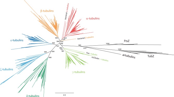
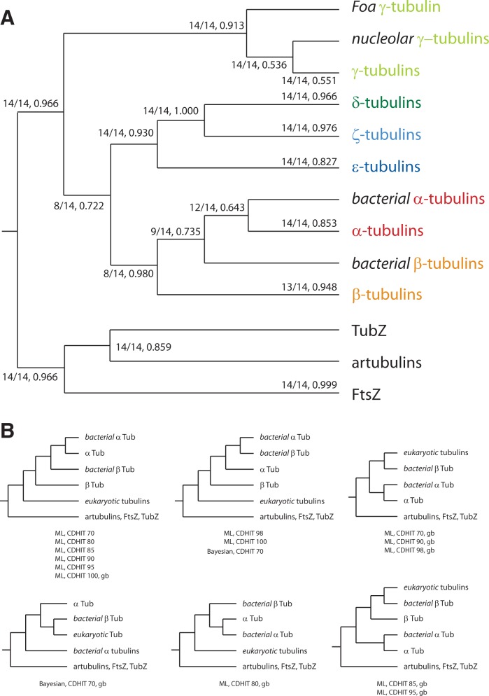
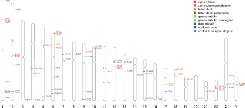
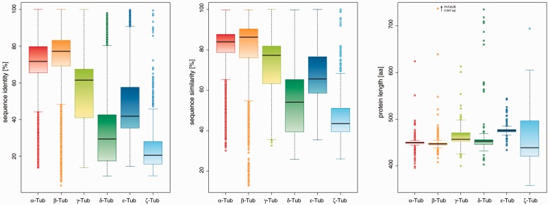
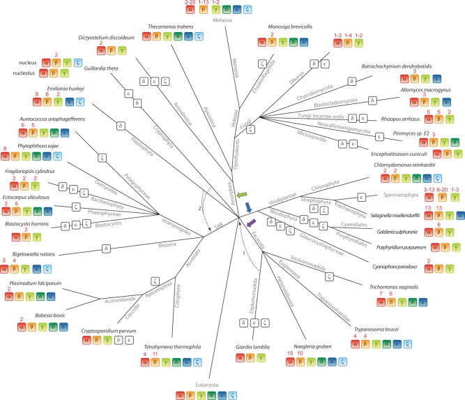
Tubulins belong to the most abundant proteins in eukaryotes providing the backbone for many cellular substructures like the mitotic and meiotic spindles, the intracellular cytoskeletal network, and the axonemes of cilia and flagella. Homologs have even been reported for archaea and bacteria. However, a taxonomically broad and whole-genome-based analysis of the tubulin protein family has never been performed, and thus, the number of subfamilies, their taxonomic distribution, and the exact grouping of the supposed archaeal and bacterial homologs are unknown. Here, we present the analysis of 3,524 tubulins from 504 species. The tubulins formed six major subfamilies, α to ζ. Species of all major kingdoms of the eukaryotes encode members of these subfamilies implying that they must have already been present in the last common eukaryotic ancestor. The proposed archaeal homologs grouped together with the bacterial TubZ proteins as sister clade to the FtsZ proteins indicating that tubulins are unique to eukaryotes. Most species contained α- and/or β-tubulin gene duplicates resulting from recent branch- and species-specific duplication events. This shows that tubulins cannot be used for constructing species phylogenies without resolving their ortholog-paralog relationships. The many gene duplicates and also the independent loss of the δ-, ε-, or ζ-tubulins, which have been shown to be part of the triplet microtubules in basal bodies, suggest that tubulins can functionally substitute each other.

Keywords: FtsZ; TubZ; artubulin; eukaryotic evolution; gene duplication; tubulin.


Figures

F<sc>ig</sc>. 1.—
Fig. 1.—
Phylogenetic tree of the tubulin protein family. Unrooted ML topology generated under the WAG + Γ model in FastTree showing branch lengths for 75 α-, 69 β-, 84 γ-, 50 δ-, 45 ε-, 32 ζ-tubulins, 2 bacterial tubulins, 1 “artubulin,” 11 bacterial FtsZ, and 8 bacterial TubZ proteins. CD-Hit (70% identity) was used to obtain a representative data set for subfamily classification and visualization. Support for the major branchings indicating the grouping of the tubulins and FtsZ family members into different subtypes is given as likelihood bootstraps (FastTree). The scale bar corresponds to estimated amino acid substitutions per site.
F<sc>ig</sc>. 2.—
Fig. 2.—
Schematic tree of the tubulin subfamilies. (A) Schematic consensus tree from 14 trees reconstructed with the ML method and based on full and reduced data sets, in which redundant sequences, divergent regions, and unique positions were removed at various stringency levels. The first number at branches denotes the number of trees supporting the respective branch followed by the median of the support values (see supplementary table S2, Supplementary Material online, for more details). (B) The small trees show the alternative topologies for the branching of the bacterial tubulins. CDHIT, application of CD-Hit with the given similarity threshold; gb, use of gblocks.
F<sc>ig</sc>. 3.—
Fig. 3.—
Chromosomal location of human tubulin genes. The human tubulin genes and pseudogenes are distributed over all chromosomes. Some genes appear in clusters of tandemly arranged gene duplicates like the α-tubulins Tub1A, Tub1B, and Tub1C on chromosome 12, the β-tubulins Tub2D and Tub2E on chromosome 2, and the γ-tubulins Tub3A and Tub3B on chromosome 17 (see also supplementary fig. S6, Supplementary Material online). The ideogram was produced with Idiographica based on the human hg19 chromosome assembly (Kin and Ono 2007).
F<sc>ig</sc>. 4.—
Fig. 4.—
Sequence conservation in tubulins. Box plots of the sequence identities (left) and similarities (middle) of all complete bacterial and eukaryotic tubulins, excluding pseudogenes. On the right, box plots of the protein lengths are shown.
F<sc>ig</sc>. 5.—
Fig. 5.—
The mutually exclusive spliced insect β-tubulin 2C genes. (A) The Diptera encode β-tubulins containing a cluster of mutually exclusive spliced exons (MXEs). This cluster most probably appeared by exon duplication in the ancestor of the Diptera, because the gene structures are conserved in other insects that diverged prior to the emergence of the Diptera. Exons and introns are represented as dark- and light-gray bars, respectively; MXEs are shown in color. The opacity of the color of the 3’ of the alternative exons corresponds to the alignment score of the alternative exon to the original one (5’-exon). (B) The structural region covered by the MXEs of the Drosophila gene is shown mapped onto the crystal structure of β-tubulin from sheep brain (PDB-ID: 3RYC) (Nawrotek et al. 2011).
F<sc>ig</sc>. 6.—
Fig. 6.—
Schematic tree of analyzed metazoans and their tubulins. Abstract representation of the phylogenetic tree of the Metazoa constructed by analyzing data from Wägele and Bartolomaeus (2014). Branches are shown at which changes in the presence of the δ-, ε-, and ζ-tubulins happened. At each leaf, one representative species of the branch is printed. Branch lengths are arbitrary. White boxes illustrate the loss of the respective tubulin subfamily member in the branch or species.
F<sc>ig</sc>. 7.—
Fig. 7.—
Evolution of the tubulin protein family across eukaryotes. The tree has been reconstructed by evaluating recent literature (Parfrey et al. 2010; Adl et al. 2012; He et al. 2014) for those eukaryotic branches that have been included in this study. However, especially the grouping of taxa that emerged close to the origin of the eukaryotes remains highly debated. Therefore, alternative branchings are also indicated in the tree. The phylogeny of the supposed supergroup Excavata is the least understood because only a few species of this branch have been completely sequenced so far. Although the grouping of the Heterolobosea, Trichomonada, and Euglenozoa into the Excavata is found in most analyses, the grouping of the Diplomonadida as separate phylum or as part of the Excavata is still debated (arrow 1; Simpson et al. 2006). The placement of the Haptophyceae and Cryptophyta to the SAR (arrow 2; Nozaki et al. 2009; Keeling 2009) is supported by some studies although most analyses are in contrast (Hampl et al. 2009; Parfrey et al. 2010). At each leaf of the tree, one representative species of the branch is printed. Branch lengths are arbitrary. The tree illustrates the presence (colored boxes) and absence (white boxes) of each tubulin subfamily in the corresponding species. The LECA must have already contained one member of each of the six subfamilies, as indicated, independent of whether the root of the eukaryotes is placed at the base of six eukaryotic supergroups as shown or whether the root is placed at alternative positions like the unikont–bikont split (green arrow; unikonts/Amorphea versus all other eukaryotes) or the photosynthetic–nonphotosynthetic split (blue arrow), or between Excavata and Neozoa (all other eukaryotes; purple arrow) (He et al. 2014). Numbers above tubulins indicate the number or range of duplicates within the species or taxon, respectively.

References

    1. Adl SM, et al. The revised classification of eukaryotes. J Eukaryot Microbiol. 2012;59:429–493. - PMC - PubMed
    1. Aylett CHS, Löwe J, Amos LA. New insights into the mechanisms ofcytomotive actin and tubulin filaments. Int Rev Cell Mol Biol. 2011;292:1–71. - PubMed
    1. Aylett CHS, Wang Q, Michie KA, Amos LA, Löwe J. Filament structure of bacterial tubulin homologue TubZ. Proc Natl Acad Sci U S A. 2010;107:19766–19771. - PMC - PubMed
    1. Breviario D, Gianì S, Morello L. Multiple tubulins: evolutionary aspects and biological implications. Plant J Cell Mol Biol. 2013;75:202–218. - PubMed
    1. Brown MW, Spiegel FW, Silberman JD. Phylogeny of the “forgotten” cellular slime mold, Fonticula alba, reveals a key evolutionary branch within Opisthokonta. Mol Biol Evol. 2009;26:2699–2709. - PubMed

LinkOut - more resources