

Preparation, characterization, and in vitro osteoblast functions of a nano-hydroxyapatite/polyetheretherketone biocomposite as orthopedic implant material
- PMID: 25170265
- PMCID: PMC4145828
- DOI: 10.2147/IJN.S67358
Preparation, characterization, and in vitro osteoblast functions of a nano-hydroxyapatite/polyetheretherketone biocomposite as orthopedic implant material
Abstract
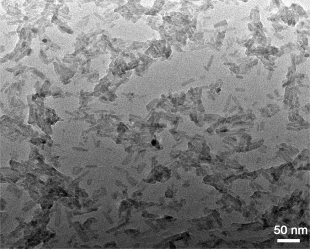
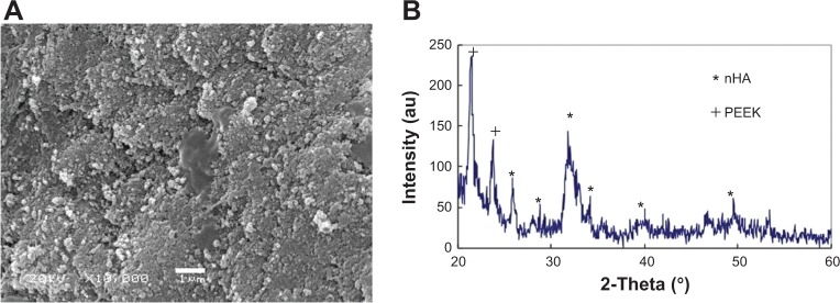
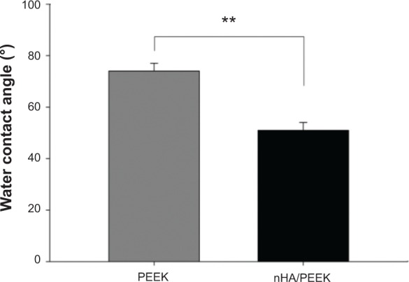
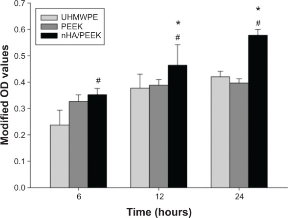
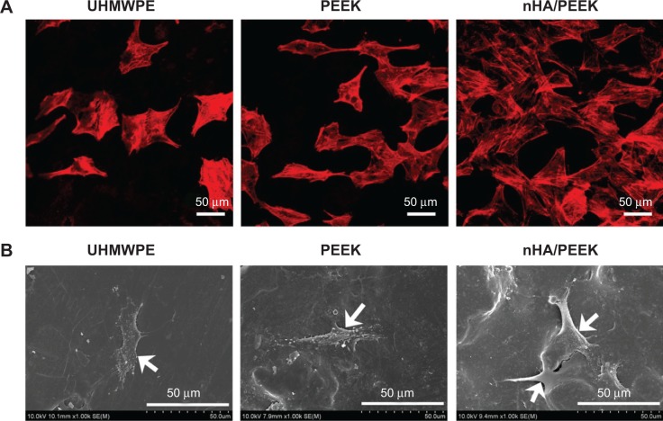
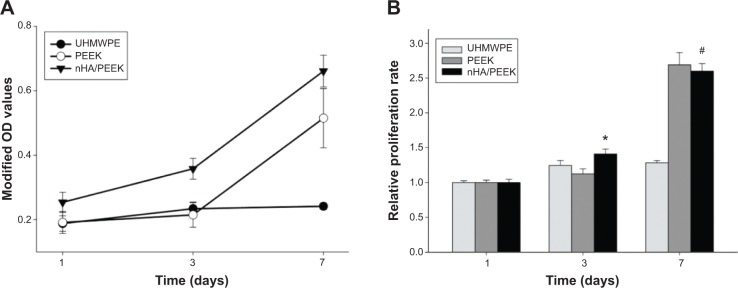
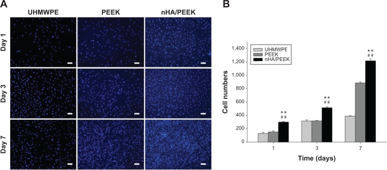
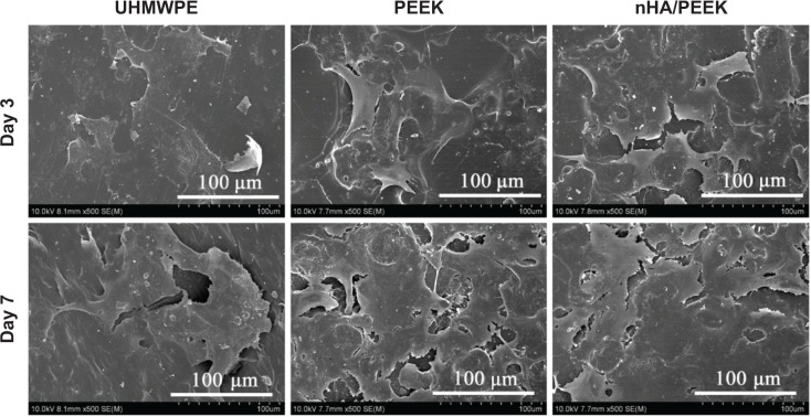
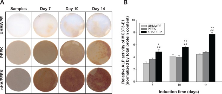
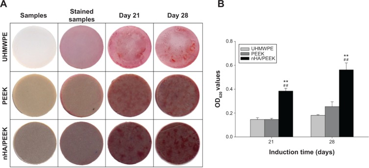
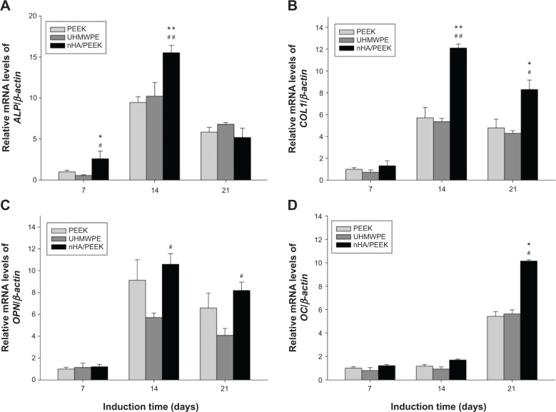
A bioactive composite was prepared by incorporating 40 wt% nano-hydroxyapatite (nHA) into polyetheretherketone (PEEK) through a process of compounding, injection, and molding. The mechanical and surface properties of the nHA/PEEK composite were characterized, and the in vitro osteoblast functions in the composite were investigated. The mechanical properties (elastic modulus and compressive strength) of the nHA/PEEK composite increased significantly, while the tensile strength decreased slightly as compared with PEEK. Further, the addition of nHA into PEEK increased the surface roughness and hydrophilicity of the nHA/PEEK composite. In cell tests, compared with PEEK and ultra-high-molecular-weight polyethylene, it was found that the nHA/PEEK composite could promote the functions of MC3T3-E1 cells, including cell attachment, spreading, proliferation, alkaline phosphatase activity, calcium nodule formation, and expression of osteogenic differentiation-related genes. Incorporation of nHA into PEEK greatly improved the bioperformance of PEEK. The nHA/PEEK composite might be a promising orthopedic implant material.
Keywords: biocomposite; nano-hydroxyapatite; orthopedic implant material; osteoblast functions; polyetheretherketone.
Figures
Similar articles
-
Effect of surface roughness on osteogenesis in vitro and osseointegration in vivo of carbon fiber-reinforced polyetheretherketone-nanohydroxyapatite composite.Int J Nanomedicine. 2015 Feb 17;10:1425-47. doi: 10.2147/IJN.S75557. eCollection 2015. Int J Nanomedicine. 2015. PMID: 25733834 Free PMC article.
-
Enhanced osteogenic activity of phosphorylated polyetheretherketone via surface-initiated grafting polymerization of vinylphosphonic acid.Colloids Surf B Biointerfaces. 2019 Jan 1;173:591-598. doi: 10.1016/j.colsurfb.2018.10.031. Epub 2018 Oct 13. Colloids Surf B Biointerfaces. 2019. PMID: 30352380
-
Preparation, characterization, cellular response and in vivo osseointegration of polyetheretherketone/nano-hydroxyapatite/carbon fiber ternary biocomposite.Colloids Surf B Biointerfaces. 2015 Dec 1;136:64-73. doi: 10.1016/j.colsurfb.2015.09.001. Epub 2015 Sep 2. Colloids Surf B Biointerfaces. 2015. PMID: 26363268
-
Biomechanical and bioactivity concepts of polyetheretherketone composites for use in orthopedic implants-a review.J Biomed Mater Res A. 2015 Nov;103(11):3689-702. doi: 10.1002/jbm.a.35480. Epub 2015 Apr 28. J Biomed Mater Res A. 2015. PMID: 25856801 Review.
-
Application of biomolecules modification strategies on PEEK and its composites for osteogenesis and antibacterial properties.Colloids Surf B Biointerfaces. 2022 Jul;215:112492. doi: 10.1016/j.colsurfb.2022.112492. Epub 2022 Apr 12. Colloids Surf B Biointerfaces. 2022. PMID: 35430485 Review.
Cited by
-
Evaluation of Cytocompatibility of PEEK-Based Composites as a Function of Manufacturing Processes.Bioengineering (Basel). 2023 Nov 17;10(11):1327. doi: 10.3390/bioengineering10111327. Bioengineering (Basel). 2023. PMID: 38002451 Free PMC article.
-
Response of Human Osteoblast to n-HA/PEEK--Quantitative Proteomic Study of Bio-effects of Nano-Hydroxyapatite Composite.Sci Rep. 2016 Mar 9;6:22832. doi: 10.1038/srep22832. Sci Rep. 2016. PMID: 26956660 Free PMC article.
-
Characterization of New PEEK/HA Composites with 3D HA Network Fabricated by Extrusion Freeforming.Molecules. 2016 May 26;21(6):687. doi: 10.3390/molecules21060687. Molecules. 2016. PMID: 27240326 Free PMC article.
-
Nanohydroxyapatite as a Biomaterial for Peripheral Nerve Regeneration after Mechanical Damage-In Vitro Study.Int J Mol Sci. 2021 Apr 24;22(9):4454. doi: 10.3390/ijms22094454. Int J Mol Sci. 2021. PMID: 33923239 Free PMC article.
-
D-RADA16-RGD-Reinforced Nano-Hydroxyapatite/Polyamide 66 Ternary Biomaterial for Bone Formation.Tissue Eng Regen Med. 2019 Jan 5;16(2):177-189. doi: 10.1007/s13770-018-0171-5. eCollection 2019 Apr. Tissue Eng Regen Med. 2019. PMID: 30989044 Free PMC article.
References
-
- McMahon RE, Wang L, Skoracki R, Mathur AB. Development of nanomaterials for bone repair and regeneration. J Biomed Mater Res B Appl Biomater. 2013;101(2):387–397. - PubMed
-
- Lee JS, Baek SD, Venkatesan J, et al. In vivo study of chitosan-natural nano hydroxyapatite scaffolds for bone tissue regeneration. Int J Biol Macromol. 2014;67:360–366. - PubMed
-
- Roeder RK, Converse GL, Kane RJ, Yue W. Hydroxyapatite-reinforced polymer biocomposites for synthetic bone substitutes. JOM. 2008;60(3):38–45.
-
- Zong C, Qian X, Tang Z, et al. Biocompatibility and bone-repairing effects: comparison between porous poly-lactic-co-glycolic acid and nano-hydroxyapatite/poly(lactic acid) scaffolds. J Biomed Nanotechnol. 2014;10(6):1091–1104. - PubMed
-
- Asti A, Gioglio L. Natural and synthetic biodegradable polymers: different scaffolds for cell expansion and tissue formation. Int J Artif Organs. 2014;37(3):187–205. - PubMed
Publication types
MeSH terms
Substances
LinkOut - more resources
Full Text Sources
Other Literature Sources
