

Characterization of ureteral stents by dual-energy computed tomography: Clinical implications
- PMID: 25170401
- PMCID: PMC4147444
- DOI: 10.4329/wjr.v6.i8.625
Characterization of ureteral stents by dual-energy computed tomography: Clinical implications
Abstract
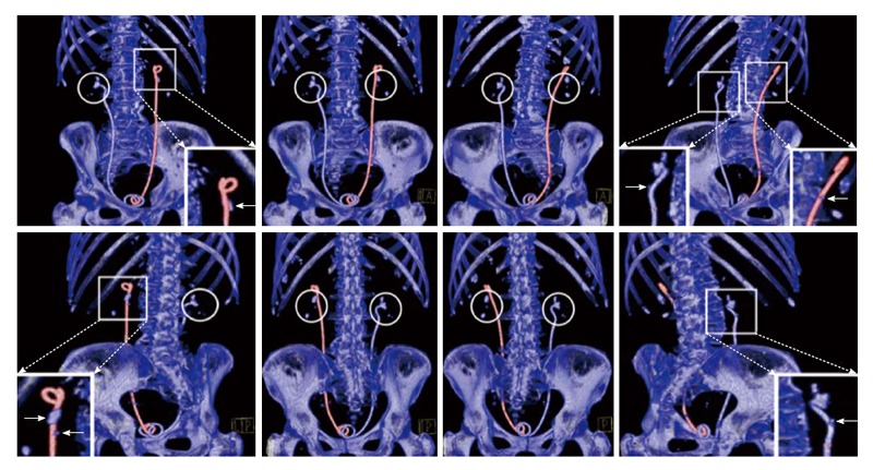
Dual-energy computed-tomography (DECT) has been suggested as the method of choice for imaging urinary calculi due to the modality's high sensitivity for detecting stones and its capability of accurately differentiating between uric-acid (UA) and non-UA (predominantly calcium) stones. The clinical significance of the latter feature relates to the differences in management of UA vs non-UA calculi. Like calculi, ureteral stents are assigned color by the dual-energy post-processing algorithm, which may lead to improved or worsened stone visualization based on the resulting stent/stone contrast. Herein we depict the case of a nephrolithiasis patient with bilateral stents, each with different color, clearly displaying the effect of stent color on stone visualization. Further, three-dimensional reconstruction of the DECT images illustrates advantages of this enhancement compared to conventional two-dimensional computed tomography. The resulting stent/stone contrast produces an unanticipated potential advantage of DECT in patients with urolithiasis and stents and may promote improved management decision-making.
Keywords: Dual-energy computed-tomography; Kidney stones; Nephrolithiasis; Ureteral stent.
Figures

References
-
- Matlaga BR, Kawamoto S, Fishman E. Dual source computed tomography: a novel technique to determine stone composition. Urology. 2008;72:1164–1168. - PubMed
-
- Hartman R, Kawashima A, Takahashi N, Silva A, Vrtiska T, Leng S, Fletcher J, McCollough C. Applications of dual-energy CT in urologic imaging: an update. Radiol Clin North Am. 2012;50:191–205, v. - PubMed
-
- Boll DT, Patil NA, Paulson EK, Merkle EM, Simmons WN, Pierre SA, Preminger GM. Renal stone assessment with dual-energy multidetector CT and advanced postprocessing techniques: improved characterization of renal stone composition--pilot study. Radiology. 2009;250:813–820. - PubMed
-
- Kulkarni NM, Eisner BH, Pinho DF, Joshi MC, Kambadakone AR, Sahani DV. Determination of renal stone composition in phantom and patients using single-source dual-energy computed tomography. J Comput Assist Tomogr. 2013;37:37–45. - PubMed
-
- Ascenti G, Siragusa C, Racchiusa S, Ielo I, Privitera G, Midili F, Mazziotti S. Stone-targeted dual-energy CT: a new diagnostic approach to urinary calculosis. AJR Am J Roentgenol. 2010;195:953–958. - PubMed
Publication types
LinkOut - more resources
Full Text Sources
Other Literature Sources
