

Development of Retinal Amacrine Cells and Their Dendritic Stratification
- PMID: 25170430
- PMCID: PMC4142557
- DOI: 10.1007/s40135-014-0048-2
Development of Retinal Amacrine Cells and Their Dendritic Stratification
Abstract
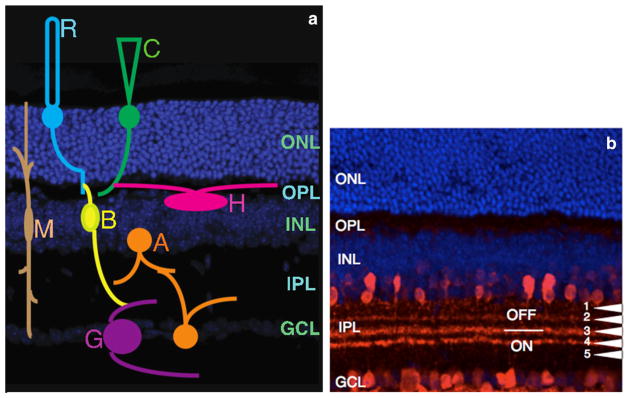
Themammalian retina containsmultiple neurons, each of which contributes differentially to visual processing. Of these retinal neurons, amacrine cells have recently come to prime light since they facilitate majority of visual processing that takes place in the retina. Amacrine cells are also the most diverse group of neurons in the retina, classified majorly based on the neurotransmitter type they express and morphology of their dendritic arbors. Currently, little is known about the molecular basis contributing to this diversity during development. Amacrine cells also contribute to most of the synapses in the inner plexiform layer and mediate visual information input from bipolar cells onto retinal ganglion cells. In this review, we will describe the current understanding of amacrine cell and cell subtype development. Furthermore, we will address the molecular basis of retinal lamination at the inner plexiform layer. Overall, our review will provide a developmental perspective of amacrine cell subtype classification and their dendritic stratification.
Keywords: Amacrine cells; Dendritic stratification; Retina; Retinogenesis; Transcription factors.
Conflict of interest statement
Figures
References
-
- Masland RH. The fundamental plan of the retina. Nat Neurosci. 2001;4(9):877–86. This article describes the diversity of retinal neurons and its association with function. - PubMed
-
- Masland RH. Neuronal diversity in the retina. Curr Opin Neurobiol. 2001;11(4):431–6. - PubMed
-
- Livesey FJ, Cepko CL. Vertebrate neural cell-fate determination: lessons from the retina. Nat Rev Neurosci. 2001;2(2):109–18. - PubMed
Grants and funding
LinkOut - more resources
Full Text Sources
Other Literature Sources