. 2014 Aug 29;9(8):e106129.
doi: 10.1371/journal.pone.0106129. eCollection 2014.

Zebrafish WNK lysine deficient protein kinase 1 (wnk1) affects angiogenesis associated with VEGF signaling

Affiliations

Zebrafish WNK lysine deficient protein kinase 1 (wnk1) affects angiogenesis associated with VEGF signaling

Ju-Geng Lai et al. PLoS One. .

Abstract

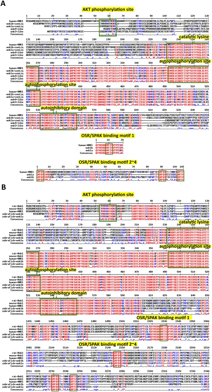
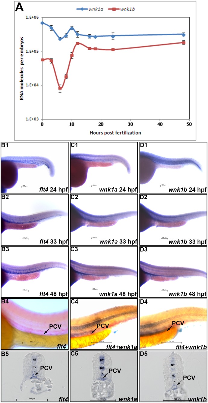
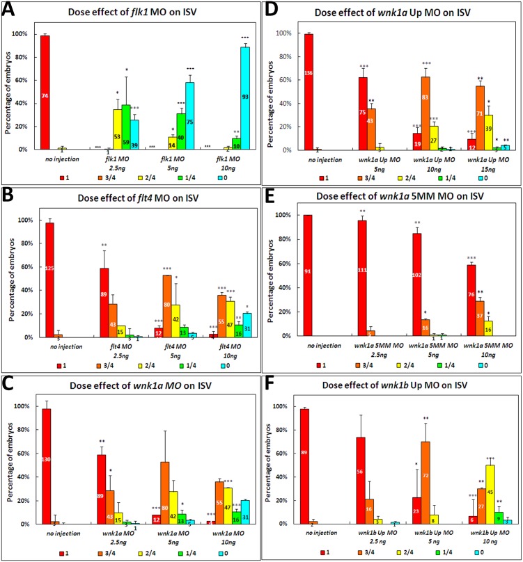
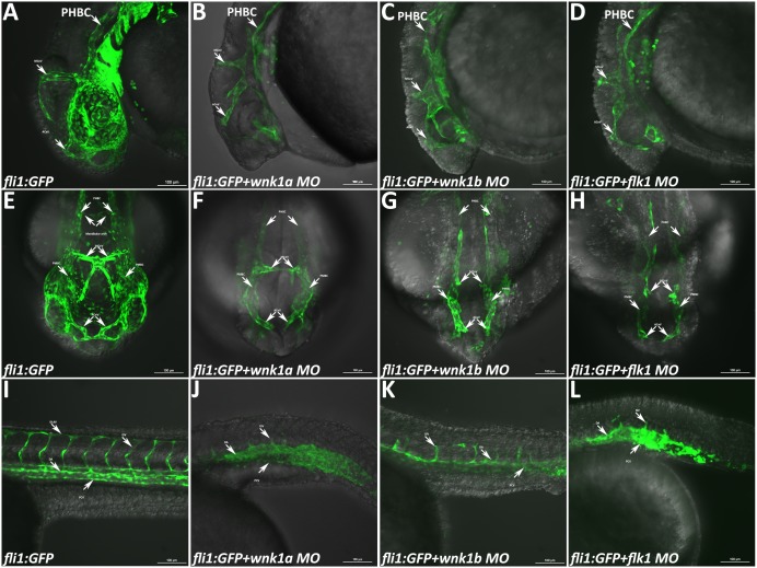
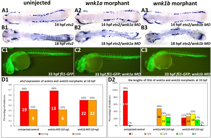
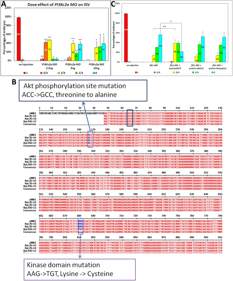
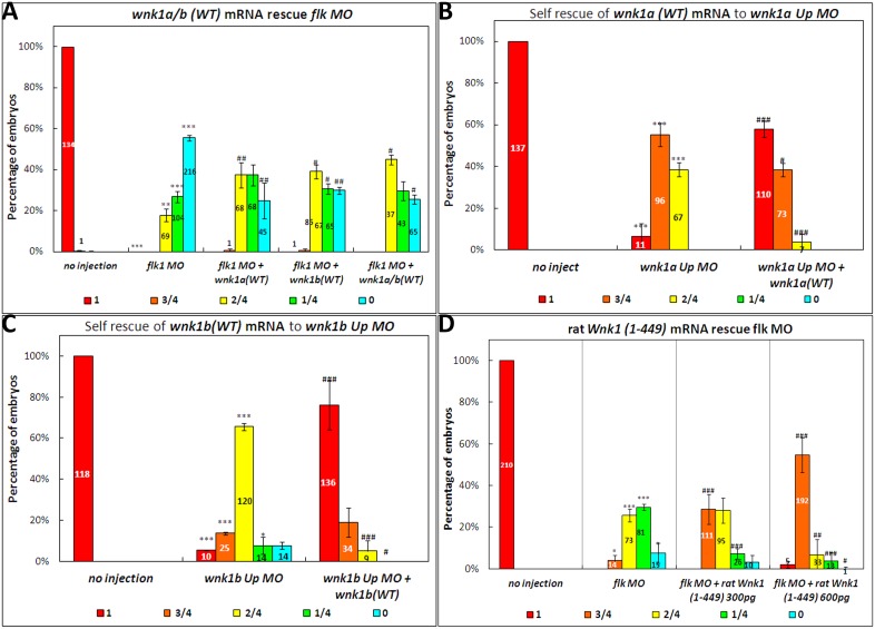
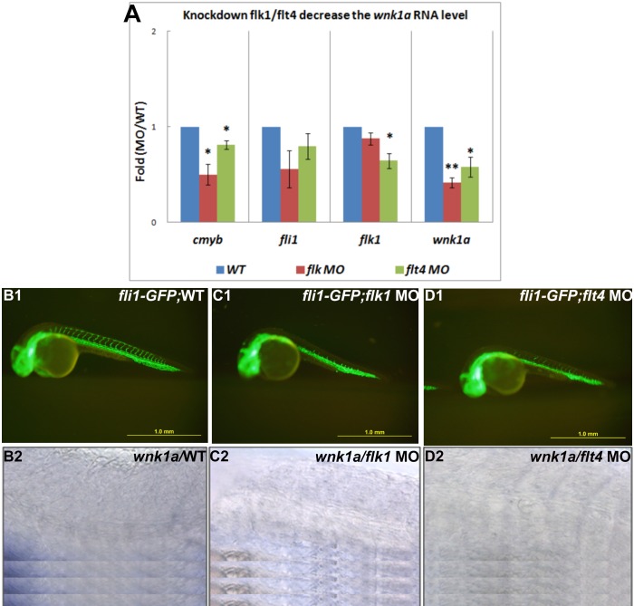
The WNK1 (WNK lysine deficient protein kinase 1) protein is a serine/threonine protein kinase with emerging roles in cancer. WNK1 causes hypertension and hyperkalemia when overexpressed and cardiovascular defects when ablated in mice. In this study, the role of Wnk1 in angiogenesis was explored using the zebrafish model. There are two zebrafish wnk1 isoforms, wnk1a and wnk1b, and both contain all the functional domains found in the human WNK1 protein. Both isoforms are expressed in the embryo at the initiation of angiogenesis and in the posterior cardinal vein (PCV), similar to fms-related tyrosine kinase 4 (flt4). Using morpholino antisense oligonucleotides against wnk1a and wnk1b, we observed that wnk1 morphants have defects in angiogenesis in the head and trunk, similar to flk1/vegfr2 morphants. Furthermore, both wnk1a and wnk1b mRNA can partially rescue the defects in vascular formation caused by flk1/vegfr2 knockdown. Mutation of the kinase domain or the Akt/PI3K phosphorylation site within wnk1 destroys this rescue capability. The rescue experiments provide evidence that wnk1 is a downstream target for Vegfr2 (vascular endothelial growth factor receptor-2) and Akt/PI3K signaling and thereby affects angiogenesis in zebrafish embryos. Furthermore, we found that knockdown of vascular endothelial growth factor receptor-2 (flk1/vegfr2) or vascular endothelial growth factor receptor-3 (flt4/vegfr3) results in a decrease in wnk1a expression, as assessed by in situ hybridization and q-RT-PCR analysis. Thus, the Vegf/Vegfr signaling pathway controls angiogenesis in zebrafish via Akt kinase-mediated phosphorylation and activation of Wnk1 as well as transcriptional regulation of wnk1 expression.


Conflict of interest statement

Competing Interests: The authors have declared that no competing interests exist.

Figures

Figure 1
Figure 1. Sequence comparison between human WNK1 and zebrafish Wnk proteins.
(A) Alignment of four zebrafish Wnks with the human WNK1 protein sequence. The WNK1 signature domain is highlighted. The AKT phosphorylation site at threonine 60 (arrow) in WNK1 is specific to Wnk1a and Wnk1b. The catalytic lysine (arrow) is located in a highly conserved region among all Wnk1s. The autophosphorylation site and the autoinhibitory domain are conserved between all four Wnk1s. The four OSR/SPAK binding motifs found in human WNK1 are specific to Wnk1a and Wnk1b. (B) Alignment of WNK1 protein sequences from mouse, rat, human and zebrafish.
Figure 2
Figure 2. Spatial and temporal expression patterns of wnk1a and wnk1b.
(A) wnk1a and wnk1b mRNA expression profiles as determined by q-RT-PCR. At least three replicates were performed, and the average number of molecules was calculated using a standard curve from a q-RT-PCR assay. The standard deviations are shown in the graph. The red and blue lines indicate the wnk1a and wnk1b expression profiles, respectively. (B∼D) Whole-mount in situ hybridization for flt4 (B) wnk1a (C), and wnk1b (D) was performed at the indicated time points. flt4 (B1∼B3), wnk1a (C1∼C3), and wnk1b (D1∼D3) mRNA expression in the tail is shown at 24, 33 and 48 hpf. Double in situ hybridization for flt4 alone (B4), wnk1a+flt4 (C4), and wnk1b+flt4 (D4) shows the co-localization of wnk1a and wnk1b with flt4 in the PCV. Expression of flt4 (B5), wnk1a (C5) and wnk1b (D5) is seen in the PCV (arrow) in sections from stained embryos. wnk1a and wnk1b are also expressed in the neural tube (NT) and notochord (NC). The dorsal aorta (*) is negative for wnk1a and wnk1b expression. Scale bar: 100 µm.
Figure 3
Figure 3. Statistical analysis of the length of intersegmental vessels in flk1, flt4, wnk1a, wnk1a 5MM, wnk1a upstream and wnk1b upstream morpholino-injected embryos.
The effects of (A) flk1 MO, (B) flt4 MO, (C) wnk1a MO, (D) wnk1a 5 base mismatch MO, (E) wnk1a upstream MO, and (F) wnk1b upstream MO on the length of intersegmental vessels. Morpholino injection has a dose-dependent effect on intersegmental vessel formation and growth. All experiments were performed at least three times, and the average ISV length was calculated along with the standard deviation, which is labeled on each bar. Red indicates that the ISVs grew to full length, orange indicates that the ISVs were 75% of the normal length, yellow indicates 50%, green indicates 25%, and light blue indicates that no ISVs were observed in the embryos. The differences between treatments were assessed using a two-tailed Student’s t-test. Significant differences between the morphants and controls are indicated (*, P<0.05; **, P<0.01; and ***, P<0.001).
Figure 4
Figure 4. Phenotype of Tg(fli1:GFP) embryos injected with various morpholinos and imaged with a confocal microscope.
(A–D) Lateral views of the heads of uninjected control embryos and wnk1a, wnk1b and flk1 morphants at 33 hpf. (E–H) Frontal views of the heads of uninjected control embryos and wnk1a, wnk1b and flk1 morphants at 33 hpf. (I–L) Lateral views of the trunk in uninjected control embryos and wnk1a, wnk1b and flk1 morphants at 33 hpf. Important vessels are indicated with arrows and labeled, with the full name given in the text. Scale bar: 100 µm.
Figure 5
Figure 5. Effect of wnk1a or wnk1b knockdown on vasculogenesis and angiogenesis in Tg(fli1:GFP) embryos.
(A, B) Whole mount in situ hybridization for etv2 at 14 and 18 hpf in uninjected control embryos (A1, B1), wnk1a morphants (A2, B2), and wnk1b morphants (A3, B3). Flat mounts of de-yolked embryos were prepared. (C) GFP fluorescence was used to assay ISV formation in uninjected control embryos (C1), wnk1a morphants (C2), and wnk1b morphants (C3) at 33 hpf. (D1) etv2 expression in uninjected control, wnk1a and wnk1b morphants as a percentage of embryos exhibit strong or expression, (D2) ISV length in wnk1a and wnk1b morphants.
Figure 6
Figure 6. Effect of knockdown of the PI3K ortholog on ISVs and the rescue effects of wild-type wnk1a, wnk1a containing an Akt phosphorylation site mutation and kinase-deficient wnk1a on flk1 morphants.
(A) The effects of pi3kc2α MO on the length of intersegmental vessels. (B) Two Thr35 mutations and one Lys206 mutation were generated using site-directed mutagenesis. The sequence of wild-type wnk1a aligned with Thr35 and Lys206 mutants, which are Akt phosphorylation site and kinase domain mutants, respectively. (C) Co-injection of wnk1a mRNA rescues flk1 morphants. Quantitative analysis of the length of the ISVs in flk1 morphants co-injected with various wnk1a mRNAs. Experiments were performed at least three times, and the number of embryos analyzed is shown in the bar graph. Red indicates that the ISVs grew to full length, orange indicates that the ISVs were 75% of the normal length, yellow indicates 50%, green indicates 25%, and light blue indicates no ISVs were observed in the embryos. The differences between treatments were assessed using a two-tailed Student’s t-test. Significant differences between the morphants and controls are indicated (*, P<0.05; **, P<0.01; and ***, P<0.001).
Figure 7
Figure 7. Effect of wild-type wnk1a and wnk1b mRNA injection on flk1, wnk1a and wnk1b morphants.
(A) Co-injection of wnk1a, wnk1b or both wnk1a and wnk1b mRNA rescues flk1 morphants. (B) Co-injection of wnk1a mRNA rescues the ISV defect caused by the wnk1a upstream MO. (C) Co-injection of wnk1b mRNA rescues the ISV defect caused by the wnk1b upstream MO. (D) Injection of rat Wnk1(1–449) rescues flk1 morphants. Experiments were performed at least three times, and the number of embryos analyzed is shown in the bar graph. Red indicates that the ISVs grew to full length, orange indicates that the ISVs were 75% of the normal length, yellow indicates 50%, green indicates 25%, and light blue indicates no ISVs were observed in the embryos. The differences between treatments were assessed using a two-tailed Student’s t-test. Significant differences between the morphants and controls are indicated (*, P<0.05; **, P<0.01; and ***, P<0.001); significant differences between co-injection of RNA with morpholino and morpholino alone are also indicated (#, P<0.05; ##, P<0.01; and ###, P<0.001).
Figure 8
Figure 8. Injections of flk1 MO and flt4 MO decrease wnk1 mRNA expression.
(A) Relative fold-change comparisons between flk1 and flt4 morphants and uninjected controls. Comparison of mRNA expression levels at 33 hpf for important transcription factors, receptors, and wnk1. Blue, red, and green bars denote mRNA expression in wild-type embryos, flk1 morphants and flt4 morphants, respectively. The x-axis indicates the expressed genes, and the y-axis shows the fold differences between the morphants and the control. The differences between treatments were assessed using a two-tailed Student’s t-test. Significant differences between the morphants and the controls are indicated (*, P<0.05 and**, P<0.01). (B1, C1, D1) The ISVs are affected in morphants compared with wild-type embryos. (B2, C2 and D2) The expression of wnk1a is reduced in the PCV in flk1 and flt4 morphants. wnk1a mRNA was detected with in situ hybridization in wild-type embryos (B2), flk1 morphants (C2) and flt4 morphants (D2).

References

    1. Baldessari D, Mione M (2008) How to create the vascular tree? (Latest) help from the zebrafish. Pharmacol Ther 118: 206–230. - PubMed
    1. Risau W (1997) Mechanisms of angiogenesis. Nature 386: 671–674. - PubMed
    1. Shibuya M (2008) Vascular endothelial growth factor-dependent and -independent regulation of angiogenesis. BMB Rep 41: 278–286. - PubMed
    1. Jiang ZY, Zhou QL, Holik J, Patel S, Leszyk J, et al. (2005) Identification of WNK1 as a substrate of Akt/protein kinase B and a negative regulator of insulin-stimulated mitogenesis in 3T3-L1 cells. J Biol Chem 280: 21622–21628. - PubMed
    1. Xu B, English JM, Wilsbacher JL, Stippec S, Goldsmith EJ, et al. (2000) WNK1, a novel mammalian serine/threonine protein kinase lacking the catalytic lysine in subdomain II. J Biol Chem 275: 16795–16801. - PubMed

Publication types

MeSH terms

Substances

LinkOut - more resources