. 2014 Aug 28;158(5):1045-1059.
doi: 10.1016/j.cell.2014.07.012.

Single-cell identity generated by combinatorial homophilic interactions between α, β, and γ protocadherins

Affiliations

Single-cell identity generated by combinatorial homophilic interactions between α, β, and γ protocadherins

Chan Aye Thu et al. Cell. .

Abstract

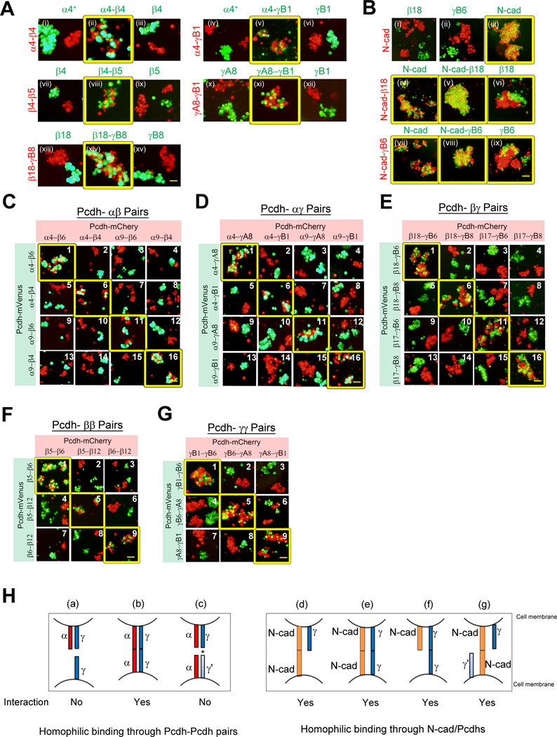
Individual mammalian neurons stochastically express distinct repertoires of α, β, and γ protocadherin (Pcdh) proteins, which function in neural circuit assembly. We report that all three subfamilies of clustered Pcdhs can engage in specific homophilic interactions, that cell surface delivery of Pcdhα isoforms requires cis interactions with other Pcdhs, and that the extracellular cadherin domain EC6 plays a critical role in this process. Examination of homophilic interactions between specific combinations of multiple Pcdh isoforms revealed that Pcdh combinatorial recognition specificities depend on the identity of all of the expressed isoforms. A single mismatched Pcdh isoform can interfere with these combinatorial homophilic interactions. A theoretical analysis reveals that assembly of Pcdh isoforms into multimeric recognition units and the observed tolerance for mismatched isoforms can generate cell surface diversity sufficient for single-cell identity. However, the competing demands of nonself discrimination and self-recognition place limitations on the mechanisms by which homophilic recognition units can function.


Figures

Figure 1
Figure 1. The Pcdh gene cluster encodes a large repertoire of cell surface recognition proteins
(A) Schematic representation of the mouse Pcdh-a, -b and -g gene clusters. Variable exons of each subtype are differentially color-coded. Pcdha and Pcdhg variable exons are joined via cis-splicing to three constant exons. An example is shown for Pcdha9. Each variable exon encodes six EC domains, a TM, and a short cytoplasmic extension. The constant exons encode the common ICD domain. (B) Schematic diagrams representing the four major subtypes of Pcdhs are shown. (C) Schematic diagram of the cell aggregation assay. mCherry-tagged Pcdh proteins are expressed in K562 cells to assay for their ability to induce cell aggregation. As shown in the examples, cells expressing mCherry alone do not aggregate, while robust cell aggregation is observed with cells expressing PcdhγC3-mCherry. (D) Survey of homophilic binding properties of all 58 Pcdh isoforms in the cell aggregation assay. Scale bar, 50 µm. (See also Figure S1B).
Figure 2
Figure 2. Pcdh-β, –γ, and C-type isoforms engage in specific homophilic interactions
(A) Schematic diagram of the binding specificity assay. Cells expressing differentially tagged Pcdh isoforms are then mixed and assayed for homophilic or heterophilic interactions. A strict homophilic interaction is indicated by mixed red-and-green co-aggregates between cells expressing only the identical isoforms and segregation of separate red and green aggregates between cells expressing different isoforms. (B) Heat map of pair-wise protein sequence identities of the EC2-EC3 domains of Pcdh isoforms and their evolutionary relationship is presented. Subsets of the isoforms within the boxed region were assayed. (See also Figure S2B). (C–E) Pairwise combinations within each subtype Pcdhβ (C), Pcdhγ (D), and C-type (E) isoforms were assayed for their binding specificity. Scale bar, 50 µm. (See also Figure S3C).
Figure 3
Figure 3. Pcdhα isoforms engage in specific homophilic interactions when delivered to the cell surface by co-expressed Pcdh-β or -γ isoforms
(A) Surface expression of mCherry-tagged Pcdh constructs bearing an extracellular c-Myc tag were shown. White arrows indicate the c-Myc staining at cell-cell contacts. Scale bar, 10 µm. (B) Cells transfected with single Pcdhα isoforms (upper panels) and cells co-transfected with Pcdhα isoforms and PcdhγB6ΔEC1 (lower panels) were assayed for aggregation. Scale bar, 50 µm. (C) Cells expressing ΔEC1-Pcdhs alone (upper panels), Pcdhα4/ΔEC1-Pcdhs (middle panels) were assayed for aggregation. Cells co-expressing Pcdhα4 and a carrier ΔEC1-Pcdhs do not interact with cells expressing only the wild-type carrier Pcdhs (lower panels). Scale bar, 50 µm. (See also Figure S3C). (D) Heat map of pairwise sequence identities of the EC2-EC3 domains of Pcdhα isoforms. The boxed region shows Pcdh-α4, -α7 and -α8, which share a high level of sequence conservation. (E–F) Cells co-expressing pairs of differentially tagged Pcdhαs and ΔEC1-Pcdhs were assayed for co-aggregation.
Figure 4
Figure 4. The role of EC6 domains in membrane delivery
(A) Mapping the minimum binding region of carrier Pcdhs. Cells expressing PcdhγB6 mutants alone (Upper panels) and with Pcdhα4 (Lower panels) were assayed for aggregation. Cell surface expression of Myc-tagged Pcdhα were shown (xv–xvi). (See also Figure S4G) (B) Schematic representation of chimeric proteins and the results of homophilic binding assays are presented. All of the chimeras bearing the EC6 domain from PcdhγC3 (yellow) mediate cell aggregation. All of the chimeras bearing the EC6 domain of Pcdhα4 (red) fail to mediate cell aggregation. Scale bar, 50 µm. (See also Figure S4B and S4H). (C) Cells expressing Pcdhα4 EC6/ICD domain deletion mutants are tested for aggregation (i–ii). Surface expression of Pcdhα constructs were shown on the right (iii–iv). (See also Figure S4I) (D) Heat map of pairwise sequence identities for EC6 domains. The EC6 domain is highly conserved in alternate Pcdhβ and Pcdhγ isoforms, but the EC6 domains of alternate Pcdhα isoforms are less conserved. (E) Multiple sequence alignment of EC6 domains of membrane-delivered Pcdhs (light gray) and non-membrane delivered Pcdhs (dark grey). Residues conserved within only one group are highlighted in blue and invariant residues in red. White arrows indicate the c-Myc staining at cell-cell contacts in A and C. Scale bar, 50 µm.
Figure 5
Figure 5. Co-expression of two distinct Pcdh isoforms generates a unique cell surface identity
(A) Cells co-expressing two distinct mCherry-tagged Pcdh isoforms were assayed for interaction with cells expressing an mVenus-tagged Pcdh isoform or identical pairs. Pcdhα4+ is efficiently membrane-delivered and it possess the EC6 domain from PcdhγC3. (See also Figures S5A, S5C, S5G) (B) Cells expressing mCherry-tagged N-cad were assayed for interaction with cells expressing single Pcdh isoform (Upper panels). Cells co-expressing pair of mCherry-tagged N-cad and Pcdh isoform were assayed for interaction with cells expressing an mVenus-tagged N-cad or Pcdh isoform (Middle and last panels). (See also Figures S5D, S5G, S6D, S6E and S6G) (C–G) Cells co-expressing different combinations of differentially tagged Pcdh/Pcdh pairs were mixed and assayed for their interaction. (See also Figure S6B) (H) Illustration of the outcome of cell-cell interaction dictated by combinatorial homophilic specificity of two distinct Pcdh isoforms (e.g a–c). Illustration of the outcome of cell-cell interaction dictated by cells co-expression N-cad and single Pcdh isoform (e.g d–g). This schematic diagram presented here does not reflect the cis-dimer and asterisk represents the non-matching Pcdhγ.
Figure 6
Figure 6. Combinatorial co-expression of multiple Pcdh isoforms generates unique cell surface identities
(A–C) Cells co-expressing an identical or a distinct set Pcdh-α, –β, and –γ isoforms (A) and with C-type isoforms (B–C) were assayed for co-aggregation. Pcdhα4+ is efficiently membrane-delivered and it possess the EC6 domain from PcdhγC3. The non-matching isoforms between two cell populations were underlined. (See also Figure S6A–C and S6G) (D) Illustration of the different behaviors of cell-cell interaction generated by combinatorial homophilic specificity of distinct sets of multiple Pcdh isoforms. (See also Figure S5G)
Figure 7
Figure 7. Probabilistic analysis of Pcdh and Dscam1 recognition
(A) Probabilities of incorrect non-self recognition between two cells as a function of the number of isoforms expressed per cell. Lines in the plot appear jagged due to the integer number of tolerated isoforms. (B) Schematic representation of recognition units for different cis-multimeric states. Two cells share one common Pcdh isoform (blue) and one distinct isoform (red and yellow). Unique multimers are shown and the number of permutations for each multimer (e.g. ×2) are given. (C) The relationships between common-isoforms and common-recognition units for different multimeric states. Vertical dotted lines mark the cases of 67% common Pcdh isoforms and show the corresponding percentage of common recognition units for monomers, dimers, trimers and tetramers. For the same percentage of common isoforms, larger multimers have a smaller percentage of common recognition unit. (D) Monte-Carlo simulations were used to estimate the average number of copies of each multimer in a single cell. For the case of 15 Pcdh isoforms expressed per cell, the average number of copies of each multimeric recognition unit generated by the stochastic assembly of Pcdh isoforms into multimers is shown as a function of the number of copies of each isoform.

References

    1. Bonn S, Seeburg PH, Schwarz MK. Combinatorial expression of alpha- and gamma-protocadherins alters their presenilin-dependent processing. Mol Cell Biol. 2007;27:4121–4132. - PMC - PubMed
    1. Chen WV, Maniatis T. Clustered protocadherins. Development. 2013;140:3297–3302. - PMC - PubMed
    1. Duguay D, Foty RA, Steinberg MS. Cadherin-mediated cell adhesion and tissue segregation: qualitative and quantitative determinants. Dev Biol. 2003;253:309–323. - PubMed
    1. Esumi S, Kakazu N, Taguchi Y, Hirayama T, Sasaki A, Hirabayashi T, Koide T, Kitsukawa T, Hamada S, Yagi T. Monoallelic yet combinatorial expression of variable exons of the protocadherin-alpha gene cluster in single neurons. Nat Genet. 2005;37:171–176. - PubMed
    1. Forbes EM, Hunt JJ, Goodhill GJ. The combinatorics of neurite self-avoidance. Neural computation. 2011;23:2746–2769. - PubMed

Publication types

LinkOut - more resources