Review
. 2015 Jan:36:90-107.
doi: 10.1016/j.yfrne.2014.08.003. Epub 2014 Aug 27.

Epigenetic regulation of female puberty

Affiliations
Review

Epigenetic regulation of female puberty

Alejandro Lomniczi et al. Front Neuroendocrinol. 2015 Jan.

Abstract

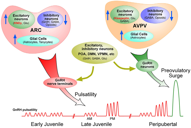
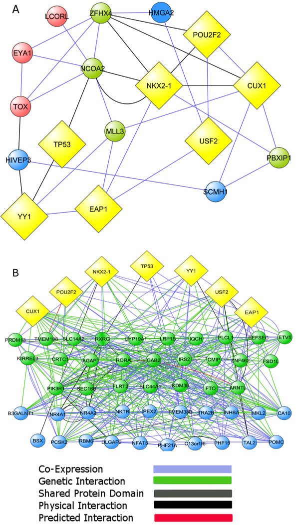
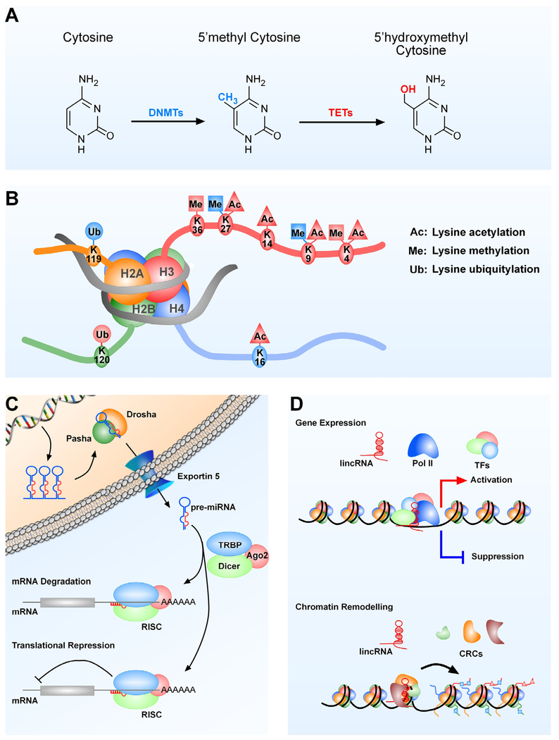
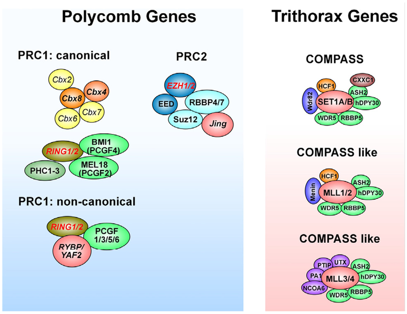
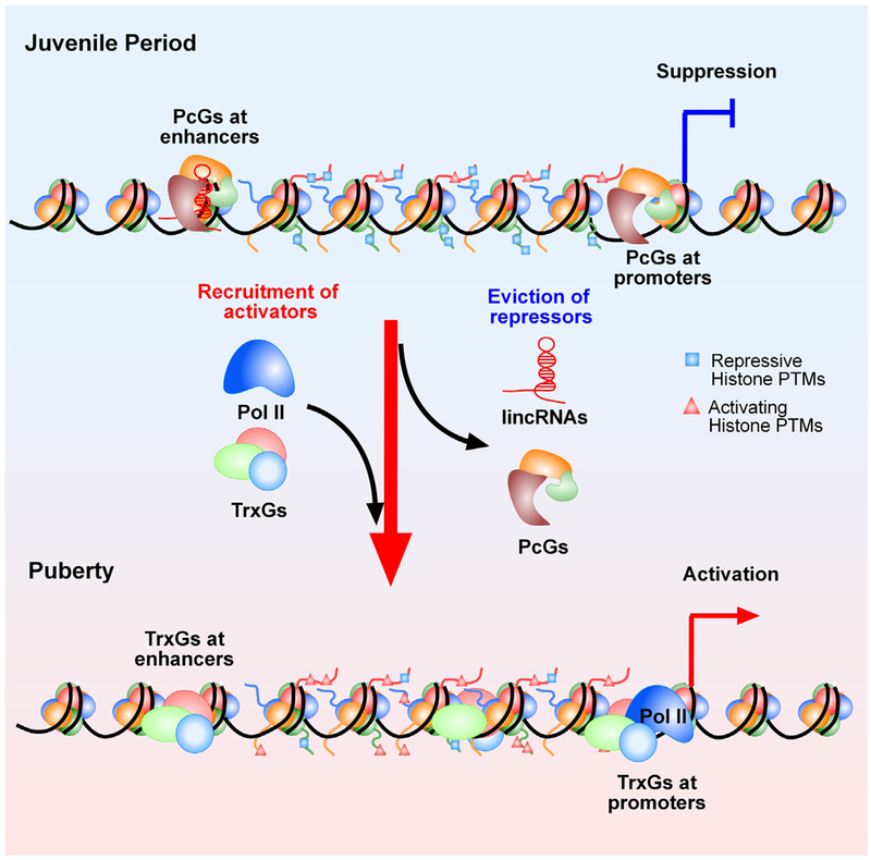
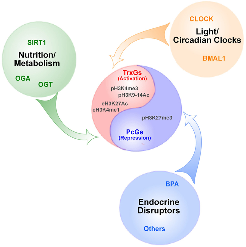
Substantial progress has been made in recent years toward deciphering the molecular and genetic underpinnings of the pubertal process. The availability of powerful new methods to interrogate the human genome has led to the identification of genes that are essential for puberty to occur. Evidence has also emerged suggesting that the initiation of puberty requires the coordinated activity of gene sets organized into functional networks. At a cellular level, it is currently thought that loss of transsynaptic inhibition, accompanied by an increase in excitatory inputs, results in the pubertal activation of GnRH release. This concept notwithstanding, a mechanism of epigenetic repression targeting genes required for the pubertal activation of GnRH neurons was recently identified as a core component of the molecular machinery underlying the central restraint of puberty. In this chapter we will discuss the potential contribution of various mechanisms of epigenetic regulation to the hypothalamic control of female puberty.

Keywords: Chromatin modifications; DNA methylation; Epigenetic regulators; Female puberty; GnRH neurons; Kisspeptin neurons; Long noncoding RNAs; Transcriptional activation; Transcriptional repression; microRNAs.


Figures

Fig. 1.
Fig. 1.. The hypothalamic control of pulsatile and surge LH release.
The hypothalamic control of puberty involves excitatory and inhibitory transsynaptic inputs to GnRH neurons, in addition to facilitatory glia-to-neuron signaling. According to this concept, the initiation of puberty involves a shift from a predominantly inhibitory (shown by downward arrows) to an excitatory mode of control (upward arrows). This shift results in diurnal activation of pulsatile GnRH release, which leads to increased LH pulsatility, the first endocrine manifestation of puberty. The change in pulsatile GnRH release results from activation of excitatory networks (neuronal and glial) operating in the ARC of the hypothalamus, with KNDy neurons playing a central role. The neuronal and glial systems involved appear to predominantly target GnRH nerve terminals at the median eminence. The preovulatory surge of gonadotropins is a later event at puberty and is triggered by activation of AVPV kisspeptin neurons responding to an elevation in circulating estrogen levels. The potential involvement of other excitatory neurons, such as those that use glutamate (Glu) and GABA acting via GABAA receptors for neurotransmission, is also indicated. However not all the excitatory or inhibitory systems regulating pulsatile GnRH release are located in the ARC or AVPV. Additional inhibitory neurons, such as those releasing GnIH are located in the dorsomedial nucleus (DMN), and groups of excitatory/inhibitory neurons are located in the medial preoptic area (POA), medial amygdala and ventral premammillary nucleus (VPMN).
Fig. 2.
Fig. 2.. Network connectivity of the most interconnected bovine puberty genes and human menarche-related genes to the central nodes of a TRG network derived from rats and nonhuman primates.
(A) The ten most connected genes of a bovine puberty gene network (Fortes et al., 2011) are first neighbors of the TRG central nodes (depicted as yellow diamonds). (B) Genes identified by GWAS as associated to the age of human menarche (Ong et al., 2009; Perry et al., 2009; Sulem et al., 2009; He et al., 2009; Elks et al., 2010; Cousminer et al., 2013; Tanikawa et al., 2013) are also highly connected to both the five original TRG central nodes (Roth et al., 2007 and to the upper-echelon transcriptional regulators TTF1/NKX2.1 and EAP1/IRF2BPL (yellow diamonds). Bovine puberty genes and menarche-related genes connected to multiple TRGs are depicted as green circles. Genes connected to at least one TRGs are shown as blue circles. Genes not connected to any TRG are represented as red circles. In both cases the connectivity is via co-expression (blue), predicted interaction (red), shared protein domain (gray), physical interactions (black), and genetic interaction (green) indicated by the GeneMANIA network construction algorithm. The thickness of each line indicates the strength of the evidence supporting that type of interaction in the Gene MANIA database. The distribution of nodes in B does not reflect a hierarchical distribution; instead it intends to emphasize the different degrees of connectivity that exists between central TRGs and menarche-related genes.
Fig. 3.
Fig. 3.. Modes of epigenetic regulation.
(A) DNA methylation. Methylation of cytosine at position 5 is carried out by DNMTs (DNMT1, DNMT3a and DNMT3b), and inactivation of this methyl group by hydroxymethylation is carried out by the TET enzymes. Names in blue color indicate repression and red color indicates activation of gene expression. (B) Histone PTMs. Only the PTMs catalyzed by the PcG and TrxG complexes (methylation, ubiquitination) or associated (acetylation) with TrxG-dependent PTMs are shown. Histone PTM in blue = PTM associated with gene repression; histone PTM in red = PTM associated with gene activation. (C) miRNAs. The pathway leading to miRNA production is outlined and the fact that miRNAs silence mRNA expression by inducing either mRNA degradation or translational repression is emphasized. (D) Long intergenic noncoding RNAs. Two mechanisms of lincRNA action are depicted. In one of them, lincRNAs modify gene expression by serving as landing pads for transcription factors that either repress or activate transcription. The other mechanism consists of lincRNAs directing the organization of chromatin states to specific genomic regions involved in gene regulation. DNMTs = DNA methyltransferases, TETs = ten eleven translocation (dioxygenase) enzymes; H = histone; Pasha = nuclear protein that is part of the microprocessor complex required for miRNA processing. Pasha associates with the RNA III enzyme Drosha. Drosha = RNA III enzyme that cleaves pri-miRNA (the primary transcript of miRNAs) to precursor (pre)-miRNA, which contains a stem-loop structure; Dicer = endoribonuclease that cleaves pre-miRNA into 20–25 mer double-stranded miRNAs; TRBP = human immunodeficiency virus transactivating response RNA-binding protein; it recruits Dicer to Ago2 for miRNA processing; Ago2 = Argonaut 2, the catalytic component of RISC; RISC = RNA induced silencing complex; Pol II = RNA polymerase 2; TF = transcription factor; CRC = chromatin remodeling complex.
Fig. 4.
Fig. 4.. Subunit composition of the PcG and COMPASS families.
The three major Polycomb repressive complexes (PRCs) are depicted. The PRC2 complex contains the histone methyltransferase enhancer of zeste homologue 1 (EZH1) or EZH2), which together with embryonic ectoderm development (EED) and suppressor of zeste 12 homolog (SUZ12) catalyzes the trimethylation of histone H3 at lysine K27 (H3K27me3). Multiple forms of the PRC1 complex exist. Canonical PRC1 complexes contain combinations of at least five Pc (Polycomb) proteins (known as chromobox proteins: CBX2, CBX4, CBX6, CBX7 and CBX8), two Psc (posterior sex comb) proteins (BMI1, also known as PCGF4 (polycomb group RING finger protein 4), MEL18 (also known as PCGF2) and one of two RING proteins, RING1 or RING 2 that provide the catalytic core to the complex because they have E3 ubiquitin ligase activity. In addition PRC1 complexes contain three polyhomeotic-like proteins (PHC1-3). Non canonical PRC1 complexes lack CBX proteins and contain instead a RING1 or RING2 protein that forms a complex with either RYBP (RING1 and YY1 binding protein) or YAF2 (YY1-associated factor) and one of four PCGF proteins different from BMI1 and MEL18 (PCGF1, 3, 5 or 6). The six known mammalian COMPASS complexes are also shown. Although all of them methylate H3K4 at lysine 4, different complexes are responsible for the mono, di or tri methylation of this amino acid (see text for details). Modified from Mohan et al. (2012).
Fig. 5.
Fig. 5.. Postulated epigenetic mechanisms controlling the onset of female puberty.
This model predicts the existence of an antagonistic (Yin-Yang) mechanism of transcriptional regulation underlying the developmental changes in expression of genes that facilitate pubertal development. According to this concept, the transcriptional activity of these genes (Kiss1, Tac2, Nell2, TTF1, others) is repressed during prepubertal development by silencing molecules, such as the PcG complex. PcG proteins catalyze the formation of a repressive chromatin structure characterized by an abundance of histone PTMs associated with gene silencing(such as H3K27me3). As puberty approaches, these “writers” of a repressive chromatin configuration are evicted from, and the content of histone repressive marks is reduced at, promoter regions controlling puberty-activating genes. Along with this change, writers of histone PTMs associated with transcriptional activation, such as H3K4me3 and H3K9, 14ac, are recruited to these regulatory regions resulting in enhanced gene expression. A strong candidate for this activational role is the TrxG activating complex, which antagonizes the silencing effect of PcG by both catalyzing the methylation of histone 3 at lysine 4 (H3K4me3, an activating histone mark) and binding to promoter DNA containing this mark. It is also envisioned that a similar relationship operates in distal enhancers regions controlling puberty-related genes. In this case, PcG deposition of the histone repressive mark H3K27me3, coupled to the presence of H3K4me1 and the absence of Pol II, define the presence of a latent enhancer. This inactive enhancer acquires an active configuration following the implementation of H3K27ac by the TrxG complex, and the recruitment of Pol II in the presence of H3K4me1 (also catalyzed by TrxG).
Fig. 6.
Fig. 6.
Different external and internal environmental stimuli (nutrition/metabolism, light/circadian clocks, endocrine disruptors) are envisioned to affect the timing and progression of puberty by altering the counteractive activity of PcG/TrxG-mediated epigenetic machinery. According to this model the different stimuli depicted can affect the time of puberty by modifying the function of either the PcG or TrxG complex or both.

References

    1. Abreu AP, Dauber A, Macedo DB, Noel SD, Brito VN, Gill JC, Cukier P, Thompson IR, Navarro VM, Gagliardi PC, Rodrigues T, Kochi C, Longui A, Beckers D, de ZF, Montenegro LR, Mendonca BB, Carroll RS, Hirschhorn JN, Latronico AC, Kaiser UB, 2013. Central precocious puberty caused by mutations in the imprinted gene MKRN3. N. Engl. J. Med 368, 2467–2475. - PMC - PubMed
    1. Ahima RS, Saper CB, Flier JS, Elmquist JK, 2000. Leptin regulation of neuroendocrine systems. Front. Neuroendocrinol 21, 263–307. - PubMed
    1. Akhtar RA, Reddy AB, Maywood ES, Clayton JD, King VM, Smith AG, Gant TW, Hastings MH, Kyriacou CP, 2002. Circadian cycling of the mouse liver transcriptome, as revealed by cDNA microarray, is driven by the suprachiasmatic nucleus. Curr. Biol 12, 540–550. - PubMed
    1. Andersson R, Gebhard C, Miguel-Escalada I, Hoof I, Bornholdt J, Boyd M, Chen Y, Zhao X, Schmidl C, Suzuki T, Ntini E, Arner E, Valen E, Li K, Schwarzfischer L, Glatz D, Raithel J, Lilje B, Rapin N, Bagger FO, Jorgensen M, Andersen PR, Bertin N, Rackham O, Burroughs AM, Baillie JK, Ishizu Y, Shimizu Y, Furuhata E, Maeda S, Negishi Y, Mungall CJ, Meehan TF, Lassmann T, Itoh M, Kawaji H, Kondo N, Kawai J, Lennartsson A, Daub O, Heutink P, Hume DA, Jensen TH, Suzuki H, Hayashizaki Y, Muller F, Forrest AR, Carninci P, Rehli M, Sandelin A, Kawaji H, Baillie JK, de Hoon MJ, Haberle V, Lassmann T, Kulakovskiy IV, Lizio M, Itoh M, Andersson R, Mungall CJ, Meehan TF, Schmeier S, Bertin N, Jorgensen M, Dimont E, Arner E, Schmid C, Schaefer U, Medvedeva YA, Plessy C, Vitezic M, Severin J, Semple CA, Ishizu Y, Young RS, Francescatto M, Alam I, Albanese D, Altschuler GM, Arakawa T, Archer JA, Arner P, Babina M, Rennie S, Balwierz PJ, Beckhouse AG, Pradhan-Bhatt S, Blake JA, Blumenthal A, Bodega B, Bonetti A, Briggs J, Brombacher F, Burroughs AM, Califano A, Cannistraci CV, Carbajo D, Chen Y, Chierici M, Ciani Y, Clevers HC, Dalla E, Davis CA, Detmar M, Diehl AD, Dohi T, Drablos F, Edge AS, Edinger M, Ekwall K, Endoh M, Enomoto H, Fagiolini M, Fairbairn L, Fang H, Farach-Carson MC, Faulkner GJ, Favorov AV, Fisher ME, Frith MC, Fujita R, Fukuda S, Furlanello C, Furuno M, Furusawa J, Geijtenbeek TB, Gibson AP, Gingeras T, Goldowitz D, Gough J, Guhl S, Guler R, Gustincich S, Ha TJ, Hamaguchi M, Hara M, Harbers M, Harshbarger J, Hasegawa A, Hasegawa Y, Hashimoto T, Herlyn M, Hitchens KJ, Ho Sui SJ, Hofmann OM, Hoof I, Hori F, Huminiecki L, Iida K, Ikawa T, Jankovic BR, Jia H, Joshi A, Jurman G, Kaczkowski B, Kai C, Kaida K, Kaiho A, Kajiyama K, Kanamori-Katayama M, Kasianov AS, Kasukawa T, Katayama S, Kato S, Kawaguchi S, Kawamoto H, Kawamura YI, Kawashima T, Kempfle JS, Kenna TJ, Kere J, Khachigian LM, Kitamura T, Klinken SP, Knox AJ, Kojima M, Kojima S, Kondo N, Koseki H, Koyasu S, Krampitz S, Kubosaki A, Kwon AT, Laros JF, Lee W, Lennartsson A, Li K, Lilje B, Lipovich L, Mackay-sim A, Manabe R, Mar JC, Marchand B, Mathelier A, Mejhert N, Meynert A, Mizuno Y, de Lima Morais DA, Morikawa H, Morimoto M, Moro K, Motakis E, Motohashi H, Mummery CL, Murata M, Nagao-Sato S, Nakachi Y, Nakahara F, Nakamura T, Nakamura Y, Nakazato K, van NE, Ninomiya N, Nishiyori H, Noma S, Nozaki T, Ogishima S, Ohkura N, Ohmiya H, Ohno H, Ohshima M, Okada-Hatakeyama M, Okazaki Y, Orlando V, Ovchinnikov DA, Pain A, Passier R, Patrikakis M, Persson H, Piazza S, Prendergast JG, Rackham OJ, Ramilowski JA, Rashid M, Ravasi T, Rizzu P, Roncador M, Roy S, Rye MB, Saijyo E, Sajantila A, Saka A, Sakaguchi S, Sakai M, Sato H, Satoh H, Savvi S, Saxena A, Schneider C, Schultes EA, Schulze-Tanzil GG, Schwegmann A, Sengstag T, Sheng G, Shimoji H, 2014. An atlas of active enhancers across human cell types and tissues. Nature 507, 455–461. - PMC - PubMed
    1. Batista PJ, Chang HY, 2013. Long noncoding RNAs: cellular address codes in development and disease. Cell 152, 1298–1307. - PMC - PubMed

Publication types

Substances