Review
. 2014 Dec:36:157-65.
doi: 10.1016/j.semcdb.2014.08.011. Epub 2014 Aug 27.

Architecture of tight junctions and principles of molecular composition

Affiliations
Review

Architecture of tight junctions and principles of molecular composition

Christina M Van Itallie et al. Semin Cell Dev Biol. 2014 Dec.

Abstract

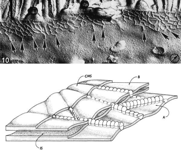
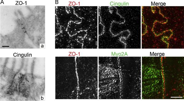
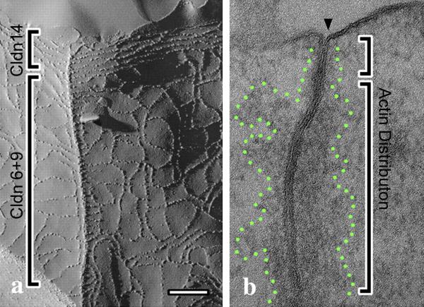
The tight junction creates an intercellular barrier limiting paracellular movement of solutes and material across epithelia. Currently many proteins have been identified as components of the tight junction and understanding their architectural organization and interactions is critical to understanding the biology of the barrier. In general the architecture can be conceptualized into compartments with the transmembrane barrier proteins (claudins, occludin, JAM-A, etc.), linked to peripheral scaffolding proteins (such as ZO-1, afadin, MAGI1, etc.) which are in turned linked to actin and microtubules through numerous linkers (cingulin, myosins, protein 4.1, etc.). Within this complex network are associated many signaling proteins that affect the barrier and broader cell functions. The PDZ domain is a commonly used motif to specifically link individual junction protein pairs. Here we review some of the key proteins defining the tight junction and general themes of their organization with the perspective that much will be learned about function by characterizing the detailed architecture and subcompartments within the junction.

Keywords: Actin; Claudin; Epithelium; Occludin; Tight junction; ZO-1.


Figures

Fig. 1
Fig. 1
Freeze fracture electron microscopic image and diagram of a tight junction. Top, FFEM image from rat jejunum showing a gradient of strand organization from the lateral to apical side of the junction. One continuous apical strand is typically followed by a network of interconnected strands and on the basal side the strands can be free and unconnected to the network. Bottom, An artist's view of a tight junction as seen in FFEM. The diagram is of two fracture planes in apposed tight junction membranes. (A) Rows of particles in one fracture plane that become a meshwork after glutaraldehyde fixation and (B) grooves, modeled in the front fibrils of this second fracture plane. IS, intercellular space and CMS, cytoplasmic membrane surface. Reproduced with permission from Staehelin, 1973.
Fig. 2
Fig. 2
Different subjunctional localization of cingulin, ZO-1, and Myo2A are revealed by immune-gold EM labeling (A) and high-resolution Structured Illumination Microscopy (SIM) (B). Cingulin (Aa) labeling is chick intestine is displaced from the membrane contacts and decorates actin filaments that radiate out from the junction contact sites. In contrast, ZO-1 (Ac) in cells is closely apposed to plasma membrane contacts corresponding to intermittent claudin-based strands. These relative positions are confirmed by immunolocalization in polarized Caco-2 cells by SIM (B) for cingulin (top), which in part co-localizes with ZO-1 but is also positions lateral to the ZO-1 signal. Myo2A (bottom) which binds cingulin, like cingulin, is positioned farther from the contacts. EM images were reproduced with permission from Stevenson, et al. 1986. SIM images produced by C.M. Van Itallie. Cells were fixed with 1% paraformaldehyde and incubated with (top) monoclonal ZO-1 (Life Technologies) and polyclonal cingulin (gift from Dr. Sandra Citi, University of Geneva) antibodies and (bottom) monoclonal ZO-1 and polyclonal non-muscle myosin 2A (Covance) antibodies; secondary antibodies were from Jackson Immunoresearch. Images were acquired on a using a GE OMX Blaze V4 Ultrafast Structured Illumination Microscope equipped with 4 sCMOS cameras using a 60X 1.42NA lens using 488 and 561 laser lines; images were acquired using Deltavision OMX software and adjusted and cropped in Adobe Photoshop. Unpublished results, bar=1 μm.
Fig. 3
Fig. 3
Two subdomains (brackets) of a tricellular (a, left) and bicellular (b, right) intercellular junction between sensory and non-sensory cells of the mouse inner ear revealed by freeze fracture and transmission EM. Left, a, Strand morphology is distinctly difference at the apical versus basal end of the junction. This correlates with restricted expression of claudin-14 to the top zone and claudin-6 and -9 to the bottom zone; in contrast, ZO-1 and occludin are expressed in both zones. Right, b, significantly more perijunctional actin is accumulated in bottom zone, outlined by green dots. Modified and reproduced with permission from Nunes, et al., 2006.

References

    1. Farquhar MG, Palade GE. Junctional complexes in various epithelia. J Cell Biol. 1963;17:375–412. - PMC - PubMed
    1. Chalcroft JP, Bullivant S. An interpretation of liver cell membrane and junction structure based on observation of freeze-fracture replicas of both sides of the fracture. J Cell Biol. 1970;47:49–60. - PMC - PubMed
    1. Claude P, Goodenough DA. Fracture faces of zonulae occludentes from "tight" and "leaky" epithelia. J Cell Biol. 1973;58:390–400. - PMC - PubMed
    1. Fromter E, Diamond J. Route of passive ion permeation in epithelia. Nat New Biol. 1972;235:9–13. - PubMed
    1. Stevenson BR, Siliciano JD, Mooseker MS, Goodenough DA. Identification of ZO-1: a high molecular weight polypeptide associated with the tight junction (zonula occludens) in a variety of epithelia. J Cell Biol. 1986;103:755–66. - PMC - PubMed

Publication types

MeSH terms