Review
. 2014 Dec;472(12):3605-22.
doi: 10.1007/s11999-014-3901-6. Epub 2014 Aug 30.

The 2014 ABJS Nicolas Andry Award: The puzzle of the thumb: mobility, stability, and demands in opposition

Affiliations
Review

The 2014 ABJS Nicolas Andry Award: The puzzle of the thumb: mobility, stability, and demands in opposition

Amy L Ladd et al. Clin Orthop Relat Res. 2014 Dec.

Abstract

Background: The paradoxical demands of stability and mobility reflect the purpose and function of the human thumb. Its functional importance is underscored when a thumb is congenitally absent, injured, or afflicted with degenerative arthritis. Prevailing literature and teaching implicate the unique shape of the thumb carpometacarpal (CMC) joint, as well as its ligament support, applied forces, and repetitive motion, as culprits causing osteoarthritis (OA). Sex, ethnicity, and occupation may predispose individuals to OA.

Questions/purposes: What evidence links ligament structure, forces, and motion to progressive CMC disease? Specifically: (1) Do unique attributes of the bony and ligamentous anatomy contribute to OA? (2) Can discrete joint load patterns be established that contribute to OA? And (3) can thumb motion that characterizes OA be measured at the fine and gross level?

Methods: We addressed the morphology, load, and movement of the human thumb, emphasizing the CMC joint in normal and arthritic states. We present comparative anatomy, gross dissections, microscopic analysis, multimodal imaging, and live-subject kinematic studies to support or challenge the current understanding of the thumb CMC joint and its predisposition to disease.

Results: The current evidence suggests structural differences and loading characteristics predispose the thumb CMC to joint degeneration, especially related to volar or central wear. The patterns of degeneration, however, are not consistently identified, suggesting influences beyond inherent anatomy, repetitive load, and abnormal motion.

Conclusions: Additional studies to define patterns of normal use and wear will provide data to better characterize CMC OA and opportunities for tailored treatment, including prevention, delay of progression, and joint arthroplasty.


Figures

Fig. 1
Fig. 1
The motion arcs of the metacarpal on the trapezium are flexion-extension and abduction-adduction. Pronation-supination represents composite rotation and translation of this triaxial joint based on morphology and muscular activity [26, 30, 37, 38, 46]. The thumb position in relation to the fingers represents a completion of the carpal arch, which places the CMC joint obliquely volar to the adjacent fingers and proximally oriented on its radial aspect. The arcs of motion thus are out of phase with the fingers. Image redrawn from CT surface rendering of a normal right hand. Published with kind permission of © S. Hegmann 2014. All Rights Reserved.
Fig. 2A–B
Fig. 2A–B
A comparison of (A) a normal human hand to (B) a silver-backed gorilla (Gorilla gorilla) hand from CT surface renderings is shown. The elderly (age 44 years) gorilla’s CMC joint is comparatively more constrained with a deeper articulating trapezial surface and more evenly matched in size compared to the human hand. Significant presence of trapezial-trapezoidal and second CMC OA is noted. Published with kind permission of © Ian Fitzgerald and Stanford University 2014. All Rights Reserved.
Fig. 3A–G
Fig. 3A–G
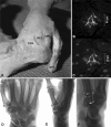
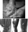
A pollicized right index finger functions like a thumb. The (A) clinical and (B) radiographic appearance before pollicization is shown. The (C, D) clinical and (EG) radiographic appearance 9 years after pollicization is also shown. The previous second metacarpal head, now the trapezium, has taken on duties of a saddle joint, with (E) change in morphology capable of (F) grasp and (G) pinch.
Fig. 4A–B
Fig. 4A–B
(A) The left trapezium metacarpal surface, viewed from above, demonstrates a slight concave curve dorsally (marked *) and concave volarly, where the beak of the metacarpal articulates (marked B). The ulnar ½ is eccentrically larger than the volar ½. This appears as a comma shape when viewed from this perspective; conversely, a right trapezium has a gentle C shape. (B) This has the same general shape of the glenoid: The left is a comma-shaped and the right C-shaped. Similar to the glenohumeral joint, the muscular bulk resides on the convex side, and more robust ligamentous support resides on the concave side. Common traumatic dislocations of the CMC joint and the glenohumeral joint occur on the sides with less musculotendinous support: Dorsal and anterior, respectively. This bony morphology and its soft tissue envelope suggest a contributing role to inherent stability. Figure 4B modified from Gray H, Lewis WH (ed). Anatomy of the Human Body. 20th ed. Philadelphia, PA: Lea & Febiger; 1918.
Fig. 5A–F
Fig. 5A–F
The dorsal ligamentous anatomy from (A) gross dissections, (B, C) immunohistochemical staining, and (DF) radiographic marking is shown. (A) In a left hand, the stout dorsal ligaments form a deltoid complex emanating from the dorsal tubercle (*), representing the dorsal radial ligament (DRL), dorsal central ligament (DCL), and posterior oblique ligament (POL). APL = abductor pollicis longus. Dorsal ligament immunofluorescent protein gene product 9.5 and 4′,6-diamidino-2-phenylindole staining demonstrate (B) a Ruffini ending and (C) nucleated collagen. These were essentially absent in all volar ligaments examined. The radiographic representation of the dorsal ligaments with (D) AP, (E) lateral, and (F) Robert’s views is shown: DRL = green; DCL = orange; POL = magenta; APL = red. Published with kind permission of © AL Ladd and E Hagert, 2014. All Rights Reserved.
Fig. 6A–D
Fig. 6A–D
The volar ligamentous anatomy from (A) gross dissections and (BD) radiographic marking is shown. (A) The volar ligaments, anterior oblique ligament (AOL) and ulnar collateral ligament (UCL), in passive extension are shown. The window between the thin ligaments is commonly found, intimal to the thenar muscles. APL = abductor pollicis longus; FCRg = the obliquely oriented flexor carpi radialis groove. (D) The radiographic representation of the volar ligaments with (B) AP, (C) lateral, and (D) Robert’s views is shown: AOL = blue; UCL = yellow; APL = red. Published with kind permission of © AL Ladd and E Hagert, 2014. All Rights Reserved.
Fig. 7
Fig. 7
The ligament and tendon location about the trapezium and metacarpal from arthroscopic specimens are illustrated as related to the common portals. Volar trapezial-metacarpal I is the same ligament as the ulnar collateral ligament (UCL) [108]. AOL = anterior oblique ligament; APL = abductor pollicis longus; EPL = extensor pollicis longus; EPB = extensor pollicis brevis. Published with kind permission of © S. Hegmann 2014. All Rights Reserved.
Fig. 8
Fig. 8
The three patterns of wear from 36 explanted surgical specimens are illustrated: the retained convex-concave saddle; the concave dish with substantial rimming osteophytes; and the cirque [103]. The cirque pattern is named for the extra lip in a glacial mountaintop formation, often seen at a headwall on a ski slope. The cirque varies in the width of the volar facet as either a small presence at the volar lip or expanding beyond the midline of the radial-ulnar axis. Published with kind permission of © S. Hegmann 2014. All Rights Reserved.
Fig. 9A–C
Fig. 9A–C
Micro-CTs of an OA trapezium viewed in the volar-dorsal (left to right) plane demonstrate the convex configuration of (A) a saddle, (B) dish, and (C) cirque. The radiodense circle represents the core made from a tap used to remove the trapezium [103]. Published with kind permission of © AL Ladd, 2014. All Rights Reserved.
Fig. 10A–D
Fig. 10A–D
Images of a trapezium are compared between (A) micro-CT to (B) histology and (C) portable flat panel CT scanner and (D) micro-CT. Toluidine blue represents preferential cartilage staining with histology. The cartilage thickness and the trabecular pattern are comparable between the different modalities. The trapezial cartilage is quite thin, and it is difficult to ascertain its thickness even in micro-CT, but it compares favorably to histology, as does trabecular patterning. The flat panel scanner has the potential to image live subjects and thus image the articulating metacarpal and adjacent joints with microscopic detail not previously attained.
Fig. 11A–B
Fig. 11A–B
(A) A CT scan is compared to (B) a false-color enhancement MRI in a cadaver with CMC OA, using the T1, T2, and proton density parameters to be reassigned as the red, green, blue channels as histograms (Beltrame). The fine trabeculae are seen in both images; the MRI demonstrates redundant dorsal-radial ligaments. Correlating scans on the MRI with the false-color assignment shows signal intensity differential on the ulnar portion of the metacarpal in the absence of obvious trabecular changes observed on CT. We hypothesize that this may represent a biochemical aberration, such as edema, abnormal crosslinking of collagen, or matrix pathology found in subchondral bone of OA.
Fig. 12A–B
Fig. 12A–B
(A) Lateral and (B) AP fluoroscopy images show a normal hand in grasp. The compression at the joint and the CMC motion is evident with load, but its precise position, translation, and rotation are not easily quantified given superimposition of structures and surfaces.
Fig. 13
Fig. 13
With markerless bone registration kinematic analysis of loaded object grasp, the thumb metacarpal undergoes ulnar translation, flexion, and abduction relative to the trapezium. With loaded key pinch (not shown), the thumb metacarpal undergoes volar translation, internal rotation, and flexion relative to the trapezium. Published with kind permission of © Brown University Orthopaedics Engineering Laboratory, 2014. All Rights Reserved.
Fig. 14
Fig. 14
A specific CMC functional coupling occurs in multiple tasks. Extension of the thumb metacarpal relative to the trapezium couples with adduction and flexion couples with abduction. Published with kind permission of © Brown University Orthopaedics Engineering Laboratory, 2014. All Rights Reserved.
Fig. 15A–B
Fig. 15A–B
Motion capture of jar opening is shown. (A) The reflective markers are placed on the thumb ray, wrist, index and small fingers, forearm, elbow, and shoulder. (B) The CMC and thumb ray demonstrate minimal abduction in a subject with OA compared to an asymptomatic control.
Fig. 16A–D
Fig. 16A–D
Still shots of the hand demonstrate (A) grasp, (B) jar opening, and (C, D) lateral pinch in an asymptomatic subject as recreated from the surface-rendered CT kinematic models. The use of slider bars and pause button permits some level of interactivity.

Comment in

References

    1. Alba DM, Moya-Sola S, Kohler M. Morphological affinities of the Australopithecus afarensis hand on the basis of manual proportions and relative thumb length. J Hum Evol. 2003;44:225–254. doi: 10.1016/S0047-2484(02)00207-5. - DOI - PubMed
    1. Almecija S, Moya-Sola S, Alba DM. Early origin for human-like precision grasping: a comparative study of pollical distal phalanges in fossil hominins. PLoS One. 2010;5:e11727. doi: 10.1371/journal.pone.0011727. - DOI - PMC - PubMed
    1. Ateshian GA, Ark JW, Rosenwasser MP, Pawluk RJ, Soslowsky LJ, Mow VC. Contact areas in the thumb carpometacarpal joint. J Orthop Res. 1995;13:450–458. doi: 10.1002/jor.1100130320. - DOI - PubMed
    1. Ateshian GA, Rosenwasser MP, Mow VC. Curvature characteristics and congruence of the thumb carpometacarpal joint: differences between female and male joints. J Biomech. 1992;25:591–607. doi: 10.1016/0021-9290(92)90102-7. - DOI - PubMed
    1. Baldwin MA, Langenderfer JE, Rullkoetter PJ, Laz PJ. Development of subject-specific and statistical shape models of the knee using an efficient segmentation and mesh-morphing approach: computer methods and programs in biomedicine. Comput Methods Programs Biomed. 2010;97:232–240. doi: 10.1016/j.cmpb.2009.07.005. - DOI - PubMed

Publication types

MeSH terms