Clinical Trial
. 2014 Oct 1;193(7):3366-77.
doi: 10.4049/jimmunol.1400346. Epub 2014 Aug 29.

CD39 and CD161 modulate Th17 responses in Crohn's disease

Affiliations
Clinical Trial

CD39 and CD161 modulate Th17 responses in Crohn's disease

Aiping Bai et al. J Immunol. .

Abstract

CD39 (ENTPD1) is expressed by subsets of pathogenic human CD4(+) T cells, such as Th17 cells. These Th17 cells are considered important in intestinal inflammation, such as seen in Crohn's disease (CD). Recently, CD161 (NKR-P1A) was shown to be a phenotypic marker of human Th17 cells. In this study, we report that coexpression of CD161 and CD39 not only identifies these cells but also promotes Th17 generation. We note that human CD4(+)CD39(+)CD161(+) T cells can be induced under stimulatory conditions that promote Th17 in vitro. Furthermore, CD4(+)CD39(+)CD161(+) cells purified from blood and intestinal tissues, from both healthy controls and patients with CD, are of the Th17 phenotype and exhibit proinflammatory functions. CD39 is coexpressed with CD161, and this association augments acid sphingomyelinase (ASM) activity upon stimulation of CD4(+) T cells. These pathways regulate mammalian target of rapamycin and STAT3 signaling to drive the Th17 phenotype. Inhibition of ASM activity by pharmacological blockers or knockdown of ASM abrogates STAT3 signaling, thereby limiting IL-17 production in CD4(+) T cells obtained from both controls and patients with active CD. Increased levels of CD39(+)CD161(+) CD4(+) T cells in blood or lamina propria are noted in patients with CD, and levels directly correlate with clinical disease activity. Hence, coexpression of CD39 and CD161 by CD4(+) T cells might serve as a biomarker to monitor Th17 responsiveness. Collectively, CD39 and CD161 modulate human Th17 responses in CD through alterations in purinergic nucleotide-mediated responses and ASM catalytic bioactivity, respectively.


Conflict of interest statement

Conflicts of interest: The authors disclose no conflicts.

Figures

FIGURE 1
FIGURE 1
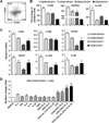
CD39 and CD161 expression patterns denote Th17 cell phenotypes in human CD4 T cell compartments. (A) Representative FACS analysis of CD39 and CD161 surface expression on CD4+ T cells of peripheral blood from healthy volunteers (n = 36). (B, C) Surface and mRNA expression of Th17-related molecules in four CD4+ T cell subsets (n = 5), as determined by FACS (B) and qRT-PCR (C), respectively. (D) Fresh isolated CD4+ T cells (n = 4) were stimulated with different cytokine cocktails in the presence of anti-CD3/CD28 T-activation Dynabeads for 18 hr. Frequencies of CD39+CD161+ CD4+ T cells were evaluated by FACS and changes are expressed relative to unstimulated controls. Data are represented as mean ± SEM of the indicated number of experiments. Percentages of gated cells are shown. *P < 0.05, **P < 0.01, ***P < 0.001.
FIGURE 2
FIGURE 2
CD4+CD39+CD161+ T cells exhibit an activated Th17 molecular signature. Subsets of CD4+ T cells were sorted by CD39 and CD161 expression by FACS, and cultured under Th17 differentiation conditions for 48 or 72 hr. (A, B) Representative flow cytometry analysis of intracellular levels of IL-17 and IFNγ at 72 hr (n = 16) (A) and gene expression at 48 hr were shown (n = 4) (B). HIF1α: hypoxia-inducible factor 1α, LDH1: lactate dehydrogenase 1, Glut1: glucose transporter 1. (C, D) Components of intracellular signaling transduction were measured at 48 hr by Western blot using probes as indicated (n = 4). PKM2: pyruvate kinase M2, ACC: acetyl-CoA carboxylase. (E) CD4+CD39+CD161+ T cells were expanded under Th17 condition for 72 hr in the presence of vehicle, 2-Deoxy-D-glucose (2-DG, a prototypical inhibitor of glycolysis), or etomoxir (an inhibitor of carnitine palmitoyltransferase 1, the rate-limiting enzyme in beta-oxidation of fatty acids). Representative flow cytometry analysis of intracellular IL-17 and IL-10 were shown (n = 4). *P < 0.05, **P < 0.01 vs. other three groups.
FIGURE 3
FIGURE 3
CD4+CD39+ CD161+ T cells are indicative of Th17 responses in Crohn’s disease (CD). (A, B) CD4+ T cells were isolated from healthy volunteers, active and inactive patients with CD using peripheral blood (n = 36, 16, or 12, respectively) or lamina propria (n = 17, 15, or 6, respectively) cells. Representative FACS analysis of subpopulations differed by CD39 and CD161 are shown (A) and expressed as a percentage of total CD4+ T cells (B). (C–E) CD4+ T cells were purified from lamina propria of patients with active CD (n = 8), then treated with PMA (50 nM) and ionomycin (500 nM) for 5 hr. This was followed by flow cytometry analysis of intracellular IL-17 and IFNγ (C, D), and CD39 and CD161 expression patterns in gated IL-17+CD4+ T cells (E). *P < 0.05, **P < 0.01, ***P < 0.001, ****P < 0.0001.
FIGURE 4
FIGURE 4
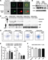
Direct association of CD39 with CD161 on CD4+ T cells and impact on purinergic signaling. (A) Membrane co-localization of CD39 and CD161 on CD4+CD39+CD161+ T cells analyzed by confocal microscopy. Activation beads adjacent to cells show reflected fluorescence. (B) CD39+CD161+ and CD39CD161 CD4+ T cells were Th17 differentiated for 12 hr or untreated (0 hr), cell lysates were subjected to co-immunoprecipitation using anti-CD39 antibody, followed by Western blotting probed for CD161 and CD39. (C–E) Hydrolysis of extracellular 14C-radiolabeled ADP by, and frequency of CD73 and ADA expression in, fresh four CD4+ T cell subsets, as determined by TLC (C) and FACS (D, E), respectively. (F) Effects of exogenous purines (i.e. ATP, AMP, and adenosine (ADO), 50 µM) on Th17 generation of CD4+CD39+CD161+ T cells post 72 hr Th17 deviation. Data were represented as mean ± SEM of three to five independent experiments. *P < .05, ns, not significant, vs. the other three groups.
FIGURE 5
FIGURE 5
Direct interaction of ASM with CD39 and CD161 mediates Th17 generation through mTOR/STAT3 signaling. (A, B) CD4+ T cells were expanded by Th17-differentiation condition, either in the presence of vehicle or imipramine (20 µM). Production of ceramide (48 hr) and intracellular levels of IL-17 and IFNγ (72 hr) were evaluated by TLC (A) and FACS (B), respectively. (C) CD39+CD161+ and CD39CD161 CD4+ T cells were stimulated with Th17-driving condition for 12 hr or unstimulated (0 hr), cell lysates were used for co-immunoprecipitation against anti-ASM antibody, followed by Western blot using probes as indicated. (D, E) TLC analysis of ceramide production by 48 hr Th17-deviated CD4+ T cell subsets (D), or CD4+CD39+CD161+ T cells stimulated with cross-linked anti-CD39 antibody (4 µg/ml), anti-CD161 antibody (4 µg/ml), or both for different times (E). Mouse anti-human IgG (4 µg/ml) served as control. (F) Effects of imipramine or rapamycin on IL-17 and IFNγ production by CD4+CD39+CD161+ T cells at 72 hr post Th17-polarization. Data were represented as mean ± SEM of three to four independent experiments. *P < 0.05 vs. the other three groups.
FIGURE 6
FIGURE 6
Knockdown of ASM blocks STAT3 signals and Th17 generation. (A) Western blotting of ASM expression in healthy blood CD4+ T cells after inhibition of ASM using lentiviral shRNAs. (B–D) Control knockdown (sh-C) and ASM knockdown (sh-1 and sh-3) healthy peripheral blood CD4+ T cells were stimulated with anti-CD3/CD28 beads under the defined Th17 differentiation conditions, followed by representative TLC analyses of ceramide at 2 hr (B), flow cytometry analysis of intracellular p-STAT3 at 3 hr (C), and intracellular IL-17 and IFNγ expression at 96 hr (D). Data were represented as mean ± SEM of three independent experiments. *P < 0.05 vs. the sh-C group.
FIGURE 7
FIGURE 7
ASM inhibition abrogates in vitro generation of Th17 cells using lamina propria CD4+ T cells purified from patients with Crohn’s disease (CD). (A–C) lamina propria CD4+ T cells isolated from patients with active disease were stimulated with anti-CD3/CD28 activation beads in the presence of vehicle or imipramine (20 µM), followed by TLC analysis of ceramide levels at 2 hr (n=3) (A), and flow cytometry determination of intracellular IL-17 and IFNγ (B, C) expression at 5 hr (n=6). *P < 0.05, **P < 0.01 vs. Vehicle.

References

    1. Constant SL, Bottomly K. Induction of Th1 and Th2 CD4+ T cell responses: the alternative approaches. Annu Rev Immunol. 1997;15:297–322. - PubMed
    1. Ouyang W, Kolls JK, Zheng Y. The biological functions of T helper 17 cell effector cytokines in inflammation. Immunity. 2008;28:454–467. - PMC - PubMed
    1. Abraham C, Cho JH. Inflammatory bowel disease. N Engl J Med. 2009;361:2066–2078. - PMC - PubMed
    1. Brand S. Crohn's disease: Th1, Th17 or both? The change of a paradigm: new immunological and genetic insights implicate Th17 cells in the pathogenesis of Crohn's disease. Gut. 2009;58:1152–1167. - PubMed
    1. Korn T, Bettelli E, Oukka M, Kuchroo VK. IL-17 and Th17 Cells. Annu Rev Immunol. 2009;27:485–517. - PubMed

Publication types

MeSH terms