Review
. 2014 Oct 1;592(19):4115-28.
doi: 10.1113/jphysiol.2014.274316. Epub 2014 Aug 28.

Corticospinal activity evoked and modulated by non-invasive stimulation of the intact human motor cortex

Affiliations
Review

Corticospinal activity evoked and modulated by non-invasive stimulation of the intact human motor cortex

Vincenzo Di Lazzaro et al. J Physiol. .

Abstract

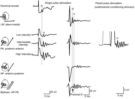
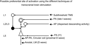
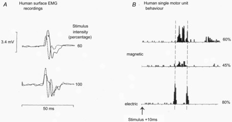
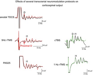
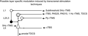
A number of methods have been developed recently that stimulate the human brain non-invasively through the intact scalp. The most common are transcranial magnetic stimulation (TMS), transcranial electric stimulation (TES) and transcranial direct current stimulation (TDCS). They are widely used to probe function and connectivity of brain areas as well as therapeutically in a variety of conditions such as depression or stroke. They are much less focal than conventional invasive methods which use small electrodes placed on or in the brain and are often thought to activate all classes of neurones in the stimulated area. However, this is not true. A large body of evidence from experiments on the motor cortex shows that non-invasive methods of brain stimulation can be surprisingly selective and that adjusting the intensity and direction of stimulation can activate different classes of inhibitory and excitatory inputs to the corticospinal output cells. Here we review data that have elucidated the action of TMS and TES, concentrating mainly on the most direct evidence available from spinal epidural recordings of the descending corticospinal volleys. The results show that it is potentially possible to test and condition specific neural circuits in motor cortex that could be affected differentially by disease, or be used in different forms of natural behaviour. However, there is substantial interindividual variability in the specificity of these protocols. Perhaps in the future it will be possible, with the advances currently being made to model the electrical fields induced in individual brains, to develop forms of stimulation that can reliably target more specific populations of neurones, and open up the internal circuitry of the motor cortex for study in behaving humans.


Figures

Figure 1
Figure 1. Descending volleys evoked by electrical and magnetic stimulation and by paired pulse magnetic stimulation
Each trace is the average of the responses to 10–25 cortical stimuli; recordings shown in the three columns have been obtained in three different subjects. Electrical anodal stimulation at threshold intensity evokes the earliest volley, termed D wave. Low intensity magnetic stimulation with a posterior–anterior (PA) induced current in the brain evokes a single descending wave with a latency about 1 ms longer than the D wave evoked by electrical stimulation, termed I1 wave. At intermediate intensity later I waves are evoked and at high intensity, an earlier small wave with the same latency as the D wave evoked by electrical anodal stimulation appears. Magnetic stimulation with a latero-medial (LM) induced current in the brain preferentially evokes D wave activity. With biphasic magnetic stimulation the earliest volley has a latency of about 0.4 ms longer than the D wave evoked by LM magnetic stimulation. Because of its longer latency, it is suggested that the D wave evoked by biphasic stimulation is initiated closer to the cell body of the PTNs than the conventional D wave evoked by LM magnetic stimulation and anodal stimulation and it is termed ‘proximal D wave’. On the right, epidural volleys evoked by test magnetic stimulus alone (continuous trace) and by test magnetic stimulus preceded by a subthreshold conditioning stimulus at 3 ms interstimulus interval (dotted trace). The test stimulus evokes multiple descending waves. There is a clear suppression of the late corticospinal volley when the test magnetic stimulus is preceded by the subthreshold conditioning stimulus.
Figure 2
Figure 2. Schematic representation of motor cortex circuits and possible preferential site of activation using the different techniques of transcranial brain stimulation
Open circles indicate excitatory neurones while filled circles indicate inhibitory neurones. This model includes a superficial inhibitory circuit composed of layer 1 (L1) neurones that have connections with layer 2 (L2) and 3 (L3) interneurones (filled circle) which inhibit the distal apical dendrites of layer 5 pyramidal neurones (Jiang et al. 2013); L2 and L3 bursting (open circle inside a dotted circle) and non-bursting excitatory interneurones projecting upon the distal apical dendrites of layer 5 pyramidal neurones; cortico-cortical axons projecting upon basal dendrites of layer 5 pyramidal neurones. It is proposed that anodal stimulation and LM magnetic stimulation activate directly the corticospinal axons of pyramidal tract neurones (PTNs) evoking a short latency wave termed D wave. The wave evoked by low intensity magnetic stimulation that appears 1–1.4 ms later than the D wave evoked by electrical anodal stimulation is suggested to be produced by monosynaptic activation of basal dendrites of PTNs by cortico-cortical axons activated by the magnetic stimulus. The late I waves evoked at higher intensities might be produced by a circuit that involves cortico-cortical axons that activate L2 and L3 bursting neurones, and in turn activate PTN apical dendrites. It is proposed that the more dispersed descending activity evoked by AP magnetic stimulation is produced by a different circuit that might include non-bursting L2 and L3 interneurones projecting upon PTN apical dendrites. It is also proposed that the longer latency D wave evoked by biphasic stimulation is initiated closer to the cell body of the PTNs at axon hillock level, rather than at some distance down the axon. It is proposed that inhibitory effects produced by a subthrehsold conditioning stimulus originate from a selective enhancement of the excitability of the GABAergic circuit originating from L1 neurones and projecting upon PTNs apical dendrites, resulting in a selective suppression of the late I waves.
Figure 3
Figure 3
A, surface EMG responses in the preactivated first dorsal interosseous muscle after two different intensities of TMS (continuous lines) and TES (dashed lines). At an intensity of 60%, the MEP to TMS has a longer latency than the response to TES; however, this difference disappears at 100%. B, comparison of TMS and TES activation of a single motor unit in the FDI muscle (same unit all traces, x-axis calibration intervals are 2.5 ms). The histograms show the number of times the unit discharged at each interval after a single TES or TMS pulse. Note that TES evokes a very early peak of increased firing plus one about 5 ms later. In contrast the earliest increase in discharge after TMS occurs about 1.5 ms later than after TES. It is followed by one or two other peaks as the intensity of TMS increases from 45–60%. These increases in firing probability are thought to be caused by arrival of EPSPs at the spinal motoneurone released by D (TES only) and I wave (both TES and TMS) volleys in the corticospinal tract. (A from Day et al. ; B from Rothwell et al. 1991).
Figure 4
Figure 4. Epidural volleys recorded in baseline conditions (black trace) and after several excitatory (red traces) and inhibitory (green traces) neuromodulation protocols
Each trace is the average of the responses to 10–25 cortical magnetic stimuli. After anodal transcranial direct current stimulation (TDCS) a D and I wave enhancement is observed. 5 Hz repetitive transcranial magnetic stimulation (rTMS) increases the amplitude of the proximal D wave and all I waves. Paired associative stimulation at 25 ms interstimulus interval (PAS25) produces a selective facilitation of late I waves with no change in I1 wave. After continuous theta burst stimulation (cTBS) the amplitude of the I1 wave is suppressed. 1 Hz rTMS produces a selective suppression of late I waves with no change in I1 wave.
Figure 5
Figure 5. Possible layer specific modulation of cortical circuits by different protocols of transcranial stimulation
It is proposed that 5 Hz subthreshold rTMS leads to a suppression of the excitability of the superficial inhibitory circuits, including superficial (L1) neurons, and that most of the protocols (iTBS, PAS25, PAS10, 1 Hz rTMS, TDCS) selectively modulate bursting cells of layer 2 and 3 that project upon PTNs and generate the late I waves. We suggest that paired pulse (Pp) rTMS might modulate the excitability of non-bursting layer 2 and 3 interneurones enhancing the non-synchronous activity that cannot be seen as a change in I wave activity. It is proposed that cTBS selectively suppresses the excitability of monosynaptic connections to PTNs. 5 Hz rTMS may produce its effects by enhancing the excitability of PTNs. Anodal TDCS together with a short lived enhancement of late I waves produces a strong and prolonged enhancement in the amplitude of the D wave, perhaps by enhancing the excitability of corticospinal axons.

References

    1. Adrian ED, Moruzzi G. Impulses in the pyramidal tract. J Physiol. 1939;97:153–199. - PMC - PubMed
    1. Alle H, Heidegger T, Krivanekova L, Ziemann U. Interactions between short-interval intracortical inhibition and short-latency afferent inhibition in human motor cortex. J Physiol. 2009;587:5163–5176. - PMC - PubMed
    1. Amassian VE, Eberle L, Maccabee PJ, Cracco RQ. Modelling magnetic coil excitation of human cerebral cortex with a peripheral nerve immersed in a brain-shaped volume conductor: the significance of fiber bending in excitation. Electroencephalogr Clin Neurophysiol. 1992;85:291–301. - PubMed
    1. Baker SN, Curio G, Lemon RN. EEG oscillations at 600 Hz are macroscopic markers for cortical spike bursts. J Physiol. 2003;550:529–534. - PMC - PubMed
    1. Barker AT, Garnham CW, Freeston IL. Magnetic nerve stimulation: the effect of waveform on efficiency, determination of neural membrane time constants and the measurement of stimulator output. Electroencephalogr Clin Neurophysiol Suppl. 1991;43:227–237. - PubMed

MeSH terms

LinkOut - more resources