. 2014 Oct;15(10):938-46.
doi: 10.1038/ni.2984. Epub 2014 Aug 31.

Neutrophils prime a long-lived effector macrophage phenotype that mediates accelerated helminth expulsion

Affiliations

Neutrophils prime a long-lived effector macrophage phenotype that mediates accelerated helminth expulsion

Fei Chen et al. Nat Immunol. 2014 Oct.

Abstract

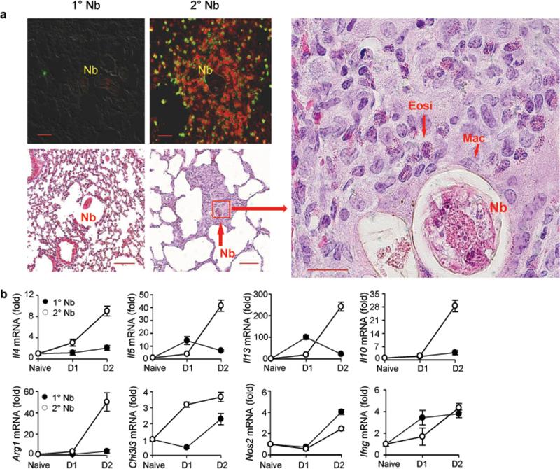
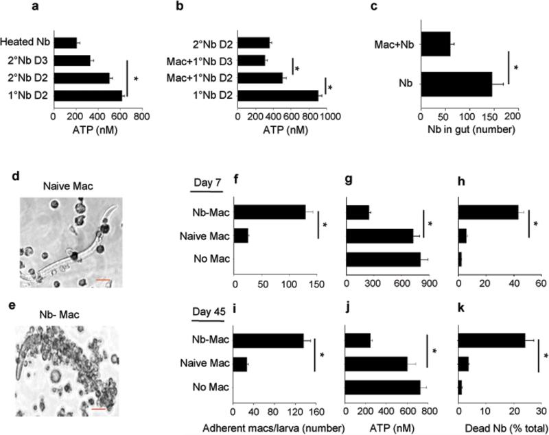
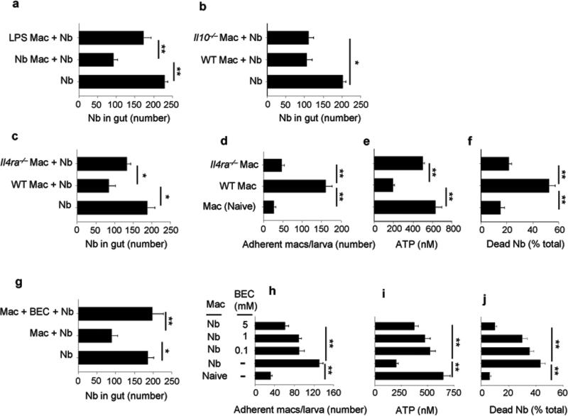
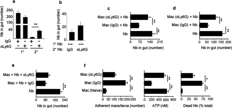
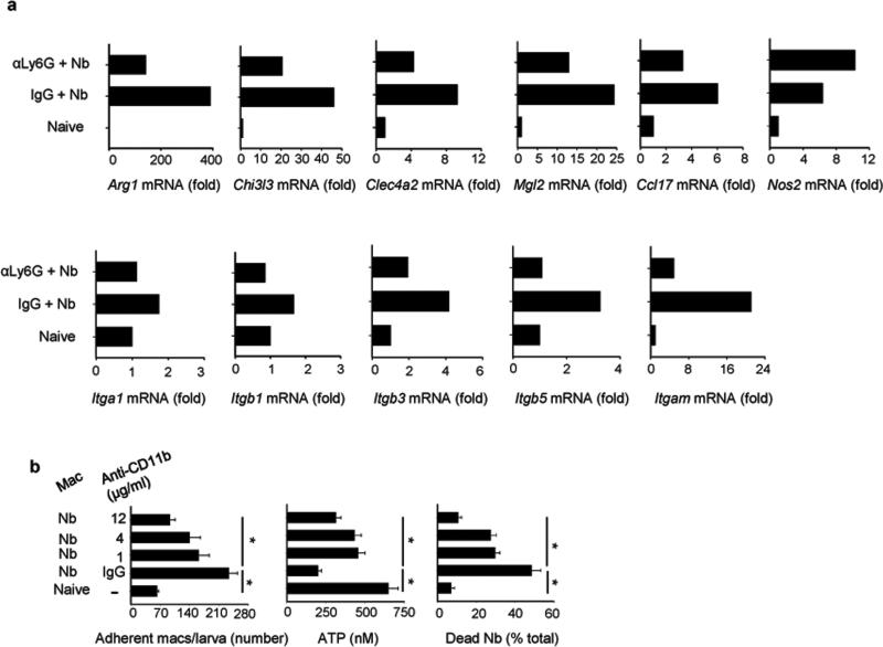
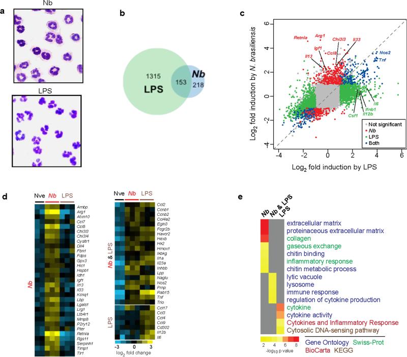
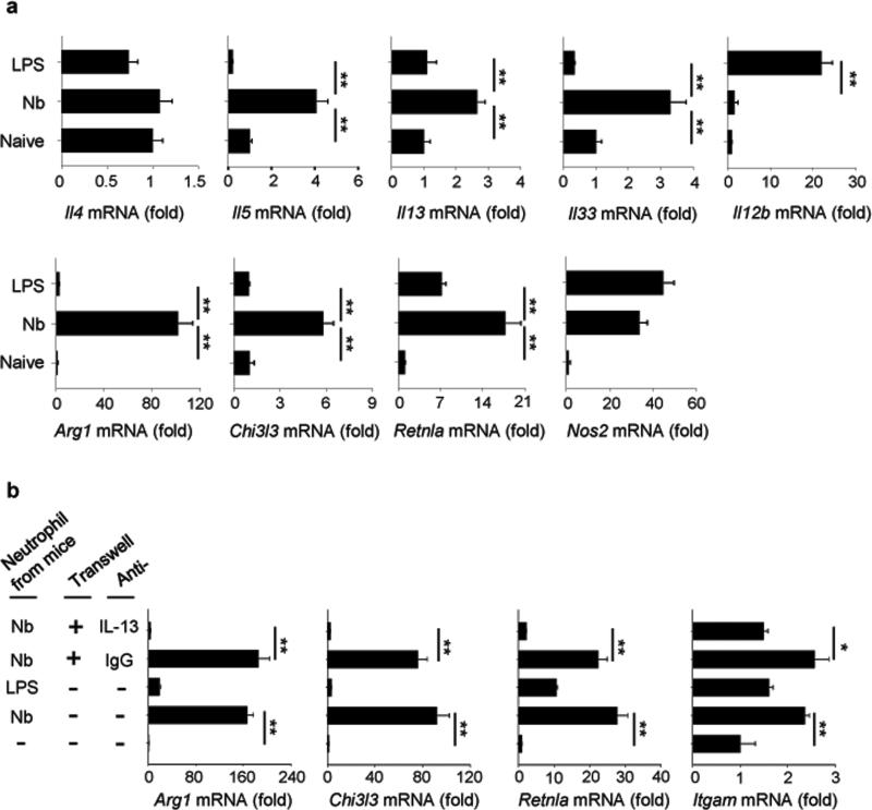
We examined the role of innate cells in acquired resistance to the natural murine parasitic nematode, Nippostrongylus brasiliensis. Macrophages obtained from lungs as late as 45 d after N. brasiliensis inoculation were able to transfer accelerated parasite clearance to naive recipients. Primed macrophages adhered to larvae in vitro and triggered increased mortality of parasites. Neutrophil depletion in primed mice abrogated the protective effects of transferred macrophages and inhibited their in vitro binding to larvae. Neutrophils in parasite-infected mice showed a distinct transcriptional profile and promoted alternatively activated M2 macrophage polarization through secretory factors including IL-13. Differentially activated neutrophils in the context of a type 2 immune response therefore prime a long-lived effector macrophage phenotype that directly mediates rapid nematode damage and clearance.


Figures

Figure 1
Figure 1. Eosinophils and macrophages surround parasitic larvae in the lung and type 2 related cytokines are upregulated shortly after secondary N. brasiliensis (Nb) inoculation
(a) Top left panels: DIC and Immunofluorescence staining of lung cryosections stained for macrophages (F4/80, PE, red) and eosinophils (MBP, Alexa Fluor488, F4/80var, yellow-green) at day 2 after primary (1°) or secondary (2°) N. brasiliensis inoculation. Scale bars 20 μm. Image is representative of five independent staining preparations of lung tissue from 3 mice infected with N. brasiliensis. Bottom panels; Representative haematoxylin and eosin (H&E) staining of formalin-fixed lung sections. Scale bars, 20 μm. H&E images are representative of five sectional layers for each mouse with each layer 20 layers removed from the other. There were 5 mice for each treatment group for two independent experiments; Nb=larvae; Mac=macrophages; Eosi=eosinophil. (b) At one or two days after primary or secondary Nb inoculation, lung tissue gene expression was determined by qPCR. Gene expression is shown as the fold increase over naive wild type (WT) controls after normalization to 18s RNA. Data shown are the mean and SEM from five individual mice per group and are representative of two independent experiments.
Figure 2
Figure 2. Macrophages from Nb-primed mice directly damage Nb larvae
(a) Helminth ATP concentrations were determined for larvae from lung tissue of mice 2 or 3 days after primary (1°) or secondary (2°) Nb inoculation. Heat killed Nb larvae were included as control (Heated Nb). (b) At day 7 after Nb inoculation, donor macrophages were electronically sorted (F4/80hiMHCIIint-hiCD11cvar) and transferred to recipient mice. Larvae were assessed for ATP concentrations at day 2 and 3 after inoculation; controls included larvae ATP concentrations after primary and secondary inoculation. (c) Experiment was performed as in (b) except that recipient mice were examined for intestinal worms at day 4 after inoculation and controls included mice not receiving macrophages. Mean and SEM from 5 mice per treatment group, and experiments were performed two times with similar results. (d-k) Lung macrophages were sorted from naive or from Nb-primed mice at day 7 (d-h) or day 45 (i-k) after inoculation, seeded to 24-well plates (2 × 10⁶ cell/well) and co-cultured with 200 exsheathed third stage larvae (L3); control group included culture with L3 only. At day 5 after culture, macrophages were microscopically imaged (scale bars, 20 μm) (d and e) and the numbers of adherent macrophages (f, i), the worm ATP concentration (g, j), and percent mortality (h, k) were determined. Mean and SEM of triplicate samples obtained from 20 larva from each well for adherent macrophages and 10 larva from each well for determination of ATP concentration; data were adjusted to per worm values. This experiment was repeated three times with similar results. *p<0.01
Figure 3
Figure 3. IL-4R signaling was required for Nb-primed macrophage-mediated resistance to Nb
WT donor mice were administered LPS or Nb infective larvae (a) or WT, Il4ra–/–, and Il10–/– donor mice were inoculated with Nb (b-c). At day 7 after inoculation electronically sorted donor macrophages were transferred to naive recipient mice, which were inoculated with Nb 2 days later. Five days post-infection gut parasites were enumerated. At day 7 after inoculation electronically sorted macrophages from WT or Il4ra–/– mice were seeded to 24-well plates (2 x 10⁶ cells/well) and co-cultured with 200 exsheathed L3 larvae. At day 5 after culture, larval adherence was determined (d), and larval ATP concentration (e) and percent mortality (f) assessed. (g) WT donor mice were inoculated with Nb and, at day 7 after inoculation, electronically sorted macrophages were transferred to naive recipient mice, with or without administered Arg1 inhibitor BEC, which were then infected with Nb 2 days later. A control group was similarly treated but did not receive donor macrophages. Five days later gut parasites were enumerated. Mean and SEM are shown for five mice/treatment group (a, b, c, g), and these experiments were repeated two times with similar results. (h-j) At day 7 after inoculation electronically sorted macrophages from WT were seeded to 24-well plates (2 × 10⁶ cells/well) and co-cultured with 200 exsheathed L3 larvae, with or without the presence of BEC at different concentrations (0.1-5 mM). At day 5 after culture, larval adherence was determined (h), and larval ATP concentration (i) and percent mortality (j) were assessed. Mean and SEM of triplicate samples (d-f, h-j) are shown for pools of 5 mice/treatment group. This experiment was repeated three times with similar results. *p<0.05, **p<0.01.
Figure 4
Figure 4. Neutrophils are required for differentiation of effector M2 macrophages after Nb primary inoculation
Parasite numbers were determined for mice administered neutrophil-depleting anti-Ly6G antibody or isotype control at days −1 and 3 after secondary inoculation (2°) (a), or at days -1, 3, and 7 after primary (1°) and days -1 and 3 after secondary (2°) inoculation (b). Donor mice were treated with anti-Ly6G antibody or isotype control on days −1 and 3 (c), or days -1, 3, and 7 (d), after inoculation. At day 7 (c), or day 41(d) after inoculation, macrophages were collected from the lung and transferred to naive recipient mice, which were inoculated 2 days later and assessed for worm expulsion from the intestine on day 5 (c) or day 4 (d) after inoculation. (e) Donor mice were sacrificed at day 7 after Nb inoculation and macrophages were isolated from the lung and transferred to naive recipient mice, which were then treated with anti-Ly6G antibody or isotype control on days −1 and 3 after Nb inoculation. Parasite numbers were assessed in gut at day 5. Data are shown as mean and SEM of 5 mice/group. (f) Nb-inoculated mice administered anti-Ly6G antibody (αLy6G) or isotype control (IgG) were sacrificed at day 7, lung macrophages were electronically sorted and co-cultured with exsheathed L3 larvae. At day 5 after culture, the number of adherent macrophages/L3 was determined and larval damage assessed by parasite ATP concentrations and percent mortality. Mean and SEM of triplicate samples obtained from pools of 5 mice/treatment group. This experiment was repeated three times with similar results. *p<0.05, **p<0.01.
Figure 5
Figure 5. Depletion of neutrophils impaired effector lung macrophage polarization after primary Nb inoculation
(a) Macrophages were electronically sorted from lung of naive mice or mice at day 7 after Nb inoculation treatment with αLy6G or isotype control (day -1, 3). Gene expression for characteristic M1 and M2 macrophage markers and integrins were analyzed by qPCR. Data shown are the mean from a pool of five mice per group (expressed as fold increase over mRNA from lungs of naive mice) and are representative of two independent experiments. (b) Flow sorted macrophages from WT mice at day 7 post infection were seeded to 24-well plates and co-cultured with exsheathed L3 larvae, with or without the presence of anti-CD11b antibody at different concentration. At day 5, larval adherence, larval ATP concentration, and percent mortality were assessed. Mean and SEM of triplicate samples obtained from pools of 5 mice per treatment group. This experiment was repeated three times with similar results. *p<0.01
Figure 6
Figure 6. Nb-primed neutrophils show a distinct gene expression profile
Mice were inoculated with Nb L3 or intratracheally administered LPS. Two days later neutrophils were purified from lung tissue of untreated controls and treatment groups using electronic cell sorting. (a) Wright-Giemsa staining of sorted CD11b+Ly6G+ cells from the lung showed characteristic homogeneous neutrophil populations in each treatment group. (b) Global intersectional overview of upregulated transcripts relative to untreated controls by microarray analysis from sorted neutrophils obtained from lungs of naive mice (Nve) or mice infected with Nb or LPS. Genes were determined to be significantly differentially regulated by Significance Analysis of Microarrays (3% FDR, 1.5-fold change) (c) Scatterplot of gene induction by LPS and/or Nb relative to untreated controls. (d) Heat map presentation of selected representative genes characteristic of polarized type 1 and type 2 immune responses. (e) Functional analysis of gene expression showing characteristic categories according to classifications from the Gene Ontology (GO) project, SwissProt, BioCarta, and KEGG and aggregated in DAVID.
Figure 7
Figure 7. Nb-primed neutrophils polarize to an alternatively activated neutrophil phenotype that promotes M2 macrophage differentiation in vitro
(a) Gene expression of characteristic immune response markers, identified by microarray, from sorted neutrophils (CD11b+Ly-6G+) analyzed by qPCR. Data shown are the mean of triplicate samples from a pool of five mice per group (expressed as fold increase over mRNA from neutrophils of naive mice) and are representative of two independent experiments. (b) Bone marrow derived macrophages (BMDM) were cultured with neutrophils from the lung of mice infected with Nb or treated with LPS. The neutrophil culture was separated from macrophages by transwell in some group, and anti-IL-13 antibody (10 μg /ml) was also added to the culture in some groups. At day 3, Arg1, Chi3l3, Retnla, and Itgam gene expression was determined by qPCR. Gene expression is presented as the ratio of treated/BMDM group without neutrophils added. Data shown are the mean and SEM of triplicate samples and repeated three times with similar results. *p<0.05, **p<0.01.

Comment in

References

    1. King CH. Health metrics for helminthic infections. Advances in parasitology. 2010;73:51–69. - PubMed
    1. Salgame P, Yap GS, Gause WC. Effect of helminth-induced immunity on infections with microbial pathogens. Nat Immunol. 2013;14:1118–1126. - PMC - PubMed
    1. Gaze S, et al. Characterising the mucosal and systemic immune responses to experimental human hookworm infection. PLoS pathogens. 2012;8:e1002520. - PMC - PubMed
    1. Anthony RM, Rutitzky LI, Urban JF, Jr., Stadecker MJ, Gause WC. Protective immune mechanisms in helminth infection. Nat Rev Immunol. 2007;7:975–987. - PMC - PubMed
    1. Gause WC, Wynn TA, Allen JE. Type 2 immunity and wound healing: evolutionary refinement of adaptive immunity by helminths. Nat Rev Immunol. 2013 - PMC - PubMed

Publication types

MeSH terms

Associated data