. 2014 Oct;128(4):505-24.
doi: 10.1007/s00401-014-1336-5. Epub 2014 Aug 31.

Aggregation-prone c9FTD/ALS poly(GA) RAN-translated proteins cause neurotoxicity by inducing ER stress

Affiliations

Aggregation-prone c9FTD/ALS poly(GA) RAN-translated proteins cause neurotoxicity by inducing ER stress

Yong-Jie Zhang et al. Acta Neuropathol. 2014 Oct.

Abstract

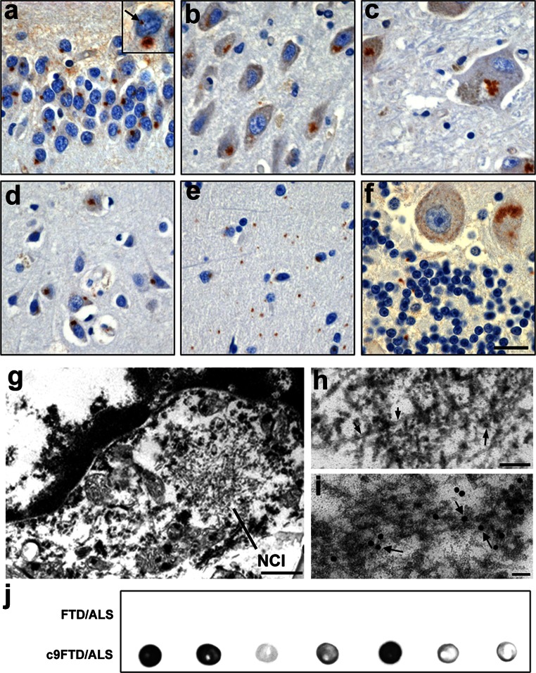
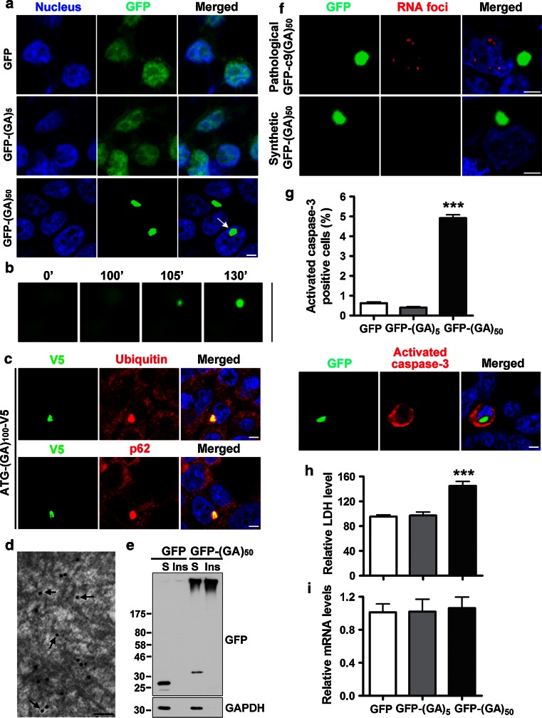
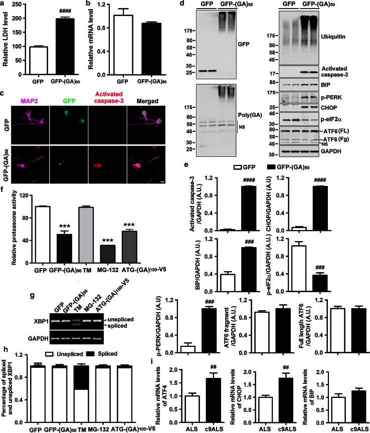
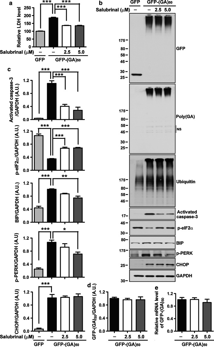
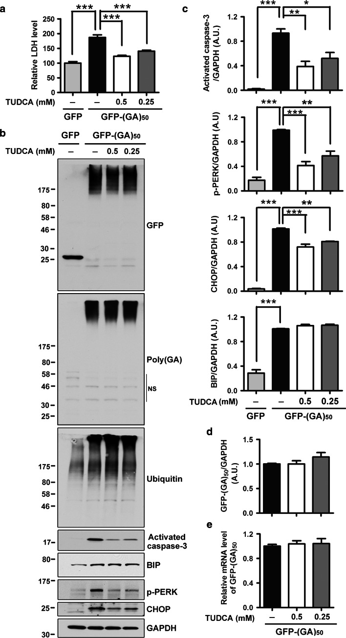
The occurrence of repeat-associated non-ATG (RAN) translation, an atypical form of translation of expanded repeats that results in the synthesis of homopolymeric expansion proteins, is becoming more widely appreciated among microsatellite expansion disorders. Such disorders include amyotrophic lateral sclerosis and frontotemporal dementia caused by a hexanucleotide repeat expansion in the C9ORF72 gene (c9FTD/ALS). We and others have recently shown that this bidirectionally transcribed repeat is RAN translated, and the "c9RAN proteins" thusly produced form neuronal inclusions throughout the central nervous system of c9FTD/ALS patients. Nonetheless, the potential contribution of c9RAN proteins to disease pathogenesis remains poorly understood. In the present study, we demonstrate that poly(GA) c9RAN proteins are neurotoxic and may be implicated in the neurodegenerative processes of c9FTD/ALS. Specifically, we show that expression of poly(GA) proteins in cultured cells and primary neurons leads to the formation of soluble and insoluble high molecular weight species, as well as inclusions composed of filaments similar to those observed in c9FTD/ALS brain tissues. The expression of poly(GA) proteins is accompanied by caspase-3 activation, impaired neurite outgrowth, inhibition of proteasome activity, and evidence of endoplasmic reticulum (ER) stress. Of importance, ER stress inhibitors, salubrinal and TUDCA, provide protection against poly(GA)-induced toxicity. Taken together, our data provide compelling evidence towards establishing RAN translation as a pathogenic mechanism of c9FTD/ALS, and suggest that targeting the ER using small molecules may be a promising therapeutic approach for these devastating diseases.


Figures

Fig. 1
Fig. 1
Neuropathology of c9RAN poly(GA) proteins in C9ORF72 repeat expansion cases. af Immunohistochemical analysis shows that poly(GA) proteins accumulate throughout the central nervous system of C9ORF72 repeat expansion carriers as neuronal cytoplasmic inclusions and neuronal intranuclear inclusions (arrow in insert, a). Regions with a particularly high burden include the dentate fascia of the hippocampus (a; Case 8), the hippocampus proper (b; CA3/2; Case 10), the anterior thalamus (c; Case 4), the frontal cortex (d; Layers IV–V; Case 7), the cerebellar molecular layer (e; Case 1) and the cerebellar internal granule cell layer (f; Case 9). Scale bar represents 25 µm (ae) and 20 µm (f). Regular electron microscopy (EM) of granule cells of the cerebellar cortex from a c9FTD-MND case shows that cytoplasmic inclusions (g) are composed of 15–17 nm filaments (arrow, h). Immuno-EM with anti-poly(GA) antibody labeled with gold particles (18 nm) reveals poly(GA) proteins localize to filaments (arrow, i). Scale bars represent 0.5 μm (g), 100 nm (h), and 50 nm (i). j Dot blot reveals that anti-poly(GA) immunoreactivity in cerebellar urea fractions is specific to c9FTD/ALS. Each dot represents one case
Fig. 2
Fig. 2
Poly(GA) proteins form inclusions and are toxic in cultured cells. a Expression of GFP-(GA)50 in cultured HEK293T cells results in the formation of cytoplasmic or nuclear (arrow) inclusions. Scale bar represents 5 µm. b Representative images of live cells demonstrating how quickly inclusions form (compare the image at 100 min to the image at 105 min). c Poly(GA) inclusions are ubiquitin and p62-positive in cultured cells. Scale bar represents 5 µm. d Immuno-electron microscopy with an anti-GFP antibody labeled with gold particles shows that cytoplasmic GFP-(GA)50 inclusions are composed of filamentous structures (arrow). Scale bar represents 100 nm. e Western blot analysis of Triton X-100 soluble (S) and insoluble (Ins) cell lysates shows that a portion of poly(GA) proteins forms high molecular weight material. f Cultured cells were made to express GFP-(GA)50, which encodes a synthetic repeat sequence (GGXGCX)50 (where X represents a random nucleotide), or GFP-c9(GA)50, in which the pathological repeat sequence (GGGGCC)50 was used. Post-transfection, cells were subjected to RNA fluorescence in situ hybridization (FISH) to visualize RNA foci. GFP-(GA)50 expression leads to the formation of poly(GA) inclusions, but transcripts from this sequence do not form RNA foci. In contrast, both RNA foci and poly(GA) inclusions are formed in cells that express GFP-c9(GA)50. Scale bar represents 5 µm. g Quantitative analysis and representative image showing that cells bearing inclusions of GFP-(GA)50 are immunoreactive for activated caspase-3, a crucial mediator of cell death. Scale bar represents 5 µm. h LDH activity in media, an indicator of cell toxicity, is increased in cells expressing GFP-(GA)50. i Transgene mRNA levels are comparable among cells expressing GFP, GFP-(GA)5 and GFP-(GA)50. Data represent the mean ± SEM from sixteen random selected fields (g) or three separate experiments (h and i). ***P < 0.001, as analyzed by one-way analysis of variance followed by Tukey’s post hoc analysis
Fig. 3
Fig. 3
Expression of poly(GA) proteins in primary neurons causes neurotoxicity accompanied by UPS impairment and ER stress. a Expression of GFP-(GA)50, but not GFP, causes toxicity, as assessed by measuring LDH activity in media. b Transgene mRNA levels are comparable in neurons expressing GFP and GFP-(GA)50. c Activated caspase-3 is observed in MAP2-positive neurons bearing GFP-(GA)50 inclusions. Scale bar represents 10 μm. Western blot (d) and densitometric analysis of blots (e) show that expression of GFP-(GA)50 in primary neurons leads to increased levels of activated caspase-3 and ubiquitinated proteins. In addition, levels of the ER stress markers, BIP, phospho-PERK and CHOP, are increased by GFP-(GA)50 expression, whereas levels of phospho-eIF2α are decreased and ATF6 levels remain unchanged. FL, Fg bands corresponding to full-length and fragmented ATF6, respectively. NS non-specific bands. f Proteasome activity assays reveal that expression of poly(GA) proteins inhibit proteasome activity. Decreased proteasome activity is also observed in neurons treated with the proteasome inhibitor MG-132, but not with the ER stress inducer, tunicamycin. RT-PCR (g) and quantitative analysis (h) show that tunicamycin induces abnormal splicing of XBP1. However, expression of poly(GA) proteins or treatment with MG-132 does not result in this alteration. i mRNA levels of ER stress markers, ATF4 and CHOP, are significantly increased in frontal cortex of ALS patients with the C9ORF72 repeat expansion. N = 8 for c9ALS and N = 11 for sporadic ALS without the C9ORF72 repeat expansion. Data represents mean ± SEM of three separate experiments (a, b, e, f, h). ## P < 0.01, ### P < 0.001 and #### P < 0.0001, as analyzed by unpaired t test. ***P < 0.001, as analyzed by one-way analysis of variance followed by Tukey’s post hoc analysis
Fig. 4
Fig. 4
Salubrinal, a selective inhibitor of eIF2α dephosphorylation, protects neurons against poly(GA)-induced ER stress and toxicity. a Salubrinal, a small molecule known to provide rescue from ER stress and associated cell death by inhibiting the dephosphorylation of eIF2α, significantly decreases LDH activity in media of neurons expressing GFP-(GA)50. Western blot (b) and densitometric analysis of blots (c) show that treatment of GFP-(GA)50-expressing neurons with salubrinal significantly inhibits caspase-3 activation, increases eIF2α phosphorylation, and decreases levels of ER stress markers, BIP and phospho-PERK. Note that salubrinal treatment does not decrease levels of ubiquitinated proteins or CHOP. Levels of GFP-(GA)50 protein and mRNA are also not changed after salubrinal treatment, as shown by Western blot (b), densitometric analysis of blot (d), and qRT-PCR (e). NS indicates non-specific bands. Data represents mean ± SEM from three separate experiments. *P < 0.05, **P < 0.01 and ***P < 0.001, as analyzed by one-way analysis of variance followed by Tukey’s post hoc analysis
Fig. 5
Fig. 5
The chemical chaperone TUDCA protects neurons against poly(GA)-induced ER stress and toxicity. a TUDCA, a chemical chaperone known to inhibit ER stress and associated downstream pathways, significantly decreases LDH activity in media of neurons expressing GFP-(GA)50. b, c Treatment of GFP-(GA)50-expressing neurons with TUDCA also significantly inhibits caspase-3 activation, and decreases levels of ER stress markers, phospho-PERK and CHOP, as shown by Western blot (b) and densitometric analysis of blots (c). Note that TUDCA treatment does not decrease levels of ubiquitinated proteins and BIP (b, c). Protein and mRNA levels of GFP-(GA)50 are not changed after TUDCA treatment (d, e). NS non-specific bands. Data represents mean ± SEM from three separate experiments. *P < 0.05, **P < 0.01 and ***P < 0.001, as analyzed by one-way analysis of variance followed by Tukey’s post hoc analysis

References

    1. Abasolo N, Torrell H, Roig B, Moyano S, Vilella E, Martorell L. RT-qPCR study on post-mortem brain samples from patients with major psychiatric disorders: reference genes and specimen characteristics. J Psychiatr Res. 2011;45(11):1411–1418. doi: 10.1016/j.jpsychires.2011.06.001. - DOI - PubMed
    1. Al-Sarraj S, King A, Troakes C, Smith B, Maekawa S, Bodi I, Rogelj B, Al-Chalabi A, Hortobagyi T, Shaw CE. p62 positive, TDP-43 negative, neuronal cytoplasmic and intranuclear inclusions in the cerebellum and hippocampus define the pathology of C9orf72-linked FTLD and MND/ALS. Acta Neuropathol. 2011;122(6):691–702. doi: 10.1007/s00401-011-0911-2. - DOI - PubMed
    1. Ash PE, Bieniek KF, Gendron TF, Caulfield T, Lin WL, Dejesus-Hernandez M, van Blitterswijk MM, Jansen-West K, Paul JW, 3rd, Rademakers R, Boylan KB, Dickson DW, Petrucelli L. Unconventional translation of C9ORF72 GGGGCC expansion generates insoluble polypeptides specific to c9FTD/ALS. Neuron. 2013;77(4):639–646. doi: 10.1016/j.neuron.2013.02.004. - DOI - PMC - PubMed
    1. Atkin JD, Farg MA, Walker AK, McLean C, Tomas D, Horne MK. Endoplasmic reticulum stress and induction of the unfolded protein response in human sporadic amyotrophic lateral sclerosis. Neurobiol Dis. 2008;30(3):400–407. doi: 10.1016/j.nbd.2008.02.009. - DOI - PubMed
    1. Belzil VV, Bauer PO, Prudencio M, Gendron TF, Stetler CT, Yan IK, Pregent L, Daughrity L, Baker MC, Rademakers R, Boylan K, Patel TC, Dickson DW, Petrucelli L. Reduced C9orf72 gene expression in c9FTD/ALS is caused by histone trimethylation, an epigenetic event detectable in blood. Acta Neuropathol. 2013;126(6):895–905. doi: 10.1007/s00401-013-1199-1. - DOI - PMC - PubMed

Publication types

MeSH terms