Review
. 2014 Dec 15:79-80:119-34.
doi: 10.1016/j.addr.2014.08.009. Epub 2014 Aug 29.

Biomechanical forces in the skeleton and their relevance to bone metastasis: biology and engineering considerations

Affiliations
Review

Biomechanical forces in the skeleton and their relevance to bone metastasis: biology and engineering considerations

Maureen E Lynch et al. Adv Drug Deliv Rev. .

Abstract

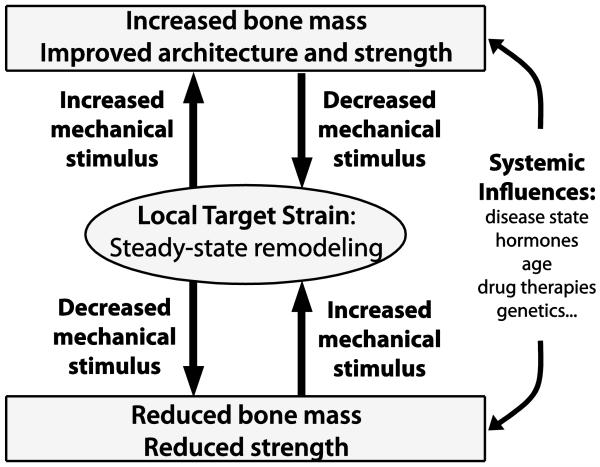
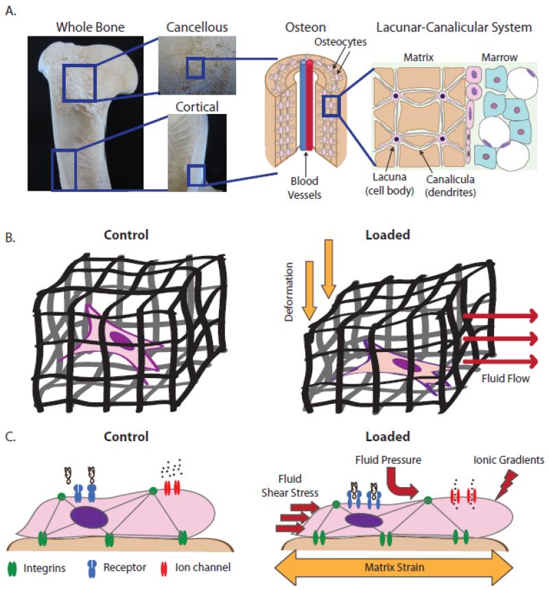
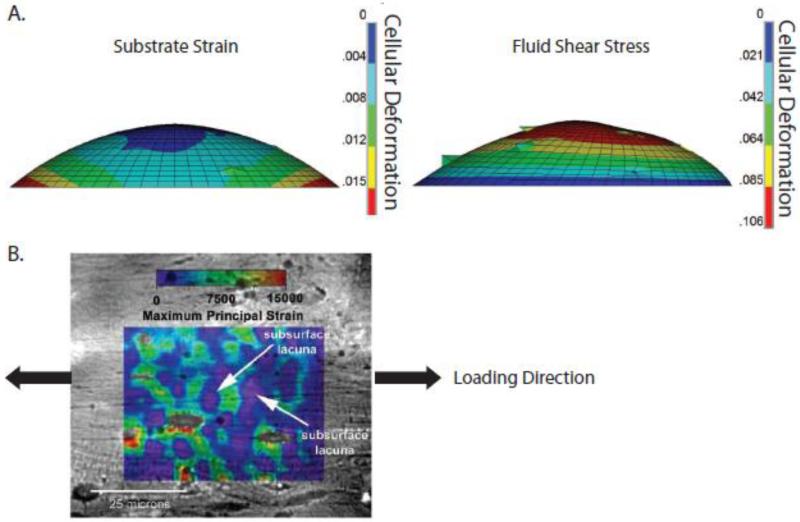
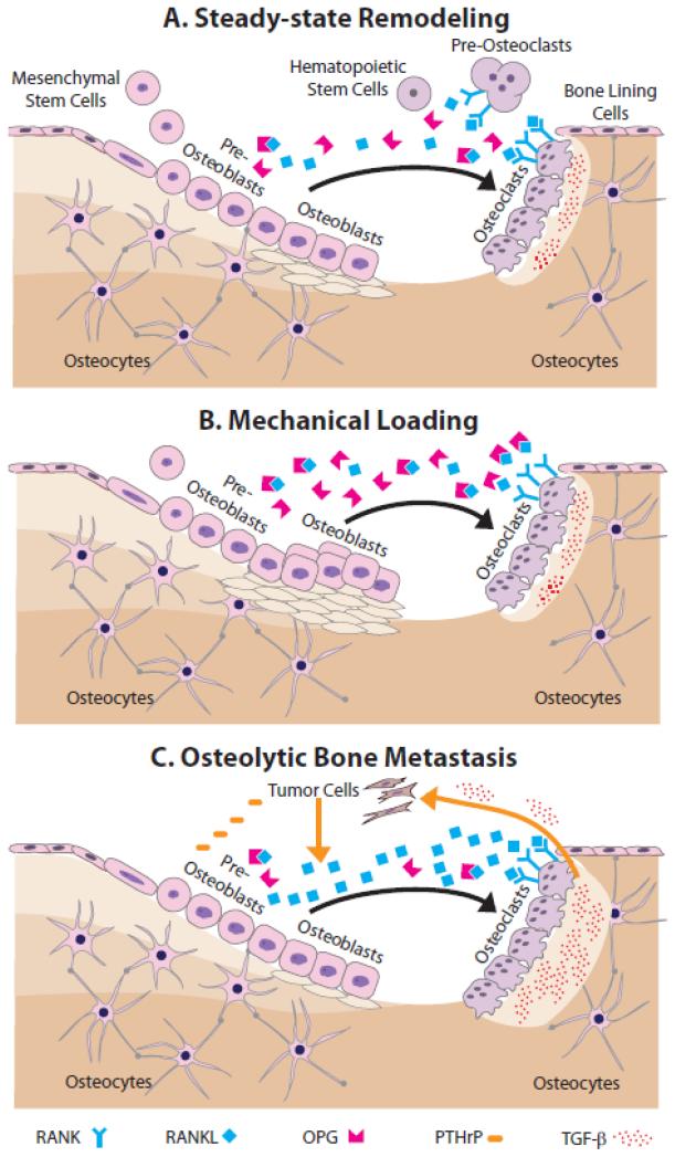
Bone metastasis represents the leading cause of breast cancer related-deaths. However, the effect of skeleton-associated biomechanical signals on the initiation, progression, and therapy response of breast cancer bone metastasis is largely unknown. This review seeks to highlight possible functional connections between skeletal mechanical signals and breast cancer bone metastasis and their contribution to clinical outcome. It provides an introduction to the physical and biological signals underlying bone functional adaptation and discusses the modulatory roles of mechanical loading and breast cancer metastasis in this process. Following a definition of biophysical design criteria, in vitro and in vivo approaches from the fields of bone biomechanics and tissue engineering that may be suitable to investigate breast cancer bone metastasis as a function of varied mechano-signaling will be reviewed. Finally, an outlook of future opportunities and challenges associated with this newly emerging field will be provided.

Keywords: Biomechanics; Bone metastasis; Breast cancer; Mechanical loading; Tissue engineering.


Figures

Figure 1
Figure 1. Schematic representation of the ‘mechanostat’ [16]
Steady-state remodeling occurs continuously within a target, non-zero strain range. Increased mechanical stimulus (e.g. exercise, reduced bone mass) increases the local strain environment and promotes osteogenesis, which then brings the local strain stimulus down to steady-state. Conversely, reduced mechanical stimulus (e.g. bed rest, microgravity) decreases the local strain environment and results in osteolysis, which then brings the local strain stimulus up to steady-state. Systemic effects, such as disease state, will alter the efficacy of the feedback loop to optimize mechanical integrity of the skeleton. Adapted from Lanyon BoneKey 2009.
Figure 2
Figure 2. Porosity within the skeleton and the cellular mechanical environment under mechanical loading
A) The skeleton contains porosity at each length scale. A whole bone is comprised of two tissue types: porous cancellous bone and compact cortical bone. The matrix of each of these two bone tissues types is comprised of a vascular porosity called the osteon (~20-40 μm). The osteon is surrounded by concentric layers of matrix containing embedded osteocytes. The cell body of the osteocyte resides in a lacuna (~15-20 μm) while its dendrites reside in canaliculae (~250-300 nm). B) Due to these levels of porosity, bone is fundamentally a fluid-filled porous matrix containing embedded cells. When the porous matrix is deformed, the fluid within is instantly pressurized, causing pressure gradients and subsequent fluid flow from high to low pressure. C) At the cellular level, cells experience substrate strain, hydrodynamic shear stress, pressure gradients, and mass transport-associated ionic gradients. These forces cause deformation of transmembrane proteins as well as changes to cytoskeletal tension, both of which alter cell signaling.
Figure 3
Figure 3. Hydrodynamic loading may increase cellular deformation relative to substrate strain
A) A computational model of individual cells undergoing substrate strain and hydrodynamic loading suggested that the effects of hydrodynamic loading dominate cellular deformation [23]. B) Macroscopic tensile strains of 1500 με applied to cortical bone specimens revealed that the local strain field is highly hetereogenous and can be several orders of magnitude greater than the applied strain, as shown by a microstructural strain field overlaid on digital micrographs. Shown here, the local perilacunar strain can reach peaks of over 15,000 με [25]. Reproduced with permission from The Journal of the Federation of American Societies for Experimental Biology and Elsevier.
Figure 4
Figure 4. Steady-state remodeling and the effects of mechanical loading and bone metastasis
A) During steady-state remodeling, mesenchymal stem cell-derived pre-osteoblasts recruit hematopoietic stem cells and induce their differentiation via RANK-RANKL signaling into large, multinucleated osteoclasts. RANK-RANKL signaling is modulated by osteoblastic secretion of OPG, which is a decoy receptor for RANKL. Mature osteoclasts remove matrix, and then apoptose. Next, active osteoblasts secrete new bone matrix, which subsequently mineralizes. After this, osteoblasts become quiescent bone lining cells, undergo apoptosis, or terminally differentiate into osteocytes. B) During mechanical loading, more mesenchymal precursors commit to the osteoblastic lineage, their differentiation and matrix deposition is enhanced, and apoptosis is inhibited. C) During breast cancer bone metastasis, tumor cells secrete a variety of osteolytic factors including PTHrP, which increases osteoblastic secretion of RANKL thus leading to greater osteoclastogenesis. Elevated resorption of the bone matrix, in turn, releases more pro-tumorigenic growth factors, most notably TGF-β, that further stimulates tumor growth.
Figure 5
Figure 5. In vivo tibial compression prevents osteolysis and secondary tumor formation
A) Schematic of compression of tumor-bearing tibiae. The box indicates region of interest for analysis in the proximal compartment. B) Representative microCT images of sham-injected (Control) and tumor-injected (Tumor) tibiae revealed that nonloaded tumor-bearing tibiae exhibited osteolytic degradation, whereas loading inhibited these adverse changes [143]. Reproduced with permission from John Wiley and Sons.
Figure 6
Figure 6. Schematics of commonly used bioreactors
A) Spinner flask: scaffolds are suspended in media that is circulated via magnetic stir bar. B) Rotating-wall vessel: the outer wall rotates relative to the stationary inner wall to circulate media. C) Direct perfusion: media is driven directly through the porous scaffold, which is housed in a cartridge that is fitted to the scaffold shape such that media cannot flow around it. D) Direct compression: scaffolds are directly compressed via a loading platen.
Figure 7
Figure 7. Exemplary 3-D studies demonstrating that interstitial flow affects cell behavior
A) Perfusion of marrow stromal cells cultured in polycaprolactone (PCL) scaffolds resulted in more uniform ECM and mineralization, as indicated by microCT analysis [212]. B) (Left) Interstitial fluid flow through a collagen gel, applied via a microfluidic device, increased overall percentage of human breast cancer cells that migrate. (Right) Additionally, this percentage increased with increasing flow rate [53]. C) Cyclic compression applied to human breast cancer cells cultured in mineral-containing poly(lactide-co-glycolide) (PLG) scaffolds reduced their expression of Runx2, a gene associated with initiating osteolysis [143]. D) Production of osteocalcin was greatest when both compression and perfusion was applied to bone marrow-derived mesenchymal stem cells cultured in spongiosa disks [222]. E) Inserting periods of rest during flow enhanced expression of osteopontin in MC3T3 pre-osteoblasts cultured in collagen-glycosaminoglycan constructs [227]. Reproduced with permission from Elsevier, John Wiley and Sons, Royal Society of Chemistry, and Mary Ann Liebert, Inc.
Figure 8
Figure 8. Computational simulations of loading-induced changes of cell behavior in 3-D porous scaffolds
A) Computational models of MC3T3 pre-osteoblasts within collagen-glycosaminoglycan (CG) scaffolds (Bottom) rendered from high-resolution scanning electron microscopy (Top) revealed that cell detachment is proportional to flow rate and inversely proportional to pore size [234]. B) Flow patterns through porous scaffolds depend on pore orientation. In polycaprolactone (PCL) scaffolds, pores are more regularly-oriented, resulting in more asymmetric flow and shear stresses. In contrast, in silk fibroin scaffolds, the pores are more irregularly-oriented, ‘conditioning’ the flow, and shear stresses are more uniform [238]. C) Perpendicular alignment of ECM fibers results in lower cellular strains (stress-shielding), while parallel alignment of the fibers increases shear stresses on cells, as demonstrated using computational fluid dynamics simulations [231]. D) When FE and CDF approaches were combined to model the effect of specific combinations of compression and perfusion on MSCs cultured within CG scaffolds, differing patterns of MSC differentiation into fibroblasts, chrondrocytes, or osteoblasts were predicted [233]. Reproduced with permission from John Wiley and Sons, Springer, and Elsevier.

Similar articles

Cited by

References

    1. www.cancer.gov.
    1. Kozlow W, Guise TA. Breast cancer metastasis to bone: mechanisms of osteolysis and implications for therapy. J Mammary Gland Biol Neoplasia. 2005;10:169–180. - PubMed
    1. Guise TA, Mohammad KS, Clines G, Stebbins EG, Wong DH, Higgins LS, Vessella R, Corey E, Padalecki S, Suva L, Chirgwin JM. Basic mechanisms responsible for osteolytic and osteoblastic bone metastases. Clin Cancer Res. 2006;12:6213s–6216s. - PubMed
    1. Weilbaecher KN, Guise TA, McCauley LK. Cancer to bone: a fatal attraction. Nat Rev Cancer. 2011;11:411–425. - PMC - PubMed
    1. Guise TA. The vicious cycle of bone metastases. J Musculoskelet Neuronal Interact. 2002;2:570–572. - PubMed

Publication types