. 2014 Aug 29:14:633.
doi: 10.1186/1471-2407-14-633.

MicroRNA-645, up-regulated in human adencarcinoma of gastric esophageal junction, inhibits apoptosis by targeting tumor suppressor IFIT2

Affiliations

MicroRNA-645, up-regulated in human adencarcinoma of gastric esophageal junction, inhibits apoptosis by targeting tumor suppressor IFIT2

Xiaoshan Feng et al. BMC Cancer. .

Abstract

Background: An increasing body of evidence indicates that miRNAs have a critical role in carcinogenesis and cancer progression; however, the role of miRNAs in the tumorigenesis of adencarcinoma of gastric esophageal junction (AGEJ) remains largely unclear.

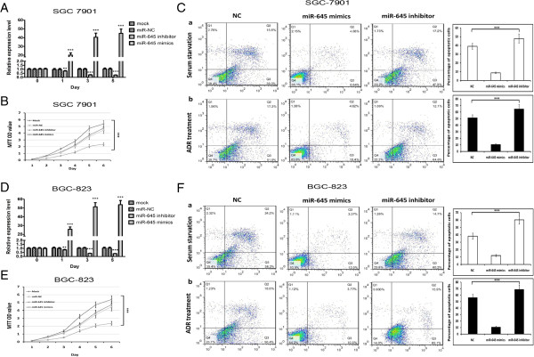
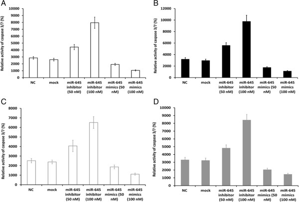
Methods: The SGC7901 and BGC-823 gastric cancer cell lines were used. The expressions of miR-645 and IFIT2 (Interferon-induced protein with tetratricopeptide repeats 2) were examined by qRT-PCR, The expressions of IFIT2 was examined by western blotting and immunohistochemistry assay. The cell apoptosis was determined by FACS. MiR-645 inhibitor, mimics and plasmid-IFIT2 transfections were performed to study the loss- and gain-function. Caspase-3/7 activity was examined by caspase-3/7 assay.

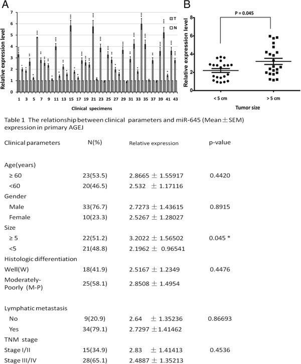
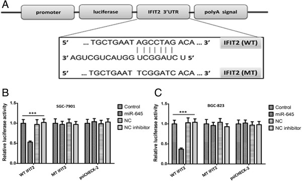
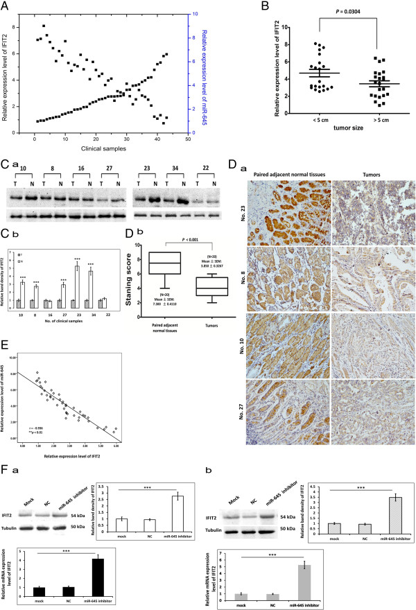
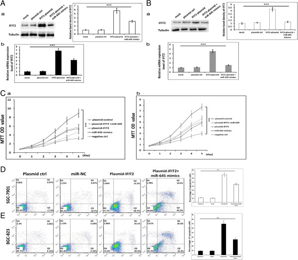
Results: In the present study, we have reported an increased expression of miR-645 in AGEJ clinical specimens compared with paired non-cancerous tissues. We also observed a significant miR-645 up-regulation in two gastric cancer (GC) cell lines, SGC7901 and BGC-823, which were used as cell models because there was no available AGEJ cell lines established to date. We found that inhibition of miR-645 could sensitize dramatically SGC7901 and BGC-823 cells to both serum starvation- and chemotherapeutic drug-induced apoptosis by up-regulating IFIT2, a mediator of apoptosis via a mitochondrial pathway, with a potential binding site for miR-645 in its mRNA's 3'UTR. Further investigation exhibited that IFIT2 expression decreases in SGC7901 and BGC-823 cells and AGEJ tissues. IFIT2 ectopic expression leads to promotion of cell apoptosis, indicating that IFIT2 may function as a suppressor in the development of AGEJ. Furthermore, inhibition of miR-645 induces up-regulation of IFIT2 and increased caspase-3/7 activity compared with control groups.

Conclusions: Our data suggest that miR-645 functions as an oncogene in human AGEJ by, at least partially through, targeting IFIT2.


Figures

Figure 1
Figure 1
Expressions of miR-645 are up-regulated in AGEJ clinical samples. A. expression of miR-645 in 43 human AGEJ clinical samples relative to the adjacent paired normal human gastric cardiac non-cancerous tissues, was measured by quantitative RT-PCR (The values indicate the mean ± SEM, n = 3, t-test,* p < 0.05, ** p < 0.01, *** p < 0.001). B. comparison of relative expression of miR-645 in human AGEJ clinical samples of different tumor size. (Two-tailed t-test,* p < 0.05).
Figure 2
Figure 2
Depletion of miR-645 promotes apoptosis of gastric cancer (GC) cells. A &D. The level of miR-645 was measured by quantitative PCR at designated time (One-way ANOVA analysis, the values indicate the mean ± SD, Figure 2 A, F =426.588, P < 0.001; Figure 2 D, F = 685.026, P < 0.001). B &E. GC cells transfected with miR-645 mimics and inhibitor subjected to MTT assay daily for 6 days (Two-way ANOVA analysis, Figure 2 B, F = 52.602, p < 0.001; Figure 2 E, F = 42.847, p < 0.001). C &F. GC cells cells transfected with miR-645 mimics and inhibitor were collected for FACS analysis after 72 h (The values indicate the mean ± SD, n = 3, One-way ANOVA analysis, Figure 2 C-a, F = 121.600, p <0.001; Figure 2 C-b, F = 250.400, p <0.001; Figure F-a, F = 194.815, p <0.001; Figure 2 F-b, F =412.741, p <0.001).
Figure 3
Figure 3
Validating the predicted binding sites between miR-645 and IFIT2. A. The schematic diagram shows the construct of Luc-IFIT2 3′UTR and Luc-IFIT2 3′Mut UTR. Both Luc-IFIT2 3′UTR and Luc-IFIT2 3′Mut UTR were cloned into a pmirGLO plasmid downstream of the firefly luciferase coding region between the PmeI and XbaI sites. B&C. SGC7901 cells (B) or BGC-823 cells (C) were co-transfected with the psiCHECK-2 constructs containing either IFIT2 3′UTR or IFIT2 3′Mut UTR and either the miR-645 inhibitor or the miR-645 mimics for 48 h. Values indicate the relative luciferase activity after normalization to Renilla luciferase activity (The values indicate the mean ± SD, n = 3, One-way ANOVA analysis, Figure 3 B, F = 283.244, Figure 3 C, F = 143.313. ***p < 0.001).
Figure 4
Figure 4
Expression of miR-645 and IFIT2 negatively correlate in AGEJ clinical samples and IFIT2 was down-regulated in AGEJ tissues compared with paired non-cancerous tissues. A. Expression of IFIT2 and miR-645 in AGEJ clinical samples were analyzed by quantitative PCR. B. comparison of relative expression of IFIT2 in human AGEJ clinical samples of different tumor size. (t-test, *p < 0.05). C. Expression of IFIT2 examined by western blotting (a, b: the values indicate the mean ± SD, normalized to tubulin, n = 3, t-test, ***p <0.001) D. a. Representative images shown are positive immunohistochemical staining of IFIT2 in human AGEJ specimens and matched adjacent normal tissues (magnification 200×). b. Staining scores of IFIT2 (t-test, ***p <0.001). E. Scatter plots showing the negative linear correlation between the mRNA expression of IFIT2 and that of miR-645 in 43 AGEJ clinical samples. F. IFIT2 and IFIT2 expression measured by western blotting in SGC7901(a) and BGC-823 cells (b). (The values indicate the mean ± SD, n = 3, One-way ANOVA analysis, ***p < 0.001).
Figure 5
Figure 5
IFIT2 mediates the function of miR-645 by promoting GC cell apoptosis. A &B. Expression of IFIT2 examined by western blotting (A: SGC7901; B, BGC-823. a, normalized to tubulin, the values indicate the mean ± SD, One-Way ANOVA analysis, for SGC7901, F = 189.307, for BGC-823, F = 85.374; b, The values indicate the mean ± SD, One-Way ANOVA analysis, for SGC7901, F = 85.374, for BGC-823, F = 219.921; ***p < 0.001). C. SGC7901 (a) and BGC-823 (b) cells subjected to MTT assay daily for 6 days (the values indicate the mean ± SD, Two-way ANOVA analysis, for SGC7901, F = 13.768, for BGC-823, F = 16.409, ***p <0.001). D &E. SGC7901 (D) and BGC-823 (E) cells were collected for FACS analysis after 72 h in the presence of ADR (0.05 μg/mL) (the values indicate the mean ± SD, n = 3, One-Way ANOVA analysis; for SGC7901, F = 361.749, for BGC-823, F = 229.952; ***p < 0.001).
Figure 6
Figure 6
Caspase-3/7 activity. Anti-apoptotic ability of SGC7901 cells and BGC-823 cells after exposure to ADR (0.2 μg/mL) or serum starvation was evaluated by caspases-3/7 activity. A &C, SGC7901 cells; B &D, BGC-823 cells. (the values indicate the mean ± SD, n = 3, One-Way ANOVA analysis; for A, F = 183.930, for B, F = 1093.797; for C, F = 1861.50, for D, F = 1483.604, ***p < 0.001).

References

    1. Vaughan TL, Davis S, Kristal A, Thomas DB. Obesity, alcohol, and tobacco as risk factors for cancers of the esophagus and gastric cardia: adenocarcinoma versus squamous cell carcinoma. Cancer Epidemiol Biomark Prev. 1995;4(2):85–92. - PubMed
    1. Chow W-H, Blot WJ, Vaughan TL, Risch HA, Gammon MD, Stanford JL, Dubrow R, Schoenberg JB, Mayne ST, Farrow DC. Body mass index and risk of adenocarcinomas of the esophagus and gastric cardia. J Natl Cancer Inst. 1998;90(2):150–155. doi: 10.1093/jnci/90.2.150. - DOI - PubMed
    1. Kubo A, Corley DA. Body mass index and adenocarcinomas of the esophagus or gastric cardia: a systematic review and meta-analysis. Cancer Epidemiol Biomark Prev. 2006;15(5):872–878. doi: 10.1158/1055-9965.EPI-05-0860. - DOI - PubMed
    1. Islami F, Sheikhattari P, Ren J, Kamangar F. Gastric atrophy and risk of oesophageal cancer and gastric cardia adenocarcinoma—a systematic review and meta-analysis. Ann Oncol. 2011;22(4):754–760. doi: 10.1093/annonc/mdq411. - DOI - PubMed
    1. Devesa SS, Blot WJ, Fraumeni JF. Changing patterns in the incidence of esophageal and gastric carcinoma in the United States. Cancer. 1998;83(10):2049–2053. doi: 10.1002/(SICI)1097-0142(19981115)83:10<2049::AID-CNCR1>3.0.CO;2-2. - DOI - PubMed
Pre-publication history
    1. The pre-publication history for this paper can be accessed here:http://www.biomedcentral.com/1471-2407/14/633/prepub

Publication types

MeSH terms