. 2014 Sep 18;55(6):868-879.
doi: 10.1016/j.molcel.2014.07.017. Epub 2014 Aug 28.

A MicroRNA precursor surveillance system in quality control of MicroRNA synthesis

Affiliations

A MicroRNA precursor surveillance system in quality control of MicroRNA synthesis

Xuhang Liu et al. Mol Cell. .

Abstract

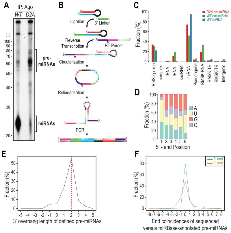
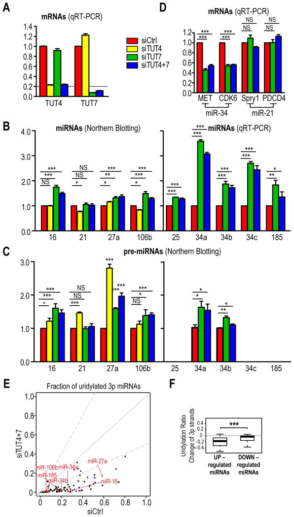
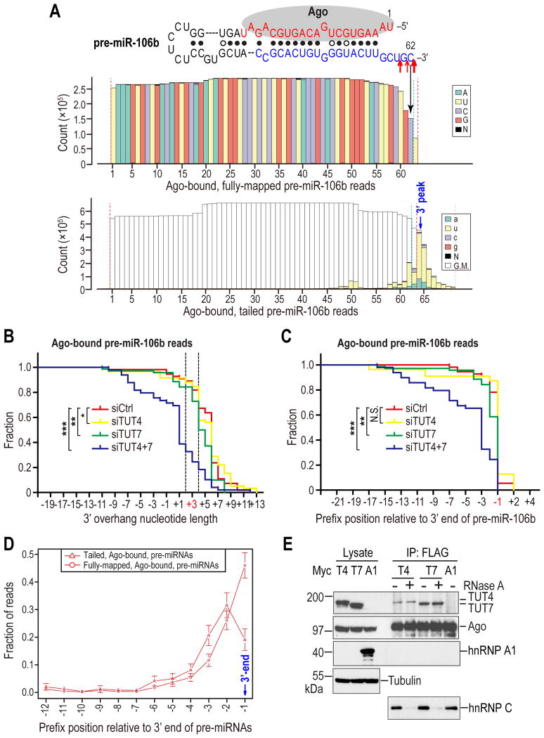
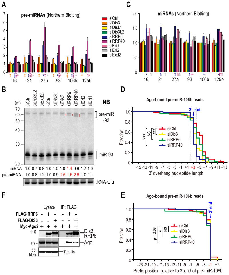
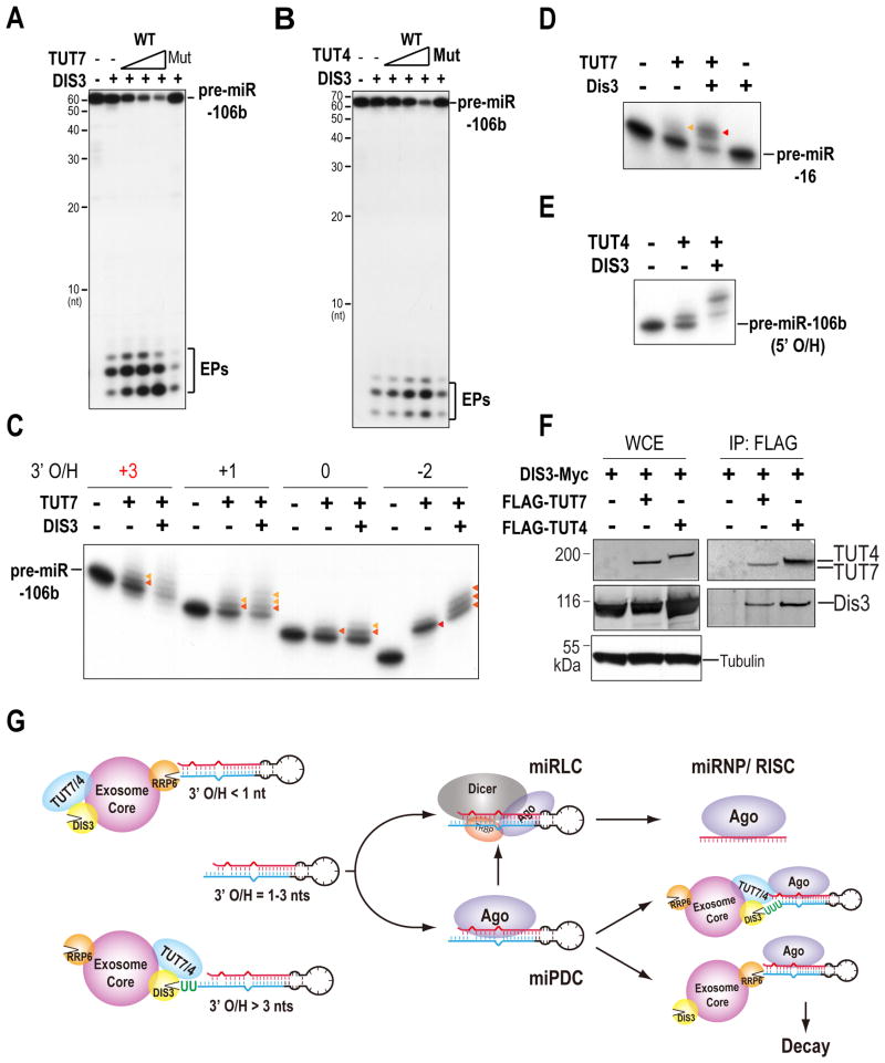
MicroRNAs (miRNAs) are essential for regulation of gene expression. Though numerous miRNAs have been identified by high-throughput sequencing, few precursor miRNAs (pre-miRNAs) are experimentally validated. Here we report a strategy for constructing high-throughput sequencing libraries enriched for full-length pre-miRNAs. We find widespread and extensive uridylation of Argonaute (Ago)-bound pre-miRNAs, which is primarily catalyzed by two terminal uridylyltransferases: TUT7 and TUT4. Uridylation by TUT7/4 not only polishes pre-miRNA 3' ends, but also facilitates their degradation by the exosome, preventing clogging of Ago with defective species. We show that the exosome exploits distinct substrate preferences of DIS3 and RRP6, its two catalytic subunits, to distinguish productive from defective pre-miRNAs. Furthermore, we identify a positive feedback loop formed by the exosome and TUT7/4 in triggering uridylation and degradation of Ago-bound pre-miRNAs. Our study reveals a pre-miRNA surveillance system that comprises TUT7, TUT4, and the exosome in quality control of miRNA synthesis.


Conflict of interest statement

Conflict of Interest: The authors declare that they have no conflict of interest.

Figures

Figure 1
Figure 1. Genome-wide profiling of Ago-bound, full-length precursor miRNAs; see also Figure S1
A. Autoradiography of 5′-end radiolabeled RNAs isolated from Ago immunoprecipitates (IPs) from Dicer-null MEFs reconstituted with wild-type (WT) or catalytically inactive (D2A) Dicer transgenes. B. Schematic of preparing pre-miRNA HITS libraries. Striped segments are complementary to solid segments with the same color. C. Genomic annotation of mapped reads. Complex: mouse complex/cluster regions. RMSK-RNA: RepeatMasker annotated RNA-repeats, such as rRNAs, tRNAs, snRNAs, etc. RMSK-TE: transposable elements, including DNA transposons, LINEs, and SINEs. RMSK-TR: tandem repeats, including micro satellite, simple and low-complexity repeats. D. Nucleotide composition for the first 6 positions of defined pre-miRNAs in the D2A library. E. 3′ overhang length distribution of defined pre-miRNAs in the D2A library. Positive and negative values indicate 3′ and 5′ overhangs respectively. The same rule applies to all other figures. F. Coincidence between annotated 5′ ends of 5p or 3′ ends of 3p miRNAs in miRBase and the defined 5′-or 3′-ends of pre-miRNAs sequenced from the D2A library. Positive and negative values indicate that the defined ends of pre-miRNAs are within and beyond the annotated ends of miRNAs in miRBase respectively.
Figure 2
Figure 2. Widespread and extensive post-transcriptional modifications in Ago-bound pre-miRNAs; see also Figure S2
A. Box plot of uridylation ratio (fraction of tailed reads) for each pre-miRNA or miRNA in each library. B. Nucleotide composition for every position of Ago-bound pre-miR-31 reads in WT pre-miRNA library. Red lightning bolt denotes Ago2-mediated cleavage site. Red dotted lines delineate the computationally defined 5′ and 3′ ends. Top panel: fully-mapped reads. Lower panel: tailed reads. Genome-matching nucleotides in tailed reads are not colored for clarity. Same depictions apply to all figures. C. Ago2-cleaved pre-miRNAs identified by 3′ cliffs in both WT and D2A pre-miRNA libraries. D, E. Nucleotide composition of RNA modifications surrounding the defined 3′ ends of 3p miRNAs (D) or pre-miRNAs (E) in WT libraries. F. Average tail length of reads with the indicated prefix (last genome-matching) positions in pre-miRNA libraries. Error bars represent 95% confidence intervals (CIs).
Figure 3
Figure 3. Uridylation of pre-miRNAs by TUT7 and TUT4 fine tunes select miRNA biogenesis; see also Figure S3
A. mRNA levels of TUT4 and TUT7 in indicated knockdown cells, measured by quantitative real-time (qRT)-PCR. The standard error of the mean is from four experiments. siCtrl, non-targeting siRNA. B, C. miRNA (B) and pre-miRNA (C) levels in indicated knockdown cells. The level of pre-miR-35 and pre-miR-185 are too low to be detected by NB. SEM is from at least three experiments. Statistical significance was calculated by one-tail t test. NS, non-significant; *, p<0.05; **, p<0.01; ***, p<0.001. Same depiction applies to all figures. D. miR-34 or miR-21 target levels in indicated knockdown cells, measured by qRT-PCR. SEM is from three experiments. Statistical significance was calculated by t test. E. Uridylation ratios of a subset of pre-miRNAs, as inferred from that of their 3p miRNAs, are dramatically diminished upon depletion of TUT4 and TUT7. Red dots indicate 3p strands of pre-miRNAs whose expression is verified by qRT-PCR or NB. Blue dots denote 3p strands of pre-let-7 family members. F. Box Plots for uridylation ratio differences of 3p strands between control and TUT4&7-depleted cells for differentially expressed (⩾ 2 fold) miRNAs. Only guide strands (more abundant species within miRNA duplexes) were used for comparison of expression levels of miRNAs. Statistical significance was calculated by Welch's t-test.
Figure 4
Figure 4. Uridylation of Ago-bound pre-miRNAs by TUT7 and TUT4 facilitates their degradation; see also Figure S4
A. Nucleotide composition for every position of Ago-bound pre-miR-106b reads in WT pre-miRNA library. Red arrows indicate 3′ cleavage sites identified from in-vitro Drosha processed pri-miR-106b products. Size of arrows reflects the clone frequency such that large, medium, and small arrows represent 10/23, 7/23, and 5/23 respectively. Red dotted lines delineate the computationally defined 5′ and 3′ ends. Blue dotted lines define the manually corrected 3′ end. B. Reverse cumulative plot of 3′ overhang lengths of pre-miR-106b reads (fully-mapped and tailed), sequenced from Ago IPs from indicated knockdown cells. The native 3′ overhang length of pre-miR-106b is 3 nts. Reads whose ends fall within the two dotted lines are optimal Dicer substrates as defined by in-vitro Dicer processing assay in Figure S4B. Statistical significance was calculated by Anderson-Darling (A-D) test. C. Reverse cumulative plot of prefix positions of pre-miR-106b reads relative to its native 3′ end (position -1), sequenced from Ago IPs from indicated knockdown cells. Statistical significance was calculated by A-D test. D. Average fraction of fully mapped or tailed reads for Ago-bound pre-miRNAs with prefix positions upstream of the defined 3′ end of pre-miRNAs. Error bars represent 95% CIs. E. Association of TUT4 and TUT7 with Ago2. Western blots of lysates and FLAG IPs from 293T cells co-expressing FLAG-Ago2 with Myc-TUT4, Myc-TUT7 or Myc-hnRNPA1. HnRNP C, which was shown to associate with Ago2 in an RNA-dependent manner, served as a positive control for RNase A digestion. Tubulin: loading control.
Figure 5
Figure 5. Quality control and turnover of pre-miRNAs by the exosome; see also Figure S5
A - C. Pre-miRNA (A) or miRNA (C) levels in indicated knockdown cells. SEM is from three independent experiments. Statistical significance was calculated by unpaired two-tail t test. Representative NB of total RNAs from indicated knockdown cells (B). Red lines denote defective (truncated) pre-miR-93. Numbers indicate fold changes of pre-miRNAs and miRNAs relative to the control knockdown. tRNA-Glu: loading control. D. Reverse cumulative plot of 3′ overhang lengths of pre-miR-106b reads (fully-mapped and tailed), sequenced from Ago IPs from indicated knockdown cells. Statistical significance was calculated by A-D test. E. Reverse cumulative plot of prefix positions of pre-miR-106b reads relative to its native 3′ end (position -1), sequenced from Ago IPs from indicated knockdown cells. Statistical significance was calculated by A-D test. F. Association of RRP6 and DIS3 with Ago2. Western blots of lysates and FLAG IPs from 293T cells co-expressing Myc-Ago2 with FLAG-RRP6 or DIS3-FLAG. Tubulin: loading control.
Figure 6
Figure 6. Distinct preferences of DIS3 and RRP6 towards pre-miRNAs; see also Figure S6
A, B. Decay assay using RRP6 and 5′ radiolabeled pre-miR-106b with progressively shortened (A) or uridylated (B) 3′ overhangs. 3′ O/H, 3′ overhang. The native 3′ overhang is marked in red. Same depiction applies to all figures. Prolonged exposure was used for comparison of the end products in panel B. C, D. Decay assay of pre-miR-106b with progressively shortened (C) or uridylated (D) 3′ overhangs using DIS3. E. Decay assay of Ago2-cleaved pre-miR-31 (ac-pre-miR-31) with or without an oligo(U) tail using DIS3. F. Decay assay of pre-miR-106b with 3′ overhangs of different lengths using a mixture of DIS3 and RRP6 (D3+R6).
Figure 7
Figure 7. A positive feedback loop constituted by TUT7, TUT4 and the exosome in uridylation and degradation of pre-miRNAs; see also Figure S7
A, B. Decay assay of pre-miR-106b using mixtures of DIS3 with wild-type (WT) or catalytically inactive (Wasmuth and Lima) TUT7 (A) and TUT4 (B). C. Uridylation assay using TUT7 and DIS3 with pre-miR-106b harboring 3′ overhangs of different lengths. D. Uridylation assay using TUT7 and DIS3 with pre-miR-16. E. Uridylation assay using TUT4 and DIS3 with pre-miR-106b with a 2-nt 5′ overhang (5′ O/H). F. Interaction of TUT4 and TUT7 with DIS3. Western blots of lysates and FLAG IPs from 293T cells co-expressing DIS3-Myc with FLAG-TUT4 or FLAG-TUT7. G. Model for a pre-miRNA surveillance network in quality control of mammalian miRNA synthesis

References

    1. Ameres SL, Horwich MD, Hung JH, Xu J, Ghildiyal M, Weng Z, Zamore PD. Target RNA-directed trimming and tailing of small silencing RNAs. Science. 2010;328:1534–1539. - PMC - PubMed
    1. Berezikov E, Robine N, Samsonova A, Westholm JO, Naqvi A, Hung JH, Okamura K, Dai Q, Bortolamiol-Becet D, Martin R, et al. Deep annotation of Drosophila melanogaster microRNAs yields insights into their processing, modification, and emergence. Genome Res. 2011;21:203–215. - PMC - PubMed
    1. Briggs MW, Burkard KT, Butler JS. Rrp6p, the yeast homologue of the human PM-Scl 100-kDa autoantigen, is essential for efficient 5.8 S rRNA 3′ end formation. J Biol Chem. 1998;273:13255–13263. - PubMed
    1. Buhler M, Spies N, Bartel DP, Moazed D. TRAMP-mediated RNA surveillance prevents spurious entry of RNAs into the Schizosaccharomyces pombe siRNA pathway. Nat Struct Mol Biol. 2008;15:1015–1023. - PMC - PubMed
    1. Burroughs AM, Kawano M, Ando Y, Daub CO, Hayashizaki Y. pre-miRNA profiles obtained through application of locked nucleic acids and deep sequencing reveals complex 5′/3′ arm variation including concomitant cleavage and polyuridylation patterns. Nucleic Acids Res. 2012;40:1424–1437. - PMC - PubMed

Publication types

MeSH terms

LinkOut - more resources