. 2014 Nov-Dec;21(6):543-54.
doi: 10.1111/xen.12124. Epub 2014 Sep 1.

Cloning and expression of porcine β1,4 N-acetylgalactosaminyl transferase encoding a new xenoreactive antigen

Affiliations
Free PMC article

Cloning and expression of porcine β1,4 N-acetylgalactosaminyl transferase encoding a new xenoreactive antigen

Guerard W Byrne et al. Xenotransplantation. 2014 Nov-Dec.
Free PMC article

Abstract

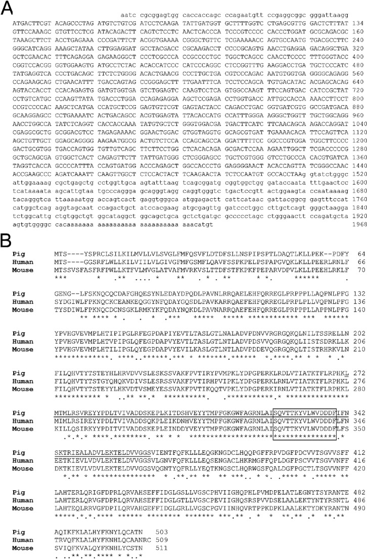
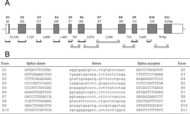
Background: Xenograft rejection of pigs organs with an engineered mutation in the GGTA-1 gene (GTKO) remains a predominantly antibody mediated process which is directed to a variety of non-Gal protein and carbohydrate antigens. We previously used an expression library screening strategy to identify six porcine endothelial cell cDNAs which encode pig antigens that bind to IgG induced after pig-to-primate cardiac xenotransplantation. One of these gene products was a glycosyltransferase with homology to the bovine β1,4 N-acetylgalactosaminyltransferase (B4GALNT2). We now characterize the porcine B4GALNT2 gene sequence, genomic organization, expression, and functional significance.

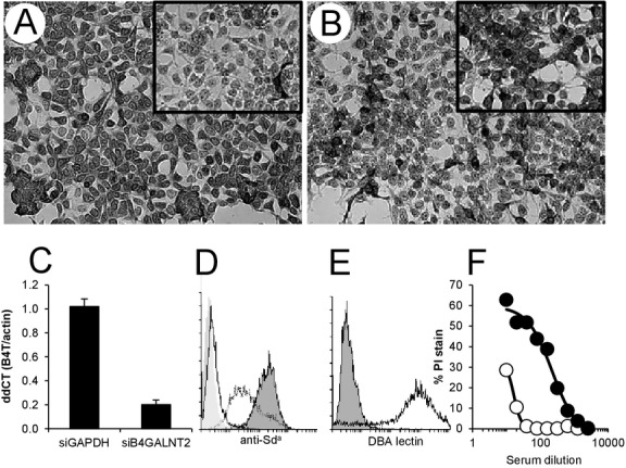
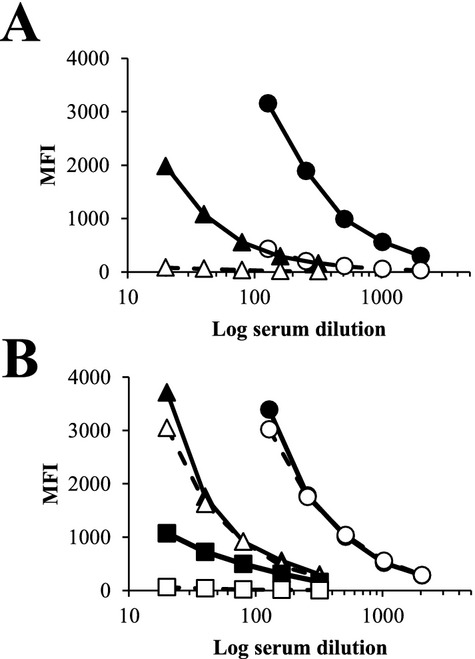
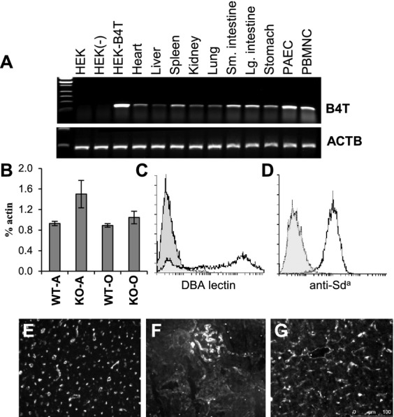
Methods: The porcine B4GALNT2 cDNA was recovered from the original library isolate, subcloned, sequenced, and used to identify a bacterial artificial chromosome (BAC) containing the entire B4GALNT2 locus from the Children's Hospital Oakland Research Institute BACPAC Resource Centre (#AC173453). PCR primers were designed to map the intron/exon genomic organization in the BAC clone. A stable human embryonic kidney (HEK) cell line expressing porcine B4GALNT2 (HEK-B4T) was produced. Expression of porcine B4GALNT2 in HEK-B4T cells was characterized by immune staining and siRNA transfection. The effects of B4GALNT2 expression in HEK-B4T cells was measured by flow cytometry and complement mediated lysis. Antibody binding to HEK and HEK-B4T cells was used to detect an induced antibody response to the B4GALNT2 produced glycan and the results were compared to GTKO PAEC specific non-Gal antibody induction. Expression of porcine B4GALNT2 in pig cells and tissues was measured by qualitative and quantitative real time reverse transcriptase PCR and by Dolichos biflorus agglutinin (DBA) tissue staining.

Results: The porcine B4GALNT2 gene shares a conserved genomic organization and encodes an open reading frame with 76 and 70% amino acid identity to the human and murine B4GALNT2 genes, respectively. The B4GALNT2 gene is expressed in porcine endothelial cells and shows a broadly distributed expression pattern. Expression of porcine B4GALNT2 in human HEK cells (HEK-B4T) results in increased binding of antibody to the B4GALNT2 enzyme, and increased reactivity with anti-Sd(a) and DBA. HEK-B4T cells show increased sensitivity to complement mediated lysis when challenged with serum from primates after pig to primate cardiac xenotransplantation. In GTKO and GTKO:CD55 cardiac xenotransplantation recipients there is a significant correlation between the induction of a non-Gal antibody, measured using GTKO PAECs, and the induction of antibodies which preferentially bind to HEK-B4T cells.

Conclusion: The functional isolation of the porcine B4GALNT2 gene from a PAEC expression library, the pattern of B4GALNT2 gene expression and its sensitization of HEK-B4T cells to antibody binding and complement mediated lysis indicates that the enzymatic activity of porcine B4GALNT2 produces a new immunogenic non-Gal glycan which contributes in part to the non-Gal immune response detected after pig-to-baboon cardiac xenotransplantation.

Keywords: B4GALNT2; antigen; cardiac xenotransplantation; immune response; porcine.


Figures

Figure 1
Figure 1
Sequence and homology comparison of porcine B4GALNT2. (A) Sequence of the porcine B4GALNT2 cDNA (GenBank: KF501048). Numbering begins with the start codon. Uppercase letters are the protein coding sequences, lower case letter are untranslated sequences. (B) Amino acid homology comparison between porcine, human and murine B4GALNT2 protein sequences. An asterisk indicates a shared amino acid identity, a line indicates a conserved amino acid substitution and a space indicates a non-conserved amino acid residue. The underlined region represents a conserved GT-A fold and the boxed region highlights the conserved divalent cation binding domain .
Figure 2
Figure 2
Schematic representation of the intron-exon organization (A) and splice junction sequences (B) of the porcine B4GALNT2 gene. (A) Shaded boxes represent exon coding sequences numbered E1–E11. Open boxes correspond to untranslated sequences present in the cDNA. The size of each exon is indicated in base pairs above each box and the intron distances are indicated below between each exon. The primer pairs (1–26, see Table1) and amplified products used to analyze the genomic structure from a BAC clone are illustrated below the genomic structure as solid bars for products covering exon/intron boundaries and open bars for products spanning entire introns. The primer numbering corresponds to the primers listed in Table1. (B) The proximal splice donor and distal splice acceptor nucleotide sequences (upper case) for each exon are shown. Adjacent intervening intron sequences (lower case) are shown. Exon numbering corresponds to (A).
Figure 3
Figure 3
Expression of B4GALNT2 protein and synthesis of the Sda antigen on HEK-B4T cells. (A) HEK-B4T cells show increased anti-human B4GALNT2 staining compared to HEK cells (insert). (B) Staining is dependent on porcine B4GALNT2 mRNA expression as HEK-B4T cells transfected with porcine B4GALNT2 siRNA show decreased staining compared to cell transfected with control GAPDH siRNA (insert in B). (C) Quantitative real-time RT-PCR analysis of B4GALNT2 RNA in HEK-B4T cells after transfection with control (siGAPDH) or porcine specific (siB4GALNT2) siRNA. Gene expression was normalized to pig beta-actin (dCT) and expressed as a ratio to B4GALNT2 expression in HEK-B4T cells without siRNA transfection (ddCT). Error bars are standard error of the mean. (D) Flow cytometry detection of anti-Sda antibody (KM694) binding to HEK (dotted line) and HEK-B4T cells (black line filled). Background is secondary antibody only binding to HEK cells (filled no line). KM694 binding to HUVECs (solid line no fill) is negative. (E) FITC conjugated Dolichos biflorus agglutinin (DBA) staining of HEK-B4T cells lines (black line) and control HEK cells (filled). (F) Complement-dependent cytotoxicity of HEK (open) and HEK-B4T (filled) cells challenged with primate serum after pig-to-primate xenotransplantation.
Figure 4
Figure 4
Serum absorption of pig-to-baboon induced antibody to HEK-B4T (B4T) cells. Sensitized baboon serum was absorbed by incubating baboon serum (1 : 10 dilution) with 1 × 107 GTKO PAECs (A) or HUVECs (B) for 1 h at 4 °C. Absorption was performed 3 times for each cell type. IgG binding was analyzed by flow cytometry in dilutions from 1 : 20 to 20 : 48. (A) Closed symbols IgG binding from whole serum to GTKO PAECs (circle) and HEK-B4T cells (triangle). Open symbols are IgG binding after absorption with GTKO PAECs. Over the dilution range, absorption reduced IgG binding by an average of 89% for GTKO PAECs and 94% for HEK-B4T cells. (B) Closed symbols IgG binding from whole serum to GTKO PAECs (circle), HEK-B4T cells (triangle) and HUVEC (square). Open symbols are IgG binding after absorption with HUVECs. Absorption reduced HUVEC reactivity on average by 96%, had a minimal effect on IgG binding to GTKO PAECs (0.05%) and only a minor reduction of IgG binding to HEK-B4T (12%) cells.
Figure 5
Figure 5
Porcine B4GALNT2 gene expression. (A) Qualitative RT-PCR analysis of porcine B4GALNT2 RNA expression in cell lines and pig tissues. The top panel represents specific expression of B4GALNT2 (B4T) and the bottom panel shows expression of porcine actin (ACTB). Samples; Human embryonic kidney cells (HEK), HEK cells transformed with an unrelated porcine cDNA (HEK-), HEK cells expressing porcine B4GALNT2 (HEK-B4T), porcine tissue samples (heart, liver, spleen, kidney, lung, stomach, and small [Sm] and large [Lg] intestine), porcine aortic endothelial cells (PAEC) and porcine peripheral blood mononuclear cells (PBMC). (B) Quantitative real time RT-PCR analysis of porcine B4GALNT2 gene expression in Gal-positive wild type (WT) and Gal-negative (KO) endothelial cells. On the x-axis the A- and O-type blood group is indicated for each Gal genotype. B4GALNT2 expression is plotted as the average percentage of βactin expression. Error bars are the standard deviation. (C) Positive FITC-DBA staining (bold line) of O-type PAECs. Background (filled) staining with FITC conjugated Solanum tuberosum agglutinin which does not bind to PAECs. (D) Negative staining of O-type PAECs with KM694 (dotted line). Background (filled) is FITC conjugated anti-mouse IgM only staining of PAECs. Positive stain (solid line) is HEK-B4T stained with KM694. (E–G) FITC conjugated DBA lectin staining of O-type GTKO pig heart (E), kidney (F) and liver (G).

References

    1. Galili U, Shohet SB, Kobrin E, et al. Man, apes, and old world monkeys differ from other mammals in the expression of alpha-Galactosyl epitopes on nucleated cells. J Biol Chem. 1988;263:17755–17762. - PubMed
    1. Byrne GW, McGregor CG. Cardiac xenotransplantation: progress and challenges. Curr Opin Organ Transplant. 2012;17:148–154. - PMC - PubMed
    1. McGregor CG, Carpentier A, Lila N, et al. Cardiac xenotransplantation technology provides materials for improved bioprosthetic heart valves. J Thorac Cardiovasc Surg. 2011;141:269–275. - PubMed
    1. Manji RA, Menkis AH, Ekser B, Cooper DK. Porcine bioprosthetic heart valves: the next generation. Am Heart J. 2012;164:177–185. - PubMed
    1. Phelps CJ, Koike C, Vaught TD, et al. Production of alpha 1,3-galactosyltransferase-deficient pigs. Science. 2003;299:411–414. - PMC - PubMed

Publication types

Substances