. 2014 Nov;23(11):1629-39.
doi: 10.1002/pro.2543. Epub 2014 Sep 25.

High-resolution crystal structures of the photoreceptor glyceraldehyde 3-phosphate dehydrogenase (GAPDH) with three and four-bound NAD molecules

Affiliations

High-resolution crystal structures of the photoreceptor glyceraldehyde 3-phosphate dehydrogenase (GAPDH) with three and four-bound NAD molecules

Bo Y Baker et al. Protein Sci. 2014 Nov.

Abstract

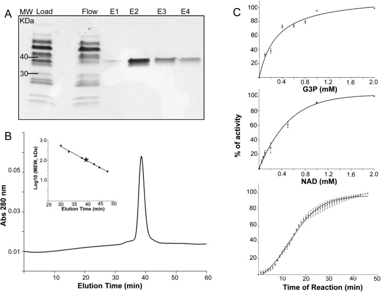
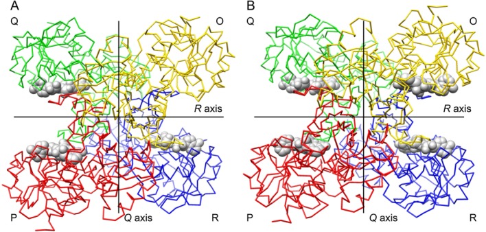
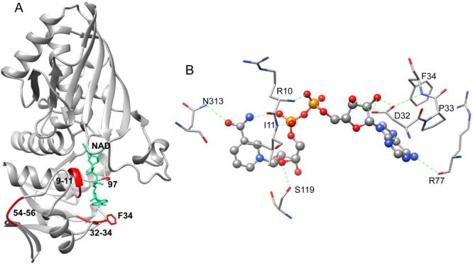
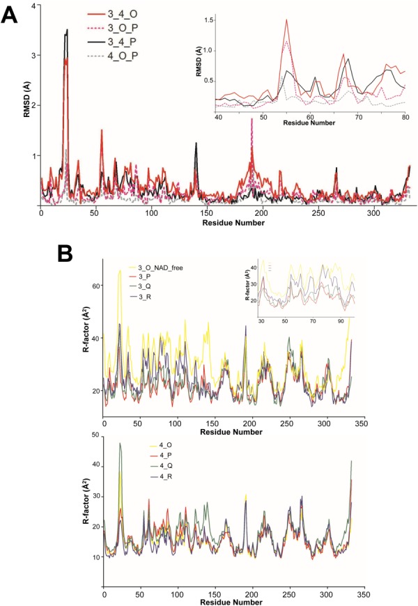
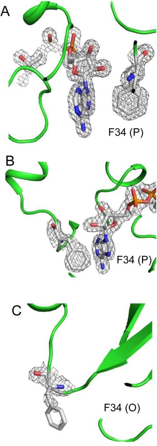
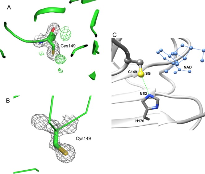
Glyceraldehyde-3-phosphate dehydrogenase (GAPDH) catalyzes the oxidative phosphorylation of d-glyceraldehyde 3-phosphate (G3P) into 1,3-diphosphoglycerate (BGP) in the presence of the NAD cofactor. GAPDH is an important drug target because of its central role in glycolysis, and nonglycolytic processes such as nuclear RNA transport, DNA replication/repair, membrane fusion and cellular apoptosis. Recent studies found that GAPDH participates in the development of diabetic retinopathy and its progression after the cessation of hyperglycemia. Here, we report two structures for native bovine photoreceptor GAPDH as a homotetramer with differing occupancy by NAD, bGAPDH(NAD)4 , and bGAPDH(NAD)3 . The bGAPDH(NAD)4 was solved at 1.52 Å, the highest resolution for GAPDH. Structural comparison of the bGAPDH(NAD)4 and bGAPDH(NAD)3 models revealed novel details of conformational changes induced by cofactor binding, including a loop region (residues 54-56). Structure analysis of bGAPDH confirmed the importance of Phe34 in NAD binding, and demonstrated that Phe34 was stabilized in the presence of NAD but displayed greater mobility in its absence. The oxidative state of the active site Cys149 residue is regulated by NAD binding, because this residue was found oxidized in the absence of dinucleotide. The distance between Cys149 and His176 decreased upon NAD binding and Cys149 remained in a reduced state when NAD was bound. These findings provide an important structural step for understanding the mechanism of GAPDH activity in vision and its pathological role in retinopathies.

Keywords: NAD; S-nitrosylation; crystal structure; diabetic retinopathy; glyceraldehyde 3-phosphate dehydrogenase; oxidization; photoreceptor; structural comparison.


Figures

Figure 1
Figure 1
Characterization of bovine GAPDH. A. SDS-PAGE analysis of bGAPDH purified from bovine ROS. B. Size exclusion profile of purified bGAPDH. The column (Superdex 200, 10/300 GL, GE Healthcare) was calibrated with protein standards (Insert). C. Enzyme activity. Purified bGAPDH (0.5 µg/100 µL) was incubated with 0.2 mM NAD and increasing concentrations of G3P (top panel), or with 1 mM G3P and increasing concentrations of NAD (middle panel). After 30 min of incubation, GAPDH activity was monitored by measuring NADH production at 340 nm. Kinetic measurements were then done over a 45 min time course in the presence of 1 mM NAD and 1 mM G3P (bottom panel).
Figure 2
Figure 2
Carbon trace representations of bovine GAPDH tetramer structures. A. Structural model of bGAPDH(NAD)3. The NAD-free subunit named as “O” is shown in yellow. B. Structural model of bGAPDH(NAD)4. This view is down the P axis. Lines show the location of the Q and R twofold molecular axes. NAD cofactors are displayed in spheres. Subunit nomenclature and coloring is as described in Ref. (26). An interactive view is available in the electronic version of the article.
Figure 3
Figure 3
NAD conformation and interactions in bGAPDH. A. Ribbon presentation of NAD binding to bGAPDH(NAD)4 (P subunit) in a typical Rossmann fold. The fluctuation regions, residues 9–11 (the Gly-rich loop), 32–34 (gate for NAD entry), 54–56 (a remote loop region), and 96 are shown in red ribbons. The view of NAD binding (shown in green) and Phe34 (shown in red) in the P subunit of bGAPDH(NAD)4 is also portrayed. B. Diagram of NAD–protein interactions in the P subunit of bGAPDH(NAD). Green dotted lines indicate hydrogen bonds formed between NAD and residues from the protein. An interactive view is available in the electronic version of the article.
Figure 4
Figure 4
Structural comparison of the NAD-free and NAD-bound subunits. (A). The RMSD of Cα atoms in paired residues from apo- and holo-subunits of bGAPDH. The subunit of O (NAD-free) from bGAPDH(NAD)3 and subunit O (NAD-bound) from bGAPDH(NAD)4 were superimposed (solid red line, labeled as 3_4_O). Subunits of O (NAD-free) and P (NAD-bound) from bGAPDH(NAD)3 were superimposed (dotted red line, labeled as 3_O_P). Subunits of P from bGAPDH(NAD)3 and bGAPDH(NAD)4 were superimposed as one control (solid black line, labeled as 3_4_P) and subunits of O and P from fully bGAPDH(NAD)4 were superimposed as another control (dotted gray line, labeled as 4_O_P). Insert shows the plotted region comprised residues 40–80. B. Comparison of B-factors of residues in the main chains of apo- and holo-subunits of bGAPDH. The averaged B-factor values of the residue from main chain atoms from the four subunits of bGAPDH(NAD)3 were plotted along with the residue number (upper panel, solid lines, labeled as 3_O (NAD_free), 3_P, 3_Q, and 3_R). Insert shows the plot region from residues 30 to 100. The averaged B-factor values of the residue from main chain atoms from the four subunits of bGAPDH(NAD)4 were plotted along with the residue number (lower panel, solid lines, labeled as 4_O, 4_P, 4_Q, and 4_R). Each subunit was presented as a solid line colored as in Figure 2.
Figure 5
Figure 5
2Fo–Fc maps of Phe34 in bGAPDH NAD-binding sites. The electron density was sufficient to cover the Phe34 residue and NAD molecule in the P subunit of both bGAPDH(NAD)4 (A) and bGAPDH(NAD)3 (B). But the electron density only weakly covered the main chain of Phe34 in the NAD-free structure, namely the O subunit of bGAPDH(NAD)3 (C). 2Fo–Fc maps of Phe34 and NAD were contoured at 1.0σ and presented as gray meshes. Figures were prepared with PyMol software.
Figure 6
Figure 6
The active site Cys149 in bGAPDH and its interaction with His176. A. Electron density of Cys149 in subunit R of bGAPDH(NAD)3. The 2Fo–Fc electron density map is presented as grey meshes contoured at 1.0σ. The green meshes represent a Fo–Fc electron density map contoured at 3.0σ. The presence of a positive density next to the SG atom of Cys149 strongly suggests an oxidative state for this residue. B. Electron density of Cys149 in subunit R of bGAPDH (NAD)4. None of the four subunits exhibited a positive density next to the SG atom of Cys149 in the Fo–Fc map. Figures were prepared with PyMol software. C. Interaction between Cys149 and His176 in subunits of bGAPDH. The distance between the SG group (shown in yellow) of Cys149 and the NE2 group (shown in blue) of His176 is indicated by a dotted green line. The adjacent NAD molecule is shown in blue ball-and-sticks.

References

    1. Seidler NW. Basic biology of GAPDH. Adv Exp Med Biol. 2013;985:1–36. - PubMed
    1. Sirover MA. New insights into an old protein: the functional diversity of mammalian glyceraldehyde-3-phosphate dehydrogenase. Biochim Biophys Acta. 1999;1432:159–184. - PubMed
    1. Sirover MA. On the functional diversity of glyceraldehyde-3-phosphate dehydrogenase: biochemical mechanisms and regulatory control. Biochim Biophys Acta. 2011;1810:741–751. - PubMed
    1. Padgett CM, Whorton AR. S-nitrosoglutathione reversibly inhibits GAPDH by S-nitrosylation. Am J Physiol. 1995;269:C739–C749. - PubMed
    1. Mohr S, Stamler JS, Brune B. Posttranslational modification of glyceraldehyde-3-phosphate dehydrogenase by S-nitrosylation and subsequent NADH attachment. J Biol Chem. 1996;271:4209–4214. - PubMed

Publication types